|
NARG1
N-alpha-acetyltransferase 15, NatA auxiliary subunit also known as gastric cancer antigen Ga19 (GA19), NMDA receptor-regulated protein 1 (NARG1), and Tbdn100 is a protein that in humans is encoded by the ''NAA15'' gene. NARG1 is the auxiliary subunit of the NatA ( Nα-acetyltransferase A) complex. This NatA complex can associate with the ribosome and catalyzes the transfer of an acetyl group to the Nα-terminal amino group of proteins as they emerge from the exit tunnel. Gene and transcripts Human NAA15 is located on chromosome 4q31.1 and contains 23 exons. Initially, 2 mRNA species were identified, of size 4.6 and 5.8 kb, both harboring the same open reading frame encoding a putative protein of 866 amino acids (~105 kDa) protein that can be detected in most human adult tissues. According to RefSeq/NCBI, only one human transcript variant exists, although 2 more isoforms are predicted. In addition to full length Naa15, an N-terminally truncated variant of Naa15 (named tubedown ... [...More Info...] [...Related Items...] OR: [Wikipedia] [Google] [Baidu] |
Tetratricopeptide
The tetratricopeptide repeat (TPR) is a structural motif. It consists of a Degeneracy (biology), degenerate 34 amino acid protein tandem repeats, tandem repeat identified in a wide variety of proteins. It is found in tandem arrays of 3–16 motifs, which form scaffolds to mediate protein–protein interactions and often the assembly of multiprotein complexes. These alpha-helix pair repeats usually protein folding, fold together to produce a single, linear solenoid protein domain, solenoid domain called a TPR domain. Proteins with such domains include the anaphase-promoting complex (APC) subunits CDC16, cdc16, CDC23, cdc23 and CDC27, cdc27, the NADPH oxidase subunit neutrophil cytosolic factor 2, p67-phox, hsp90-binding immunophilins, transcription factors, the protein kinase R (PKR), the major receptor for peroxisomal matrix protein import PEX5, protein arginine methyltransferase 9 (PRMT9), and mitochondrial import proteins. Structure The structure of the PPP5C, PP5 protein w ... [...More Info...] [...Related Items...] OR: [Wikipedia] [Google] [Baidu] |
Protein
Proteins are large biomolecules and macromolecules that comprise one or more long chains of amino acid residues. Proteins perform a vast array of functions within organisms, including catalysing metabolic reactions, DNA replication, responding to stimuli, providing structure to cells and organisms, and transporting molecules from one location to another. Proteins differ from one another primarily in their sequence of amino acids, which is dictated by the nucleotide sequence of their genes, and which usually results in protein folding into a specific 3D structure that determines its activity. A linear chain of amino acid residues is called a polypeptide. A protein contains at least one long polypeptide. Short polypeptides, containing less than 20–30 residues, are rarely considered to be proteins and are commonly called peptides. The individual amino acid residues are bonded together by peptide bonds and adjacent amino acid residues. The sequence of amino acid residue ... [...More Info...] [...Related Items...] OR: [Wikipedia] [Google] [Baidu] |
Hsp70
The 70 kilodalton heat shock proteins (Hsp70s or DnaK) are a family of conserved ubiquitously expressed heat shock proteins. Proteins with similar structure exist in virtually all living organisms. Intracellularly localized Hsp70s are an important part of the cell's machinery for protein folding, performing chaperoning functions, and helping to protect cells from the adverse effects of physiological stresses. Additionally, membrane-bound Hsp70s have been identified as a potential target for cancer therapies and their extracellularly localized counterparts have been identified as having both membrane-bound and membrane-free structures. Discovery Members of the Hsp70 family are very strongly upregulated by heat stress and toxic chemicals, particularly heavy metals such as arsenic, cadmium, copper, mercury, etc. Heat shock was originally discovered by Ferruccio Ritossa in the 1960s when a lab worker accidentally boosted the incubation temperature of Drosophila (fruit flies). When ... [...More Info...] [...Related Items...] OR: [Wikipedia] [Google] [Baidu] |
Asplenia
Asplenia refers to the absence of normal spleen function and is associated with some serious infection risks. Hyposplenism is used to describe reduced ('hypo-') splenic functioning, but not as severely affected as with asplenism. ''Functional'' asplenia occurs when splenic tissue is present but does not work well (e.g. sickle-cell disease, polysplenia) -such patients are managed as if asplenic-, while in ''anatomic'' asplenia, the spleen itself is absent. Causes Congenital * Congenital asplenia is rare. There are two distinct types of genetic disorders: heterotaxy syndromeOnline Mendelian Inheritance in Man. OMIM entry 208530: Right atrial isomerism; RAI. Johns Hopkins University/ref> and isolated congenital asplenia.Online Mendelian Inheritance in Man. Johns Hopkins UniversityOMIM entry 271400: Asplenia, isolated congenital; ICAS./ref> * polysplenia Acquired Acquired asplenia occurs for several reasons: * Following splenectomy due to splenic rupture from trauma or because of ... [...More Info...] [...Related Items...] OR: [Wikipedia] [Google] [Baidu] |
Hydronephrosis
Hydronephrosis describes hydrostatic dilation of the renal pelvis and calyces as a result of obstruction to urine flow downstream. Alternatively, hydroureter describes the dilation of the ureter, and hydronephroureter describes the dilation of the entire upper urinary tract (both the renal pelvicalyceal system and the ureter). Signs and symptoms The signs and symptoms of hydronephrosis depend upon whether the obstruction is acute or chronic, partial or complete, unilateral or bilateral. Hydronephrosis that occurs acutely with sudden onset (as caused by a kidney stone) can cause intense pain in the flank area (between the hips and ribs) known as a renal colic. Historically, this type of pain has been described as "Dietl's crisis". Conversely, hydronephrosis that develops gradually over time will generally cause either a dull discomfort or no pain. Nausea and vomiting may also occur. An obstruction that occurs at the urethra or bladder outlet can cause pain and pressure result ... [...More Info...] [...Related Items...] OR: [Wikipedia] [Google] [Baidu] |
Anomalous Pulmonary Venous Connection
Anomalous pulmonary venous connection (or anomalous pulmonary venous drainage or anomalous pulmonary venous return) is a congenital defect of the pulmonary veins. Total anomalous pulmonary venous connection ''Total anomalous pulmonary venous connection'', also known as total ''anomalous pulmonary venous return'', is a rare cyanotic congenital heart defect in which all four pulmonary veins are malpositioned and make anomalous connections to the systemic venous circulation. (Normally, pulmonary veins return oxygenated blood from the lungs to the left atrium where it can then be pumped to the rest of the body). A patent foramen ovale, patent ductus arteriosus or an atrial septal defect ''must'' be present, or else the condition is fatal due to a lack of systemic blood flow. In some cases, it can be detected prenatally. There are four variants: Supracardiac (50%): blood drains to one of the innominate veins (brachiocephalic veins) or the superior vena cava; Cardiac (20%), where ... [...More Info...] [...Related Items...] OR: [Wikipedia] [Google] [Baidu] |
Dextrocardia
Dextrocardia (from Latin ''dextro'', meaning "right hand side," and Greek ''kardia'', meaning "heart") is a rare congenital condition in which the apex of the heart is located on the right side of the body, rather than the more typical placement towards the left. There are two main types of dextrocardia: dextrocardia of embryonic arrest (also known as isolated dextrocardia) and dextrocardia ''situs inversus''. Dextrocardia ''situs inversus'' is further divided. Classification Dextrocardia of embryonic arrest In this form of dextrocardia, the heart is simply placed further right in the thorax than is normal. It is commonly associated with severe defects of the heart and related abnormalities including pulmonary hypoplasia. Dextrocardia situs solitus Dextrocardia refers to a heart positioned in the right side of the chest. Situs solitus describes viscera that are in the normal position, with the stomach on the left side. Dextrocardia situs inversus Dextrocardia situs inversus ... [...More Info...] [...Related Items...] OR: [Wikipedia] [Google] [Baidu] |
Heterotaxy
Situs ambiguus is a rare congenital defect in which the major visceral organs are distributed abnormally within the chest and abdomen. Clinically heterotaxy spectrum generally refers to any defect of Left-right asymmetry and arrangement of the visceral organs; however, classical heterotaxy requires multiple organs to be affected. This does not include the congenital defect situs inversus, which results when arrangement of all the organs in the abdomen and chest are mirrored, so the positions are opposite the normal placement. Situs inversus is the mirror image of situs solitus, which is normal asymmetric distribution of the abdominothoracic visceral organs. Situs ambiguus can also be subdivided into left-isomerism and right isomerism based on the defects observed in the spleen, lungs and atria of the heart. Individuals with situs inversus or situs solitus do not experience fatal dysfunction of their organ systems, as general anatomy and morphology of the abdominothoracic organ-vess ... [...More Info...] [...Related Items...] OR: [Wikipedia] [Google] [Baidu] |
Congenital Heart Disease
A congenital heart defect (CHD), also known as a congenital heart anomaly and congenital heart disease, is a defect in the structure of the heart or great vessels that is present at birth. A congenital heart defect is classed as a cardiovascular disease. Signs and symptoms depend on the specific type of defect. Symptoms can vary from none to life-threatening. When present, symptoms may include rapid breathing, bluish skin (cyanosis), poor weight gain, and feeling tired. CHD does not cause chest pain. Most congenital heart defects are not associated with other diseases. A complication of CHD is heart failure. The cause of a congenital heart defect is often unknown. Risk factors include certain infections during pregnancy such as rubella, use of certain medications or drugs such as alcohol or tobacco, parents being closely related, or poor nutritional status or obesity in the mother. Having a parent with a congenital heart defect is also a risk factor. A number of genetic condition ... [...More Info...] [...Related Items...] OR: [Wikipedia] [Google] [Baidu] |
Hsp40
In molecular biology, chaperone DnaJ, also known as Hsp40 (heat shock protein 40 kD), is a molecular chaperone protein. It is expressed in a wide variety of organisms from bacteria to humans. Function Molecular chaperones are a diverse family of proteins that function to protect proteins from irreversible aggregation during synthesis and in times of cellular stress. The bacterial molecular chaperone DnaK is an enzyme that couples cycles of ATP binding, hydrolysis, and ADP release by an N-terminal ATP-hydrolyzing domain to cycles of sequestration and release of unfolded proteins by a C-terminal substrate binding domain. Dimeric GrpE is the co-chaperone for DnaK, and acts as a nucleotide exchange factor, stimulating the rate of ADP release 5000-fold. DnaK is itself a weak ATPase; ATP hydrolysis by DnaK is stimulated by its interaction with another co-chaperone, DnaJ. Thus the co-chaperones DnaJ and GrpE are capable of tightly regulating the nucleotide-bound and substrate-bound st ... [...More Info...] [...Related Items...] OR: [Wikipedia] [Google] [Baidu] |
Endoplasmic Reticulum
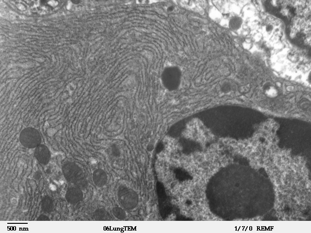
The endoplasmic reticulum (ER) is, in essence, the transportation system of the eukaryotic cell, and has many other important functions such as protein folding. It is a type of organelle made up of two subunits – rough endoplasmic reticulum (RER), and smooth endoplasmic reticulum (SER). The endoplasmic reticulum is found in most eukaryotic cells and forms an interconnected network of flattened, membrane-enclosed sacs known as cisternae (in the RER), and tubular structures in the SER. The membranes of the ER are continuous with the outer nuclear membrane. The endoplasmic reticulum is not found in red blood cells, or spermatozoa. The two types of ER share many of the same proteins and engage in certain common activities such as the synthesis of certain lipids and cholesterol. Different types of cells contain different ratios of the two types of ER depending on the activities of the cell. RER is found mainly toward the nucleus of cell and SER towards the cell membrane or plasma ... [...More Info...] [...Related Items...] OR: [Wikipedia] [Google] [Baidu] |


