|
Mental Nerve
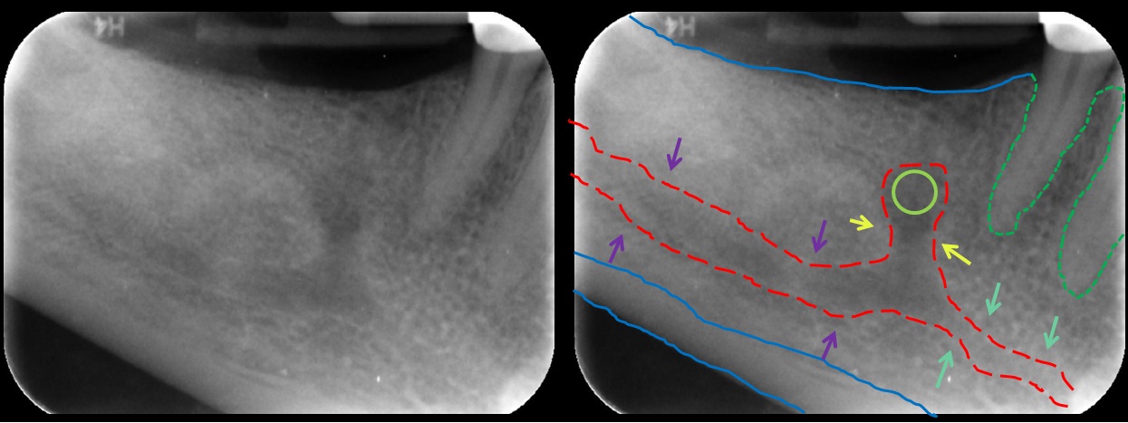
The mental nerve is a sensory nerve of the face. It is a branch of the posterior trunk of the inferior alveolar nerve, itself a branch of the mandibular nerve (CN V3), itself a branch of the trigeminal nerve (CN V). It provides sensation to the front of the chin and the lower lip, as well as the gums of the anterior mandibular (lower) teeth. It can be blocked with local anaesthesia for procedures on the chin, lower lip, and mucous membrane of the inner cheek. Problems with the nerve cause chin numbness. Structure The mental nerve is a branch of the posterior trunk of the inferior alveolar nerve. This is a branch of the mandibular nerve (CN V3), itself a branch of the trigeminal nerve (CN V). It emerges from the mental foramen in the mandible. It divides into three branches beneath the depressor anguli oris muscle. One branch descends to the skin of the chin. Two branches ascend to the skin and mucous membrane of the lower lip. These branches communicate freely with the facial ne ... [...More Info...] [...Related Items...] OR: [Wikipedia] [Google] [Baidu] |
Maxillary Nerve
In neuroanatomy, the maxillary nerve (V) is one of the three branches or divisions of the trigeminal nerve, the fifth (CN V) cranial nerve. It comprises the principal functions of sensation from the maxilla, nasal cavity, sinuses, the palate and subsequently that of the mid-face, and is intermediate, both in position and size, between the ophthalmic nerve and the mandibular nerve.Illustrated Anatomy of the Head and Neck, Fehrenbach and Herring, Elsevier, 2012, page 180 Structure It begins at the middle of the trigeminal ganglion as a flattened plexiform band then it passes through the lateral wall of the cavernous sinus. It leaves the skull through the foramen rotundum, where it becomes more cylindrical in form, and firmer in texture. After leaving foramen rotundum it gives two branches to the pterygopalatine ganglion. It then crosses the pterygopalatine fossa, inclines lateralward on the back of the maxilla, and enters the orbit through the inferior orbital fissure. It t ... [...More Info...] [...Related Items...] OR: [Wikipedia] [Google] [Baidu] |
Elsevier
Elsevier () is a Dutch academic publishing company specializing in scientific, technical, and medical content. Its products include journals such as '' The Lancet'', ''Cell'', the ScienceDirect collection of electronic journals, '' Trends'', the '' Current Opinion'' series, the online citation database Scopus, the SciVal tool for measuring research performance, the ClinicalKey search engine for clinicians, and the ClinicalPath evidence-based cancer care service. Elsevier's products and services also include digital tools for data management, instruction, research analytics and assessment. Elsevier is part of the RELX Group (known until 2015 as Reed Elsevier), a publicly traded company. According to RELX reports, in 2021 Elsevier published more than 600,000 articles annually in over 2,700 journals; as of 2018 its archives contained over 17 million documents and 40,000 e-books, with over one billion annual downloads. Researchers have criticized Elsevier for its high profit m ... [...More Info...] [...Related Items...] OR: [Wikipedia] [Google] [Baidu] |
Masseter Muscle
In human anatomy, the masseter is one of the muscles of mastication. Found only in mammals, it is particularly powerful in herbivores to facilitate chewing of plant matter. The most obvious muscle of mastication is the masseter muscle, since it is the most superficial and one of the strongest. Structure The masseter is a thick, somewhat quadrilateral muscle, consisting of three heads, superficial, deep and coronoid. The fibers of superficial and deep heads are continuous at their insertion. Superficial head The superficial head, the larger, arises by a thick, tendinous aponeurosis from the temporal process of the zygomatic bone, and from the anterior two-thirds of the inferior border of the zygomatic arch. Its fibers pass inferior and posterior, to be inserted into the angle of the mandible and inferior half of the lateral surface of the ramus of the mandible. Deep head The deep head is much smaller, and more muscular in texture. It arises from the posterior third of the low ... [...More Info...] [...Related Items...] OR: [Wikipedia] [Google] [Baidu] |
Temporalis Muscle
In anatomy, the temporalis muscle, also known as the temporal muscle, is one of the muscles of mastication (chewing). It is a broad, fan-shaped convergent muscle on each side of the head that fills the temporal fossa, superior to the zygomatic arch so it covers much of the temporal bone.Illustrated Anatomy of the Head and Neck, Fehrenbach and Herring, Elsevier, 2012, page 98''Temporal'' refers to the head's temples. Structure In humans, the temporalis muscle arises from the temporal fossa and the deep part of temporal fascia. This is a very broad area of attachment. It passes medial to the zygomatic arch. It forms a tendon which inserts onto the coronoid process of the mandible, with its insertion extending into the retromolar fossa posterior to the most distal mandibular molar.Human Anatomy, Jacobs, Elsevier, 2008, page 194 In other mammals, the muscle usually spans the dorsal part of the skull all the way up to the medial line. There, it may be attached to a sagittal cr ... [...More Info...] [...Related Items...] OR: [Wikipedia] [Google] [Baidu] |
Surgery
Surgery ''cheirourgikē'' (composed of χείρ, "hand", and ἔργον, "work"), via la, chirurgiae, meaning "hand work". is a medical specialty that uses operative manual and instrumental techniques on a person to investigate or treat a pathological condition such as a disease or injury, to help improve bodily function, appearance, or to repair unwanted ruptured areas. The act of performing surgery may be called a surgical procedure, operation, or simply "surgery". In this context, the verb "operate" means to perform surgery. The adjective surgical means pertaining to surgery; e.g. surgical instruments or surgical nurse. The person or subject on which the surgery is performed can be a person or an animal. A surgeon is a person who practices surgery and a surgeon's assistant is a person who practices surgical assistance. A surgical team is made up of the surgeon, the surgeon's assistant, an anaesthetist, a circulating nurse and a surgical technologist. Surgery usually ... [...More Info...] [...Related Items...] OR: [Wikipedia] [Google] [Baidu] |
Local Anesthesia
Local anesthesia is any technique to induce the absence of sensation in a specific part of the body, generally for the aim of inducing local analgesia, that is, local insensitivity to pain, although other local senses may be affected as well. It allows patients to undergo surgical and dental procedures with reduced pain and distress. In many situations, such as cesarean section, it is safer and therefore superior to general anesthesia. The following terms are often used interchangeably: * ''Local anesthesia'', in a strict sense, is anesthesia of a small part of the body such as a tooth or an area of skin. * ''Regional anesthesia'' is aimed at anesthetizing a larger part of the body such as a leg or arm. * ''Conduction anesthesia'' encompasses a great variety of local and regional anesthetic techniques. Medical A local anesthetic is a drug that causes reversible local anesthesia and a loss of nociception. When it is used on specific nerve pathways ( nerve block), effects suc ... [...More Info...] [...Related Items...] OR: [Wikipedia] [Google] [Baidu] |
Facial Nerve
The facial nerve, also known as the seventh cranial nerve, cranial nerve VII, or simply CN VII, is a cranial nerve that emerges from the pons of the brainstem, controls the muscles of facial expression, and functions in the conveyance of taste sensations from the anterior two-thirds of the tongue. The nerve typically travels from the pons through the facial canal in the temporal bone and exits the skull at the stylomastoid foramen. It arises from the brainstem from an area posterior to the cranial nerve VI (abducens nerve) and anterior to cranial nerve VIII (vestibulocochlear nerve). The facial nerve also supplies preganglionic parasympathetic fibers to several head and neck ganglia. The facial and intermediate nerves can be collectively referred to as the nervus intermediofacialis. The path of the facial nerve can be divided into six segments: # intracranial (cisternal) segment # meatal (canalicular) segment (within the internal auditory canal) # labyrinthine segment (i ... [...More Info...] [...Related Items...] OR: [Wikipedia] [Google] [Baidu] |
Depressor Anguli Oris Muscle
The depressor anguli oris muscle (triangularis muscle) is a facial muscle. It originates from the mandible and inserts into the angle of the mouth. It is associated with frowning, as it depresses the corner of the mouth. Structure The depressor anguli oris arises from the lateral surface of the mandible. Its fibres then converge. It is inserted by a narrow fasciculus into the angle of the mouth. At its origin, it is continuous with the platysma muscle, and at its insertion with the orbicularis oris muscle and risorius muscle. Some of its fibers are directly continuous with those of the levator anguli oris muscle, and others are occasionally found crossing from the muscle of one side to that of the other; these latter fibers constitute the transverse muscle of the chin. The depressor anguli oris muscle receives its blood supply from a branch of the facial artery. Nerve supply The depressor anguli oris muscle is supplied by the marginal mandibular branch of the facial ... [...More Info...] [...Related Items...] OR: [Wikipedia] [Google] [Baidu] |
Saunders (imprint)
Saunders is an American academic publisher based in the United States. It is currently an imprint (trade name), imprint of Elsevier. Formerly independent, the W. B. Saunders company was acquired by CBS in 1968, who added it to their publishing division Henry Holt and Company, Holt, Rinehart & Winston. When CBS left the publishing field in 1986, it sold the academic publishing units to Harcourt (publisher), Harcourt Brace Jovanovich. Harcourt was acquired by Reed Elsevier in 2001. . Northern Illinois University Libraries. Retrieved May 2, 2015. W. B. Saunders published the Kinsey Reports and Dorland's medical reference works. Elsevier still sells the latter under the Saunders imprint. References External links * Book publishing companies based in Pennsylvania El ...[...More Info...] [...Related Items...] OR: [Wikipedia] [Google] [Baidu] |
Human Mandible
In anatomy, the mandible, lower jaw or jawbone is the largest, strongest and lowest bone in the human facial skeleton. It forms the lower jaw and holds the lower teeth in place. The mandible sits beneath the maxilla. It is the only movable bone of the skull (discounting the ossicles of the middle ear). It is connected to the temporal bones by the temporomandibular joints. The bone is formed in the fetus from a fusion of the left and right mandibular prominences, and the point where these sides join, the mandibular symphysis, is still visible as a faint ridge in the midline. Like other symphyses in the body, this is a midline articulation where the bones are joined by fibrocartilage, but this articulation fuses together in early childhood.Illustrated Anatomy of the Head and Neck, Fehrenbach and Herring, Elsevier, 2012, p. 59 The word "mandible" derives from the Latin word ''mandibula'', "jawbone" (literally "one used for chewing"), from '' mandere'' "to chew" and ''-bula' ... [...More Info...] [...Related Items...] OR: [Wikipedia] [Google] [Baidu] |
Mental Foramen
The mental foramen is one of two foramina (openings) located on the anterior surface of the mandible. It is part of the mandibular canal. It transmits the terminal branches of the inferior alveolar nerve and the mental vessels. Structure The mental foramen is located on the anterior surface of the mandible. It is directly below the commisure of the lips, and the tendon of depressor labii inferioris muscle. It is at the end of the mandibular canal, which begins at the mandibular foramen on the posterior surface of the mandible. It transmits the terminal branches of the inferior alveolar nerve (the mental nerve), the mental artery, and the mental vein. Variation The mental foramen descends slightly in toothless individuals. The mental foramen is in line with the longitudinal axis of the 2nd premolar in 63% of people. It generally lies at the level of the vestibular fornix and about a finger's breadth above the inferior border of the mandible. In the general populatio ... [...More Info...] [...Related Items...] OR: [Wikipedia] [Google] [Baidu] |
Mucous Membrane
A mucous membrane or mucosa is a membrane that lines various cavities in the body of an organism and covers the surface of internal organs. It consists of one or more layers of epithelial cells overlying a layer of loose connective tissue. It is mostly of endodermal origin and is continuous with the skin at body openings such as the eyes, eyelids, ears, inside the nose, inside the mouth, lips, the genital areas, the urethral opening and the anus. Some mucous membranes secrete mucus, a thick protective fluid. The function of the membrane is to stop pathogens and dirt from entering the body and to prevent bodily tissues from becoming dehydrated. Structure The mucosa is composed of one or more layers of epithelial cells that secrete mucus, and an underlying lamina propria of loose connective tissue. The type of cells and type of mucus secreted vary from organ to organ and each can differ along a given tract. Mucous membranes line the digestive, respiratory and reproductive ... [...More Info...] [...Related Items...] OR: [Wikipedia] [Google] [Baidu] |

.jpg)

