|
McMurray's Test
The McMurray test, also known as the McMurray circumduction test is used to evaluate individuals for tears in the meniscus of the knee. A tear in the meniscus may cause a pedunculated tag of the meniscus which may become jammed between the joint surfaces. To perform the test, the knee is held by one hand, which is placed along the joint line, and flexed to complete flexion while the foot is held by the sole (of the foot) with the other hand. The examiner then rotates the leg internally while extending the knee to 90 degrees of flexion. If a "thud" or "click" is felt along with pain, this constitutes a "positive McMurray test" for a tear in the posterior portion of the lateral meniscus. Likewise, external rotation of the leg can be applied to test the posterior portion of the medial meniscus. The McMurray test is named after Thomas Porter McMurray, a British orthopedic surgeon from the late nineteenth and early twentieth century who was the first to describe this test. The descript ... [...More Info...] [...Related Items...] OR: [Wikipedia] [Google] [Baidu] |
Meniscus (anatomy)
A meniscus is a crescent-shaped fibrocartilaginous anatomical structure that, in contrast to an articular disc, only partly divides a joint cavity.Platzer (2004), p 208 In humans they are present in the knee, wrist, acromioclavicular, sternoclavicular, and temporomandibular joints; in other animals they may be present in other joints. Generally, the term "meniscus" is used to refer to the cartilage of the knee, either to the lateral or medial meniscus. Both are cartilaginous tissues that provide structural integrity to the knee when it undergoes tension and torsion. The menisci are also known as "semi-lunar" cartilages, referring to their half-moon, crescent shape. The term "meniscus" is from the Ancient Greek word (), meaning "crescent". Structure The menisci of the knee are two pads of fibrocartilaginous tissue which serve to disperse friction in the knee joint between the lower leg (tibia) and the thigh (femur). They are concave on the top and flat on the bottom, articula ... [...More Info...] [...Related Items...] OR: [Wikipedia] [Google] [Baidu] |
Knee
In humans and other primates, the knee joins the thigh with the leg and consists of two joints: one between the femur and tibia (tibiofemoral joint), and one between the femur and patella (patellofemoral joint). It is the largest joint in the human body. The knee is a modified hinge joint, which permits flexion and extension as well as slight internal and external rotation. The knee is vulnerable to injury and to the development of osteoarthritis. It is often termed a ''compound joint'' having tibiofemoral and patellofemoral components. (The fibular collateral ligament is often considered with tibiofemoral components.) Structure The knee is a modified hinge joint, a type of synovial joint, which is composed of three functional compartments: the patellofemoral articulation, consisting of the patella, or "kneecap", and the patellar groove on the front of the femur through which it slides; and the medial and lateral tibiofemoral articulations linking the femur, or thigh bone ... [...More Info...] [...Related Items...] OR: [Wikipedia] [Google] [Baidu] |
Flexion
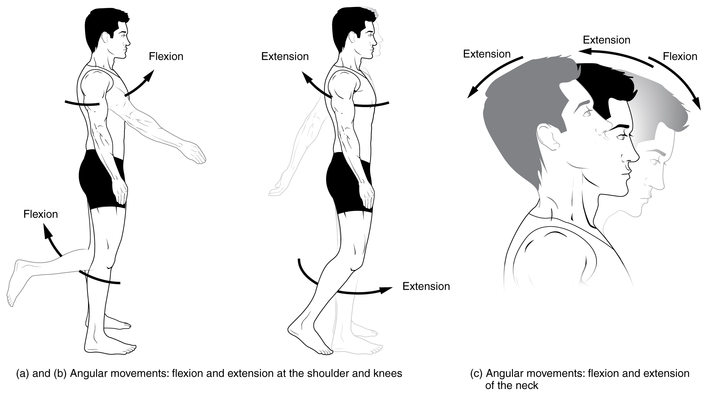
Motion, the process of movement, is described using specific anatomical terms. Motion includes movement of organs, joints, limbs, and specific sections of the body. The terminology used describes this motion according to its direction relative to the anatomical position of the body parts involved. Anatomists and others use a unified set of terms to describe most of the movements, although other, more specialized terms are necessary for describing unique movements such as those of the hands, feet, and eyes. In general, motion is classified according to the anatomical plane it occurs in. ''Flexion'' and ''extension'' are examples of ''angular'' motions, in which two axes of a joint are brought closer together or moved further apart. ''Rotational'' motion may occur at other joints, for example the shoulder, and are described as ''internal'' or ''external''. Other terms, such as ''elevation'' and ''depression'', describe movement above or below the horizontal plane. Many anatomica ... [...More Info...] [...Related Items...] OR: [Wikipedia] [Google] [Baidu] |
Internal Rotation
Motion, the process of movement, is described using specific anatomical terms. Motion includes movement of organs, joints, limbs, and specific sections of the body. The terminology used describes this motion according to its direction relative to the anatomical position of the body parts involved. Anatomists and others use a unified set of terms to describe most of the movements, although other, more specialized terms are necessary for describing unique movements such as those of the hands, feet, and eyes. In general, motion is classified according to the anatomical plane it occurs in. ''Flexion'' and ''extension'' are examples of ''angular'' motions, in which two axes of a joint are brought closer together or moved further apart. ''Rotational'' motion may occur at other joints, for example the shoulder, and are described as ''internal'' or ''external''. Other terms, such as ''elevation'' and ''depression'', describe movement above or below the horizontal plane. Many anatomica ... [...More Info...] [...Related Items...] OR: [Wikipedia] [Google] [Baidu] |
Lateral Meniscus
The lateral meniscus (external semilunar fibrocartilage) is a fibrocartilaginous band that spans the lateral side of the interior of the knee joint. It is one of two meniscus (anatomy), menisci of the knee, the other being the medial meniscus. It is nearly circular and covers a larger portion of the articular surface than the medial. It can occasionally be injured or torn by twisting the knee or applying direct force, as seen in contact sports. Structure The lateral meniscus is grooved laterally for the tendon of the popliteus, which separates it from the fibular collateral ligament. Its anterior end is attached in front of the intercondyloid eminence of the tibia, lateral to, and behind, the anterior cruciate ligament, with which it blends; the posterior end is attached behind the intercondyloid eminence of the tibia and in front of the posterior end of the medial meniscus. The anterior attachment of the lateral meniscus is twisted on itself so that its free margin looks backwa ... [...More Info...] [...Related Items...] OR: [Wikipedia] [Google] [Baidu] |
External Rotation
Motion, the process of movement, is described using specific anatomical terms. Motion includes movement of organs, joints, limbs, and specific sections of the body. The terminology used describes this motion according to its direction relative to the anatomical position of the body parts involved. Anatomists and others use a unified set of terms to describe most of the movements, although other, more specialized terms are necessary for describing unique movements such as those of the hands, feet, and eyes. In general, motion is classified according to the anatomical plane it occurs in. ''Flexion'' and ''extension'' are examples of ''angular'' motions, in which two axes of a joint are brought closer together or moved further apart. ''Rotational'' motion may occur at other joints, for example the shoulder, and are described as ''internal'' or ''external''. Other terms, such as ''elevation'' and ''depression'', describe movement above or below the horizontal plane. Many anatomica ... [...More Info...] [...Related Items...] OR: [Wikipedia] [Google] [Baidu] |
Medial Meniscus
The medial meniscus is a fibrocartilage semicircular band that spans the knee joint medially, located between the medial condyle of the femur and the medial condyle of the tibia.Platzer (2004), p 208 It is also referred to as the internal semilunar fibrocartilage. The medial meniscus has more of a crescent shape while the lateral meniscus is more circular. The anterior aspects of both menisci are connected by the transverse ligament. It is a common site of injury, especially if the knee is twisted. Structure The meniscus attaches to the tibia via coronary ligaments. Its anterior end, thin and pointed, is attached to the anterior intercondyloid fossa of the tibia, in front of the anterior cruciate ligament; Its posterior end is fixed to the posterior intercondyloid fossa of the tibia, between the attachments of the lateral meniscus and the posterior cruciate ligament. It is fused with the tibial collateral ligament which makes it far less mobile than the lateral meniscus. The ... [...More Info...] [...Related Items...] OR: [Wikipedia] [Google] [Baidu] |
Thomas Porter McMurray
Thomas Porter McMurray (5 December 1887, Belfast – 16 November 1949, London) was a British orthopaedic surgeon remembered for describing the McMurray test. Biography Thomas McMurray graduated from Queen's University, Belfast in 1910, and took up a house job in Liverpool under Sir Robert Jones. He served as a captain in the Royal Army Medical Corps in France for a short time, returning to the Alder Hey Military Hospital in Liverpool in 1914. He continued his training under Robert Jones, becoming lecturer at the Department of Orthopaedic Surgery at the University of Liverpool from 1924 to 1938, and succeeding Robert Jones as director of orthopaedics. He became the first professor of orthopaedic surgery in Liverpool in 1938. He was president of the British Orthopaedic Society and the Liverpool Medical Institution, and president-elect of the British Medical Association. His dexterity as a surgeon was noted; he was able to remove a meniscus in five minutes, and disarticulate a ... [...More Info...] [...Related Items...] OR: [Wikipedia] [Google] [Baidu] |


