|
Lateral Meniscus
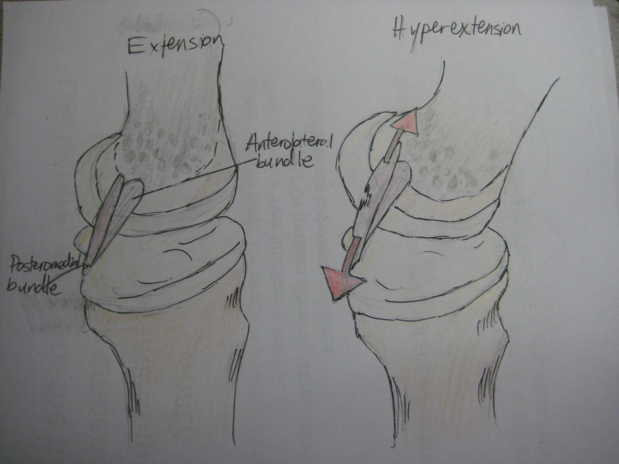
The lateral meniscus (external semilunar fibrocartilage) is a fibrocartilaginous band that spans the lateral side of the interior of the knee joint. It is one of two meniscus (anatomy), menisci of the knee, the other being the medial meniscus. It is nearly circular and covers a larger portion of the articular surface than the medial. It can occasionally be injured or torn by twisting the knee or applying direct force, as seen in contact sports. Structure The lateral meniscus is grooved laterally for the tendon of the popliteus, which separates it from the fibular collateral ligament. Its anterior end is attached in front of the intercondyloid eminence of the tibia, lateral to, and behind, the anterior cruciate ligament, with which it blends; the posterior end is attached behind the intercondyloid eminence of the tibia and in front of the posterior end of the medial meniscus. The anterior attachment of the lateral meniscus is twisted on itself so that its free margin looks backwa ... [...More Info...] [...Related Items...] OR: [Wikipedia] [Google] [Baidu] |
Tibia
The tibia (; ), also known as the shinbone or shankbone, is the larger, stronger, and anterior (frontal) of the two bones in the leg below the knee in vertebrates (the other being the fibula, behind and to the outside of the tibia); it connects the knee with the ankle. The tibia is found on the medial side of the leg next to the fibula and closer to the median plane. The tibia is connected to the fibula by the interosseous membrane of leg, forming a type of fibrous joint called a syndesmosis with very little movement. The tibia is named for the flute ''tibia''. It is the second largest bone in the human body, after the femur. The leg bones are the strongest long bones as they support the rest of the body. Structure In human anatomy, the tibia is the second largest bone next to the femur. As in other vertebrates the tibia is one of two bones in the lower leg, the other being the fibula, and is a component of the knee and ankle joints. The ossification or formation of the bone ... [...More Info...] [...Related Items...] OR: [Wikipedia] [Google] [Baidu] |
Fasciculus
''Fasciculus vesanus'' is an extinct species of stem-group ctenophores known from the Burgess Shale of British Columbia, Canada. It is dated to and belongs to middle Cambrian strata. The species is remarkable for its two sets of long and short comb rows, not seen in similar form elsewhere in the fossil record or among modern species. See also *''Ctenorhabdotus capulus'' *''Xanioascus canadensis'' Maotianshan shales ctenophores **''Maotianoascus octonarius'' **''Sinoascus paillatus'' **''Stromatoveris psygmoglena ''Stromatoveris psygmoglena'' is a genus of basal petalonam from the Chengjiang deposits of Yunnan that was originally aligned with the fossil ''Charnia'' (strictly, the Charniomorpha) from the Ediacara biota. However, such an affinity is devel ...'' References External links * Prehistoric ctenophore genera Burgess Shale animals Monotypic ctenophore genera Fossil taxa described in 1978 Cambrian genus extinctions {{Ctenophore-stub ... [...More Info...] [...Related Items...] OR: [Wikipedia] [Google] [Baidu] |
Meniscal Cartilage Replacement Therapy
Left knee-joint from behind, showing interior ligaments Meniscal cartilage replacement therapy is surgical replacement of the meniscus of the knee as a treatment for where the meniscus is so damaged that it would otherwise need to be removed. Anatomy The meniscus is a C-shaped piece of fibrocartilage located at the peripheral aspect of the knee joint that offers lubrication and nutrition to the joint. Each knee has two menisci, medial and lateral, whose purpose is to provide space between the tibia and the femur, preventing friction and allowing for the diffusion of articular cartilage. The majority of the meniscus has no blood supply. As a result, if the meniscus is damaged, from trauma or with age, it is unable to undergo the body’s normal healing process. Therefore, a torn piece can begin to move inside the joint, get caught between the bones, and cause pain, swelling, and decreased mobility. However, recent research has called into question whether many meniscus tears ... [...More Info...] [...Related Items...] OR: [Wikipedia] [Google] [Baidu] |
Discoid Meniscus
Discoid meniscus is a rare human anatomic variant that usually affects the lateral meniscus of the knee. Usually a person with this anomaly has no complaints; however, it may present as pain, swelling, or a snapping sound heard from the affected knee. Strong suggestive findings on magnetic resonance imaging includes a thickened meniscal body seen on more than two contiguous sagittal slices. Description The Watanabe classification of discoid lateral meniscus is: (A) Incomplete, (B) Complete, and C) Wrisberg-ligament variant Normally, the meniscus is a thin crescent-shaped piece of cartilage that lies between the weight bearing joint surfaces of the femur and the tibia. It is attached to the lining of the knee joint along its periphery and serves to absorb about a third of the impact load that the joint cartilage surface sees and also provides some degree of stabilization for the knee. There are two menisci in the knee joint, with one on the outside (away from midline) being the ... [...More Info...] [...Related Items...] OR: [Wikipedia] [Google] [Baidu] |
Arthroscopically
Arthroscopy (also called arthroscopic or keyhole surgery) is a minimally invasive surgical procedure on a joint in which an examination and sometimes treatment of damage is performed using an arthroscope, an endoscope that is inserted into the joint through a small incision. Arthroscopic procedures can be performed during ACL reconstruction. The advantage over traditional open surgery is that the joint does not have to be opened up fully. For knee arthroscopy only two small incisions are made, one for the arthroscope and one for the surgical instruments to be used in the knee cavity. This reduces recovery time and may increase the rate of success due to less trauma to the connective tissue. It has gained popularity due to evidence of faster recovery times with less scarring, because of the smaller incisions. Irrigation fluid (most commonly 'normal' saline) is used to distend the joint and make a surgical space. The surgical instruments are smaller than traditional instruments ... [...More Info...] [...Related Items...] OR: [Wikipedia] [Google] [Baidu] |
McMurray's Test
The McMurray test, also known as the McMurray circumduction test is used to evaluate individuals for tears in the meniscus of the knee. A tear in the meniscus may cause a pedunculated tag of the meniscus which may become jammed between the joint surfaces. To perform the test, the knee is held by one hand, which is placed along the joint line, and flexed to complete flexion while the foot is held by the sole (of the foot) with the other hand. The examiner then rotates the leg internally while extending the knee to 90 degrees of flexion. If a "thud" or "click" is felt along with pain, this constitutes a "positive McMurray test" for a tear in the posterior portion of the lateral meniscus. Likewise, external rotation of the leg can be applied to test the posterior portion of the medial meniscus. The McMurray test is named after Thomas Porter McMurray, a British orthopedic surgeon from the late nineteenth and early twentieth century who was the first to describe this test. The descript ... [...More Info...] [...Related Items...] OR: [Wikipedia] [Google] [Baidu] |
Posterior Cruciate Ligament
The posterior cruciate ligament (PCL) is a ligament in each knee of humans and various other animals. It works as a counterpart to the anterior cruciate ligament (ACL). It connects the posterior intercondylar area of the tibia to the medial condyle of the femur. This configuration allows the PCL to resist forces pushing the tibia posteriorly relative to the femur. The PCL and ACL are intracapsular ligaments because they lie deep within the knee joint. They are both isolated from the fluid-filled synovial cavity, with the synovial membrane wrapped around them. The PCL gets its name by attaching to the posterior portion of the tibia. The PCL, ACL, MCL, and LCL are the four main ligaments of the knee in primates. Structure The PCL is located within the knee joint where it stabilizes the articulating bones, particularly the femur and the tibia, during movement. It originates from the lateral edge of the medial femoral condyle and the roof of the intercondyle notch then stretches ... [...More Info...] [...Related Items...] OR: [Wikipedia] [Google] [Baidu] |
Femur
The femur (; ), or thigh bone, is the proximal bone of the hindlimb in tetrapod vertebrates. The head of the femur articulates with the acetabulum in the pelvic bone forming the hip joint, while the distal part of the femur articulates with the tibia (shinbone) and patella (kneecap), forming the knee joint. By most measures the two (left and right) femurs are the strongest bones of the body, and in humans, the largest and thickest. Structure The femur is the only bone in the upper leg. The two femurs converge medially toward the knees, where they articulate with the proximal ends of the tibiae. The angle of convergence of the femora is a major factor in determining the femoral-tibial angle. Human females have thicker pelvic bones, causing their femora to converge more than in males. In the condition ''genu valgum'' (knock knee) the femurs converge so much that the knees touch one another. The opposite extreme is ''genu varum'' (bow-leggedness). In the general populatio ... [...More Info...] [...Related Items...] OR: [Wikipedia] [Google] [Baidu] |
Medial Condyle Of Femur
The medial condyle is one of the two projections on the lower extremity of femur, the other being the lateral condyle. The medial condyle is larger than the lateral (outer) condyle due to more weight bearing caused by the centre of mass being medial to the knee. On the posterior surface of the condyle the linea aspera The linea aspera ( la, rough line) is a ridge of roughened surface on the posterior surface of the shaft of the femur. It is the site of attachments of muscles and the intermuscular septum. Its margins diverge above and below. The linea asper ... (a ridge with two lips: medial and lateral; running down the posterior shaft of the femur) turns into the medial and lateral supracondylar ridges, respectively. The outermost protrusion on the medial surface of the medial condyle is referred to as the "medial epicondyle" and can be palpated by running fingers medially from the patella with the knee in flexion. It is important to take into consideration the differen ... [...More Info...] [...Related Items...] OR: [Wikipedia] [Google] [Baidu] |
Wrisberg
Heinrich August Wrisberg (20 June 1739 – 29 March 1808) was an anatomist. He also published under the Latinized version of his name as Henricus Augustus Wrisberg. Education He obtained his MD in 1763 at the University of Göttingen with a thesis entitled: ''De Respiratione Prima Nervo Phrenico Et Calore Animali: Pavca Disserit Et Simvl Vicarias Anatomiam Profitendi Operas Ad Diem XXIV. Octobris Aperiendas Indicit.'' Career He was a professor of medicine and obstetrics. Wrisberg studied the sympathetic nervous system and described the Wrisberg ganglion of the cardiac plexus. He also wrote a text on hernias. The cuneiform cartilages In the human larynx, the cuneiform cartilages (from Latin: ''cunei'', "wedge-shaped"; also known as cartilages of Wrisberg) are two small, elongated pieces of yellow elastic cartilage, placed one on either side, in the aryepiglottic fold. The ... are sometimes called the "Wrisberg cartilages". There are two nerves known as the nerve of Wris ... [...More Info...] [...Related Items...] OR: [Wikipedia] [Google] [Baidu] |
Intercondyloid Eminence
The intercondylar area is the separation between the medial and lateral condyle on the upper extremity of the tibia. The anterior and posterior cruciate ligaments and the menisci attach to the intercondylar area. The intercondyloid eminence is composed of the medial and lateral intercondylar tubercles, and divides the intercondylar area into an anterior and a posterior area. Structure Anterior area The anterior intercondylar area (or anterior intercondyloid fossa) is an area on the tibia, a bone in the lower leg. Together with the posterior intercondylar area it makes up the intercondylar area. The intercondylar area is the separation between the medial and lateral condyle located toward the proximal portion of the tibia. The intercondylar eminence composed of the medial and lateral intercondylar tubercle divides the intercondylar area into anterior and posterior part. The anterior intercondylar area is the location where the anterior cruciate ligament attaches to the ti ... [...More Info...] [...Related Items...] OR: [Wikipedia] [Google] [Baidu] |
Meniscus (anatomy)
A meniscus is a crescent-shaped fibrocartilaginous anatomical structure that, in contrast to an articular disc, only partly divides a joint cavity.Platzer (2004), p 208 In humans they are present in the knee, wrist, acromioclavicular, sternoclavicular, and temporomandibular joints; in other animals they may be present in other joints. Generally, the term "meniscus" is used to refer to the cartilage of the knee, either to the lateral or medial meniscus. Both are cartilaginous tissues that provide structural integrity to the knee when it undergoes tension and torsion. The menisci are also known as "semi-lunar" cartilages, referring to their half-moon, crescent shape. The term "meniscus" is from the Ancient Greek word (), meaning "crescent". Structure The menisci of the knee are two pads of fibrocartilaginous tissue which serve to disperse friction in the knee joint between the lower leg (tibia) and the thigh (femur). They are concave on the top and flat on the bottom, articula ... [...More Info...] [...Related Items...] OR: [Wikipedia] [Google] [Baidu] |


