|
MUC9
Oviduct-specific glycoprotein also known as oviductal glycoprotein (OGP) or estrogen-dependent oviduct protein, oviductin or mucin-9 is a protein that in humans is encoded by the ''OVGP1'' gene. Function Oviduct-specific glycoprotein is a large, carbohydrate-rich, epithelial glycoprotein with numerous O-glycosylation sites located within threonine, serine, and proline-rich tandem repeats. The gene is similar to members of the mucin and the glycosyl hydrolase 18 gene families. Regulation of expression may be estrogen-dependent. Gene expression and protein secretion occur during late follicular development through early cleavage-stage embryonic development. The protein is secreted from non-ciliated oviductal epithelial cells and associates with ovulated oocytes, blastomeres, and spermatozoon acrosomal regions. Beyond the oviduct, OVGP1 is detected in the mouse ovary, testis and epididymis suggesting its roles beyond fertilization. It is not detected in the mouse uterus, cervix, ... [...More Info...] [...Related Items...] OR: [Wikipedia] [Google] [Baidu] |
Mucin
Mucins () are a family of high molecular weight, heavily glycosylated proteins (glycoconjugates) produced by epithelial tissues in most animals. Mucins' key characteristic is their ability to form gels; therefore they are a key component in most gel-like secretions, serving functions from lubrication to cell signalling to forming chemical barriers. They often take an inhibitory role. Some mucins are associated with controlling mineralization, including nacre formation in mollusks, calcification in echinoderms and bone formation in vertebrates. They bind to pathogens as part of the immune system. Overexpression of the mucin proteins, especially MUC1, is associated with many types of cancer. Although some mucins are membrane-bound due to the presence of a hydrophobic membrane-spanning domain that favors retention in the plasma membrane, most mucins are secreted as principal components of mucus by mucous membranes or are secreted to become a component of saliva. Genes Human muci ... [...More Info...] [...Related Items...] OR: [Wikipedia] [Google] [Baidu] |
Protein
Proteins are large biomolecules and macromolecules that comprise one or more long chains of amino acid residues. Proteins perform a vast array of functions within organisms, including catalysing metabolic reactions, DNA replication, responding to stimuli, providing structure to cells and organisms, and transporting molecules from one location to another. Proteins differ from one another primarily in their sequence of amino acids, which is dictated by the nucleotide sequence of their genes, and which usually results in protein folding into a specific 3D structure that determines its activity. A linear chain of amino acid residues is called a polypeptide. A protein contains at least one long polypeptide. Short polypeptides, containing less than 20–30 residues, are rarely considered to be proteins and are commonly called peptides. The individual amino acid residues are bonded together by peptide bonds and adjacent amino acid residues. The sequence of amino acid residue ... [...More Info...] [...Related Items...] OR: [Wikipedia] [Google] [Baidu] |
Oviduct
The oviduct in mammals, is the passageway from an ovary. In human females this is more usually known as the Fallopian tube or uterine tube. The eggs travel along the oviduct. These eggs will either be fertilized by spermatozoa to become a zygote, or will degenerate in the body. Normally, these are paired structures, but in birds and some cartilaginous fishes, one or the other side fails to develop (together with the corresponding ovary), and only one functional oviduct can be found. Except in teleosts, the oviduct is not directly in contact with the ovary. Instead, the most anterior portion ends in a funnel-shaped structure called the infundibulum, which collects eggs as they are released by the ovary into the body cavity. The only female vertebrates to lack oviducts are the jawless fishes. In these species, the single fused ovary releases eggs directly into the body cavity. The fish eventually extrudes the eggs through a small genital pore towards the rear of the body. Fish and ... [...More Info...] [...Related Items...] OR: [Wikipedia] [Google] [Baidu] |
Endometrium
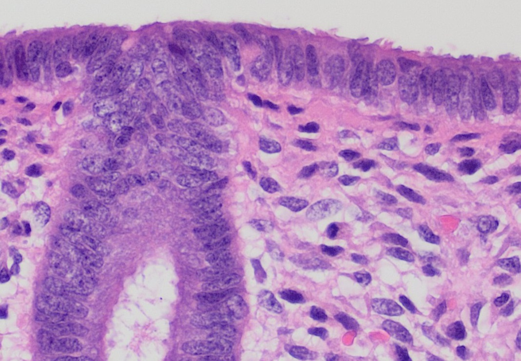
The endometrium is the inner epithelial layer, along with its mucous membrane, of the mammalian uterus. It has a basal layer and a functional layer: the basal layer contains stem cells which regenerate the functional layer. The functional layer thickens and then is shed during menstruation in humans and some other mammals, including apes, Old World monkeys, some species of bat, the elephant shrew and the Cairo spiny mouse. In most other mammals, the endometrium is reabsorbed in the estrous cycle. During pregnancy, the glands and blood vessels in the endometrium further increase in size and number. Vascular spaces fuse and become interconnected, forming the placenta, which supplies oxygen and nutrition to the embryo and fetus.Blue Histology - Female Reproductive System . School ... [...More Info...] [...Related Items...] OR: [Wikipedia] [Google] [Baidu] |
Epididymis
The epididymis (; plural: epididymides or ) is a tube that connects a testicle to a vas deferens in the male reproductive system. It is a single, narrow, tightly-coiled tube in adult humans, in length. It serves as an interconnection between the multiple efferent ducts at the rear of a testicle (proximally), and the vas deferens (distally). Anatomy The epididymis is situated posterior and somewhat lateral to the testis. The epididymis is invested completely by the tunica vaginalis (which is continuous with the tunica vaginalis covering the testis). The epididymis can be divided into three main regions: * The head ( la, caput). The head of the epididymis receives spermatozoa via the efferent ducts of the mediastinium of the testis at the superior pole of the testis. The head is characterized histologically by a thick epithelium with long stereocilia (described below) and a little smooth muscle. It is involved in absorbing fluid to make the sperm more concentrated. The concentrat ... [...More Info...] [...Related Items...] OR: [Wikipedia] [Google] [Baidu] |
Testis
A testicle or testis (plural testes) is the male reproductive gland or gonad in all bilaterians, including humans. It is homologous to the female ovary. The functions of the testes are to produce both sperm and androgens, primarily testosterone. Testosterone release is controlled by the anterior pituitary luteinizing hormone, whereas sperm production is controlled both by the anterior pituitary follicle-stimulating hormone and gonadal testosterone. Structure Appearance Males have two testicles of similar size contained within the scrotum, which is an extension of the abdominal wall. Scrotal asymmetry, in which one testicle extends farther down into the scrotum than the other, is common. This is because of the differences in the vasculature's anatomy. For 85% of men, the right testis hangs lower than the left one. Measurement and volume The volume of the testicle can be estimated by palpating it and comparing it to ellipsoids of known sizes. Another method is to use caliper ... [...More Info...] [...Related Items...] OR: [Wikipedia] [Google] [Baidu] |
Ovary
The ovary is an organ in the female reproductive system that produces an ovum. When released, this travels down the fallopian tube into the uterus, where it may become fertilized by a sperm. There is an ovary () found on each side of the body. The ovaries also secrete hormones that play a role in the menstrual cycle and fertility. The ovary progresses through many stages beginning in the prenatal period through menopause. It is also an endocrine gland because of the various hormones that it secretes. Structure The ovaries are considered the female gonads. Each ovary is whitish in color and located alongside the lateral wall of the uterus in a region called the ovarian fossa. The ovarian fossa is the region that is bounded by the external iliac artery and in front of the ureter and the internal iliac artery. This area is about 4 cm x 3 cm x 2 cm in size.Daftary, Shirish; Chakravarti, Sudip (2011). Manual of Obstetrics, 3rd Edition. Elsevier. pp. 1-16. . The ovari ... [...More Info...] [...Related Items...] OR: [Wikipedia] [Google] [Baidu] |
Acrosome
The acrosome is an organelle that develops over the anterior (front) half of the head in the spermatozoa (sperm cells) of many animals including humans. It is a cap-like structure derived from the Golgi apparatus. In placental mammals the acrosome contains degradative enzymes (including hyaluronidase and acrosin). These enzymes break down the outer membrane of the ovum, called the zona pellucida, allowing the haploid nucleus in the sperm cell to join with the haploid nucleus in the ovum. This shedding of the acrosome, or acrosome reaction, can be stimulated ''in vitro'' by substances a sperm cell may encounter naturally such as progesterone or follicular fluid, as well as the more commonly used calcium ionophore A23187. This can be done to serve as a positive control when assessing the acrosome reaction of a sperm sample by flow cytometry or fluorescence microscopy. This is usually done after staining with a fluoresceinated lectin such as FITC-PNA, FITC-PSA, FITC-ConA, or flu ... [...More Info...] [...Related Items...] OR: [Wikipedia] [Google] [Baidu] |
Blastomere
In biology, a blastomere is a type of cell produced by cell division (cleavage) of the zygote after fertilization; blastomeres are an essential part of blastula formation, and blastocyst formation in mammals. Human blastomere characteristics In humans, blastomere formation begins immediately following fertilization and continues through the first week of embryonic development. About 90 minutes after fertilization, the zygote divides into two cells. The two-cell blastomere state, present after the zygote first divides, is considered the earliest mitotic product of the fertilized oocyte. These mitotic divisions continue and result in a grouping of cells called blastomeres. During this process, the total size of the embryo does not increase, so each division results in smaller and smaller cells. When the zygote contains 16 to 32 blastomeres it is referred to as a morula. These are the preliminary stages in the embryo beginning to form. Once this begins, microtubules within the mor ... [...More Info...] [...Related Items...] OR: [Wikipedia] [Google] [Baidu] |
Oocyte
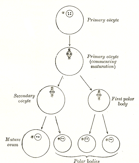
An oocyte (, ), oöcyte, or ovocyte is a female gametocyte or germ cell involved in reproduction. In other words, it is an immature ovum, or egg cell. An oocyte is produced in a female fetus in the ovary during female gametogenesis. The female germ cells produce a primordial germ cell (PGC), which then undergoes mitosis, forming oogonia. During oogenesis, the oogonia become primary oocytes. An oocyte is a form of genetic material that can be collected for cryoconservation. Formation The formation of an oocyte is called oocytogenesis, which is a part of oogenesis. Oogenesis results in the formation of both primary oocytes during fetal period, and of secondary oocytes after it as part of ovulation. Characteristics Cytoplasm Oocytes are rich in cytoplasm, which contains yolk granules to nourish the cell early in development. Nucleus During the primary oocyte stage of oogenesis, the nucleus is called a germinal vesicle. The only normal human type of secondary oocyte has t ... [...More Info...] [...Related Items...] OR: [Wikipedia] [Google] [Baidu] |
Ovarian Follicle
An ovarian follicle is a roughly spheroid cellular aggregation set found in the ovaries. It secretes hormones that influence stages of the menstrual cycle. At the time of puberty, women have approximately 200,000 to 300,000 follicles, each with the potential to release an egg cell (ovum) at ovulation for fertilization. These eggs are developed once every menstrual cycle with around 450–500 being ovulated during a woman's reproductive lifetime. Structure Ovarian follicles are the basic units of female reproductive biology. Each of them contains a single oocyte (immature ovum or egg cell). These structures are periodically initiated to grow and develop, culminating in ovulation of usually a single competent oocyte in humans. They also consist of granulosa cells and theca of follicle. Oocyte Once a month, one of the ovaries releases a mature egg (ovum), known as an oocyte. The nucleus of such an oocyte is called a '' germinal vesicle (see picture).'' Cumulus oophorus Cumulu ... [...More Info...] [...Related Items...] OR: [Wikipedia] [Google] [Baidu] |
Gene
In biology, the word gene (from , ; "...Wilhelm Johannsen coined the word gene to describe the Mendelian units of heredity..." meaning ''generation'' or ''birth'' or ''gender'') can have several different meanings. The Mendelian gene is a basic unit of heredity and the molecular gene is a sequence of nucleotides in DNA that is transcribed to produce a functional RNA. There are two types of molecular genes: protein-coding genes and noncoding genes. During gene expression, the DNA is first copied into RNA. The RNA can be directly functional or be the intermediate template for a protein that performs a function. The transmission of genes to an organism's offspring is the basis of the inheritance of phenotypic traits. These genes make up different DNA sequences called genotypes. Genotypes along with environmental and developmental factors determine what the phenotypes will be. Most biological traits are under the influence of polygenes (many different genes) as well as gen ... [...More Info...] [...Related Items...] OR: [Wikipedia] [Google] [Baidu] |


