|
Myostatin Inhibitors
Myostatin (also known as growth differentiation factor 8, abbreviated GDF8) is a protein that in humans is encoded by the ''MSTN'' gene. Myostatin is a myokine that is produced and released by myocytes and acts on muscle cells to inhibit muscle growth. Myostatin is a secreted growth differentiation factor that is a member of the TGF beta protein family. Myostatin is assembled and produced in skeletal muscle before it is released into the blood stream. Most of the data regarding the effects of myostatin comes from studies performed on mice. Animals either lacking myostatin or treated with substances that block the activity of myostatin have significantly more muscle mass. Furthermore, individuals who have mutations in both copies of the myostatin gene have significantly more muscle mass and are stronger than normal. There is hope that studies into myostatin may have therapeutic application in treating muscle wasting diseases such as muscular dystrophy. Discovery and sequencing ... [...More Info...] [...Related Items...] OR: [Wikipedia] [Google] [Baidu] |
MSTN Location
Myostatin (also known as growth differentiation factor 8, abbreviated GDF8) is a protein that in humans is encoded by the ''MSTN'' gene. Myostatin is a myokine that is produced and released by myocytes and acts on muscle cells to inhibit muscle growth. Myostatin is a secreted growth differentiation factor that is a member of the TGF beta protein family. Myostatin is assembled and produced in skeletal muscle before it is released into the blood stream. Most of the data regarding the effects of myostatin comes from studies performed on mice. Animals either lacking myostatin or treated with substances that block the activity of myostatin have significantly more muscle mass. Furthermore, individuals who have mutations in both copies of the myostatin gene have significantly more muscle mass and are stronger than normal. There is hope that studies into myostatin may have therapeutic application in treating muscle wasting diseases such as muscular dystrophy. Discovery and sequencing ... [...More Info...] [...Related Items...] OR: [Wikipedia] [Google] [Baidu] |
ACVR1B
Activin receptor type-1B is a protein that in humans is encoded by the ''ACVR1B'' gene. ACVR1B or ALK-4 acts as a transducer of activin or activin-like ligands (e.g., inhibin) signals. Activin binds to either ACVR2A or ACVR2B and then forms a complex with ACVR1B. These go on to recruit the R-SMADs SMAD2 or SMAD3. ACVR1B also transduces signals of nodal, GDF-1, and Vg1; however, unlike activin, they require other coreceptor molecules such as the protein Cripto. Function Activins are dimeric growth and differentiation factors which belong to the transforming growth factor-beta (TGF-beta) superfamily of structurally related signaling proteins. Activins signal through a heteromeric complex of receptor serine kinases which include at least two type I (I and IB) and two type II (II and IIB) receptors. These receptors are all transmembrane proteins, composed of a ligand-binding extracellular domain with a cysteine-rich region, a transmembrane domain, and a cytoplasmic domain wit ... [...More Info...] [...Related Items...] OR: [Wikipedia] [Google] [Baidu] |
Belgian Blue
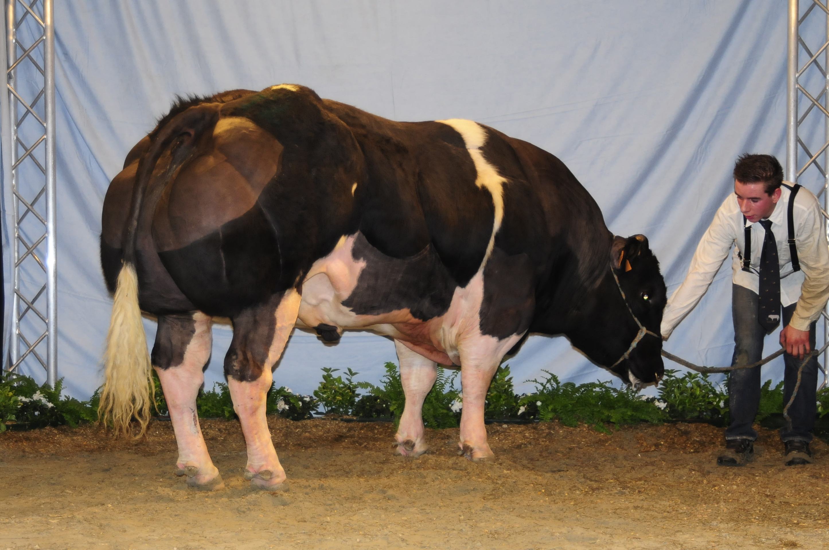
The Belgian Blue (french: 'Blanc-Bleu Belge', nl, 'Belgisch Witblauw', both literally meaning "Belgian White-Blue") is a breed of beef cattle from Belgium. It may also be known as the , or nl, dikbil, label=none (literally "fat buttocks" in Dutch). Alternative names for this breed include Belgian Blue-White; Belgian White and Blue Pied; Belgian White Blue; Blue; and Blue Belgian. The Belgian Blue's extremely lean, hyper-sculpted, ultra-muscular physique is termed " double-muscling". The double-muscling phenotype is a heritable condition resulting in an increased number of muscle fibres (hyperplasia), instead of the (normal) enlargement of ''individual'' muscle fibres (hypertrophy). This particular trait is shared with another breed of cattle known as Piedmontese. Both of these breeds have an increased ability to convert feed into lean muscle, which causes these particular breeds' meat to have a reduced fat content and reduced tenderness. The Belgian Blue is named after i ... [...More Info...] [...Related Items...] OR: [Wikipedia] [Google] [Baidu] |
Nucleotide
Nucleotides are organic molecules consisting of a nucleoside and a phosphate. They serve as monomeric units of the nucleic acid polymers – deoxyribonucleic acid (DNA) and ribonucleic acid (RNA), both of which are essential biomolecules within all life-forms on Earth. Nucleotides are obtained in the diet and are also synthesized from common nutrients by the liver. Nucleotides are composed of three subunit molecules: a nucleobase, a five-carbon sugar (ribose or deoxyribose), and a phosphate group consisting of one to three phosphates. The four nucleobases in DNA are guanine, adenine, cytosine and thymine; in RNA, uracil is used in place of thymine. Nucleotides also play a central role in metabolism at a fundamental, cellular level. They provide chemical energy—in the form of the nucleoside triphosphates, adenosine triphosphate (ATP), guanosine triphosphate (GTP), cytidine triphosphate (CTP) and uridine triphosphate (UTP)—throughout the cell for the many cellular func ... [...More Info...] [...Related Items...] OR: [Wikipedia] [Google] [Baidu] |
Protein Biosynthesis
Protein biosynthesis (or protein synthesis) is a core biological process, occurring inside cells, balancing the loss of cellular proteins (via degradation or export) through the production of new proteins. Proteins perform a number of critical functions as enzymes, structural proteins or hormones. Protein synthesis is a very similar process for both prokaryotes and eukaryotes but there are some distinct differences. Protein synthesis can be divided broadly into two phases - transcription and translation. During transcription, a section of DNA encoding a protein, known as a gene, is converted into a template molecule called messenger RNA (mRNA). This conversion is carried out by enzymes, known as RNA polymerases, in the nucleus of the cell. In eukaryotes, this mRNA is initially produced in a premature form (pre-mRNA) which undergoes post-transcriptional modifications to produce mature mRNA. The mature mRNA is exported from the cell nucleus via nuclear pores to the cytoplasm of ... [...More Info...] [...Related Items...] OR: [Wikipedia] [Google] [Baidu] |
Muscle Hypertrophy
Muscle hypertrophy or muscle building involves a hypertrophy or increase in size of skeletal muscle through a growth in size of its component cells. Two factors contribute to hypertrophy: sarcoplasmic hypertrophy, which focuses more on increased muscle glycogen storage; and myofibrillar hypertrophy, which focuses more on increased myofibril size. It is the most major part of the bodybuilding-related activities. Hypertrophy stimulation A range of stimuli can increase the volume of muscle cells. These changes occur as an adaptive response that serves to increase the ability to generate force or resist fatigue in anaerobic conditions. Strength training Strength training (resistance training) causes neural and muscular adaptations which increase the capacity of an athlete to exert force through voluntary muscular contraction: After an initial period of neuro-muscular adaptation, the muscle tissue expands by creating sarcomeres (contractile elements) and increasing non-contractil ... [...More Info...] [...Related Items...] OR: [Wikipedia] [Google] [Baidu] |
Myoblasts
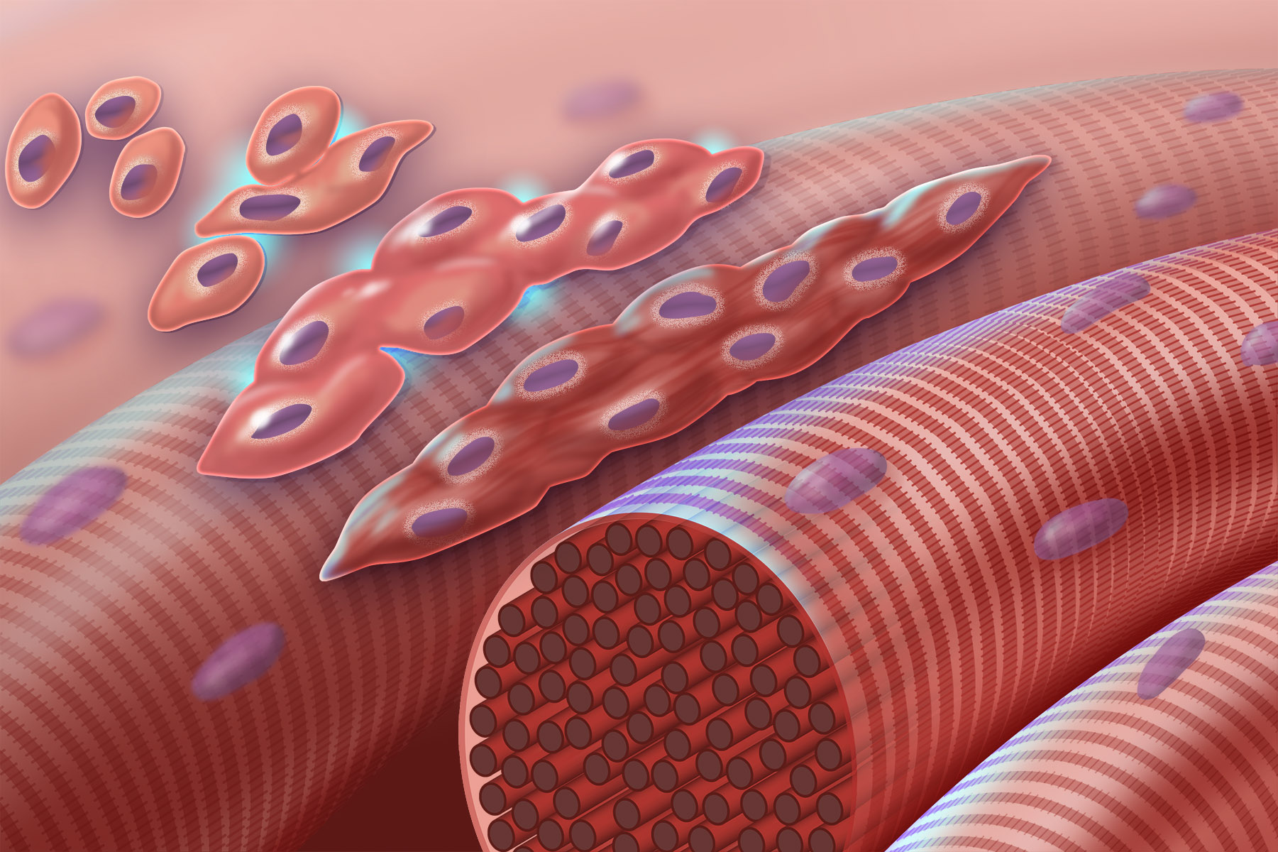
Myogenesis is the formation of skeletal muscular tissue, particularly during embryonic development. Muscle fibers generally form through the fusion of precursor myoblasts into multinucleated fibers called ''myotubes''. In the early development of an embryo, myoblasts can either proliferate, or differentiate into a myotube. What controls this choice in vivo is generally unclear. If placed in cell culture, most myoblasts will proliferate if enough fibroblast growth factor (FGF) or another growth factor is present in the medium surrounding the cells. When the growth factor runs out, the myoblasts cease division and undergo terminal differentiation into myotubes. Myoblast differentiation proceeds in stages. The first stage, involves cell cycle exit and the commencement of expression of certain genes. The second stage of differentiation involves the alignment of the myoblasts with one another. Studies have shown that even rat and chick myoblasts can recognise and align with one an ... [...More Info...] [...Related Items...] OR: [Wikipedia] [Google] [Baidu] |
Regulation Of Gene Expression
Regulation of gene expression, or gene regulation, includes a wide range of mechanisms that are used by cells to increase or decrease the production of specific gene products (protein or RNA). Sophisticated programs of gene expression are widely observed in biology, for example to trigger developmental pathways, respond to environmental stimuli, or adapt to new food sources. Virtually any step of gene expression can be modulated, from Transcriptional regulation, transcriptional initiation, to RNA processing, and to the post-translational modification of a protein. Often, one gene regulator controls another, and so on, in a gene regulatory network. Gene regulation is essential for viruses, prokaryotes and eukaryotes as it increases the versatility and adaptability of an organism by allowing the cell to express protein when needed. Although as early as 1951, Barbara McClintock showed interaction between two genetic loci, Activator (''Ac'') and Dissociator (''Ds''), in the color f ... [...More Info...] [...Related Items...] OR: [Wikipedia] [Google] [Baidu] |
SMAD3
Mothers against decapentaplegic homolog 3 also known as SMAD family member 3 or SMAD3 is a protein that in humans is encoded by the SMAD3 gene. SMAD3 is a member of the SMAD family of proteins. It acts as a mediator of the signals initiated by the transforming growth factor beta (TGF-β) superfamily of cytokines, which regulate cell proliferation, differentiation and death. Based on its essential role in TGF beta signaling pathway, SMAD3 has been related with tumor growth in cancer development. Gene The human SMAD3 gene is located on chromosome 15 on the cytogenic band at 15q22.33. The gene is composed of 9 exons over 129,339 base pairs. It is one of several human homologues of a gene that was originally discovered in the fruit fly ''Drosophila melanogaster''. The expression of SMAD3 has been related to the mitogen-activated protein kinase (MAPK/ERK pathway), particularly to the activity of mitogen-activated protein kinase kinase-1 (MEK1). Studies have demonstrated that inhi ... [...More Info...] [...Related Items...] OR: [Wikipedia] [Google] [Baidu] |
SMAD2
Mothers against decapentaplegic homolog 2 also known as SMAD family member 2 or SMAD2 is a protein that in humans is encoded by the ''SMAD2'' gene. MAD homolog 2 belongs to the SMAD, a family of proteins similar to the gene products of the ''Drosophila'' gene 'mothers against decapentaplegic' (Mad) and the ''C. elegans'' gene Sma. SMAD proteins are signal transducers and transcriptional modulators that mediate multiple signaling pathways. Function SMAD2 mediates the signal of the transforming growth factor (TGF)-beta, and thus regulates multiple cellular processes, such as cell proliferation, apoptosis, and differentiation. This protein is recruited to the TGF-beta receptors through its interaction with the SMAD anchor for receptor activation (SARA) protein. In response to TGF-beta signal, this protein is phosphorylated by the TGF-beta receptors. The phosphorylation induces the dissociation of this protein with SARA and the association with the family member SMAD4. The ass ... [...More Info...] [...Related Items...] OR: [Wikipedia] [Google] [Baidu] |
SMAD (protein)
Smads (or SMADs) comprise a family of structurally similar proteins that are the main signal transducers for receptors of the transforming growth factor beta (TGF-B) superfamily, which are critically important for regulating cell development and growth. The abbreviation refers to the homologies to the ''Caenorhabditis elegans'' SMA ("small" worm phenotype) and MAD family ("Mothers Against Decapentaplegic") of genes in Drosophila. There are three distinct sub-types of Smads: receptor-regulated Smads ( R-Smads), common partner Smads (Co-Smads), and inhibitory Smads ( I-Smads). The eight members of the Smad family are divided among these three groups. Trimers of two receptor-regulated SMADs and one co-SMAD act as transcription factors that regulate the expression of certain genes. Sub-types The R-Smads consist of Smad1, Smad2, Smad3, Smad5 and Smad8/9, and are involved in direct signaling from the TGF-B receptor. Smad4 is the only known human Co-Smad, and has the role of partneri ... [...More Info...] [...Related Items...] OR: [Wikipedia] [Google] [Baidu] |


