|
SMAD (protein)
Smads (or SMADs) comprise a family of structurally similar proteins that are the main signal transducers for receptors of the transforming growth factor beta (TGF-B) superfamily, which are critically important for regulating cell development and growth. The abbreviation refers to the homologies to the ''Caenorhabditis elegans'' SMA ("small" worm phenotype) and MAD family ("Mothers Against Decapentaplegic") of genes in Drosophila. There are three distinct sub-types of Smads: receptor-regulated Smads ( R-Smads), common partner Smads (Co-Smads), and inhibitory Smads ( I-Smads). The eight members of the Smad family are divided among these three groups. Trimers of two receptor-regulated SMADs and one co-SMAD act as transcription factors that regulate the expression of certain genes. Sub-types The R-Smads consist of Smad1, Smad2, Smad3, Smad5 and Smad8/9, and are involved in direct signaling from the TGF-B receptor. Smad4 is the only known human Co-Smad, and has the role of partne ... [...More Info...] [...Related Items...] OR: [Wikipedia] [Google] [Baidu] |
Protein
Proteins are large biomolecules and macromolecules that comprise one or more long chains of amino acid residues. Proteins perform a vast array of functions within organisms, including catalysing metabolic reactions, DNA replication, responding to stimuli, providing structure to cells and organisms, and transporting molecules from one location to another. Proteins differ from one another primarily in their sequence of amino acids, which is dictated by the nucleotide sequence of their genes, and which usually results in protein folding into a specific 3D structure that determines its activity. A linear chain of amino acid residues is called a polypeptide. A protein contains at least one long polypeptide. Short polypeptides, containing less than 20–30 residues, are rarely considered to be proteins and are commonly called peptides. The individual amino acid residues are bonded together by peptide bonds and adjacent amino acid residues. The sequence of amino acid resid ... [...More Info...] [...Related Items...] OR: [Wikipedia] [Google] [Baidu] |
Mothers Against Decapentaplegic
Mothers against decapentaplegic is a protein from the SMAD family that was discovered in ''Drosophila''. During ''Drosophila'' research, it was found that a mutation in the gene in the mother repressed the gene decapentaplegic in the embryo. The phrase "Mothers against" was added as a humorous take-off on organizations opposing various issues e.g. Mothers Against Drunk Driving (MADD); and based on a tradition of such unusual naming within the gene research community. Several human homologues are known: * Mothers against decapentaplegic homolog 1 * Mothers against decapentaplegic homolog 2 * Mothers against decapentaplegic homolog 3 * Mothers against decapentaplegic homolog 4 * Mothers against decapentaplegic homolog 5 * Mothers against decapentaplegic homolog 6 * Mothers against decapentaplegic homolog 7 * Mothers against decapentaplegic homolog 9 Mothers against decapentaplegic homolog 9 also known as SMAD9, SMAD8, and MADH6 is a protein that in humans is encoded by th ... [...More Info...] [...Related Items...] OR: [Wikipedia] [Google] [Baidu] |
SiRNA
Small interfering RNA (siRNA), sometimes known as short interfering RNA or silencing RNA, is a class of double-stranded RNA at first non-coding RNA molecules, typically 20-24 (normally 21) base pairs in length, similar to miRNA, and operating within the RNA interference (RNAi) pathway. It interferes with the expression of specific genes with complementary nucleotide sequences by degrading mRNA after transcription, preventing translation. Text was copied from this source, which is available under Creative Commons Attribution 4.0 International License Structure Naturally occurring siRNAs have a well-defined structure that is a short (usually 20 to 24- bp) double-stranded RNA (dsRNA) with phosphorylated 5' ends and hydroxylated 3' ends with two overhanging nucleotides. The Dicer enzyme catalyzes production of siRNAs from long dsRNAs and small hairpin RNAs. siRNAs can also be introduced into cells by transfection. Since in principle any gene can be knocked down by a ... [...More Info...] [...Related Items...] OR: [Wikipedia] [Google] [Baidu] |
Luciferase
Luciferase is a generic term for the class of oxidative enzymes that produce bioluminescence, and is usually distinguished from a photoprotein. The name was first used by Raphaël Dubois who invented the words '' luciferin'' and ''luciferase'', for the substrate and enzyme, respectively. Both words are derived from the Latin word ''lucifer'', meaning "lightbearer", which in turn is derived from the Latin words for "light" (''lux)'' and "to bring or carry" (''ferre)''.Luciferases are widely used in biotechnology, for bioluminescence imaging microscopy and as reporter genes, for many of the same applications as fluorescent proteins. However, unlike fluorescent proteins, luciferases do not require an external light source, but do require addition of luciferin, the consumable substrate. Examples A variety of organisms regulate their light production using different luciferases in a variety of light-emitting reactions. The majority of studied luciferases have been found in anim ... [...More Info...] [...Related Items...] OR: [Wikipedia] [Google] [Baidu] |
Cytostasis
Cytostasis (cyto – cell; stasis – stoppage) is the inhibition of cell growth and multiplication. Cytostatic refers to a cellular component or medicine that inhibits cell division. Cytostasis is an important prerequisite for structured multicellular organisms. Without regulation of cell growth and division only unorganized heaps of cells would be possible. Chemotherapy of cancer, treatment of skin diseases and treatment of infections are common use cases of cytostatic drugs. Active hygienic products generally contain cytostatic substances. Cytostatic mechanisms and drugs generally occur together with cytotoxic ones. Activators Nitric oxide – activated macrophages produce large amounts of nitric oxide (NO), which induces both cytostasis and cytotoxicity to tumor cells both ''in vitro'' and ''in vivo''. Nitric oxide-induced cytostasis targets ribonucleotide reductase by rapid and reversible inhibition. However, other studies show there could be other targets that are respon ... [...More Info...] [...Related Items...] OR: [Wikipedia] [Google] [Baidu] |
Serine/threonine Kinase
A serine/threonine protein kinase () is a kinase enzyme, in particular a protein kinase, that phosphorylates the OH group of the amino-acid residues serine or threonine, which have similar side chains. At least 350 of the 500+ human protein kinases are serine/threonine kinases (STK). In enzymology, the term ''serine/threonine protein kinase'' describes a class of enzymes in the family of transferases, that transfer phosphates to the oxygen atom of a serine or threonine side chain in proteins. This process is called phosphorylation. Protein phosphorylation in particular plays a significant role in a wide range of cellular processes and is a very important posttranslational modification. The chemical reaction performed by these enzymes can be written as :ATP + a protein \rightleftharpoons ADP + a phosphoprotein Thus, the two substrates of this enzyme are ATP and a protein, whereas its two products are ADP and phosphoprotein. The systematic name of this enzyme class is ... [...More Info...] [...Related Items...] OR: [Wikipedia] [Google] [Baidu] |
WW Domain
The WW domain, (also known as the rsp5-domain or WWP repeating motif) is a modular protein domain that mediates specific interactions with protein ligands. This domain is found in a number of unrelated signaling and structural proteins and may be repeated up to four times in some proteins. Apart from binding preferentially to proteins that are proline-rich, with particular proline-motifs, PP-P- PY, some WW domains bind to phosphoserine- phosphothreonine-containing motifs. Structure and ligands The WW domain is one of the smallest protein modules, composed of only 40 amino acids, which mediates specific protein-protein interactions with short proline-rich or proline-containing motifs. Named after the presence of two conserved tryptophans (W), which are spaced 20-22 amino acids apart within the sequence, the WW domain folds into a meandering triple-stranded beta sheet. The identification of the WW domain was facilitated by the analysis of two splice isoforms of YAP gen ... [...More Info...] [...Related Items...] OR: [Wikipedia] [Google] [Baidu] |
Proteasome
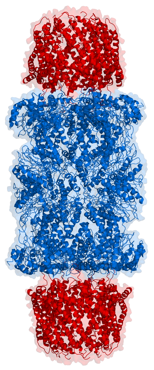
Proteasomes are protein complexes which degrade unneeded or damaged proteins by proteolysis, a chemical reaction that breaks peptide bonds. Enzymes that help such reactions are called proteases. Proteasomes are part of a major mechanism by which cells regulate the concentration of particular proteins and degrade misfolded proteins. Proteins are tagged for degradation with a small protein called ubiquitin. The tagging reaction is catalyzed by enzymes called ubiquitin ligases. Once a protein is tagged with a single ubiquitin molecule, this is a signal to other ligases to attach additional ubiquitin molecules. The result is a ''polyubiquitin chain'' that is bound by the proteasome, allowing it to degrade the tagged protein. The degradation process yields peptides of about seven to eight amino acids long, which can then be further degraded into shorter amino acid sequences and used in synthesizing new proteins. Proteasomes are found inside all eukaryotes and archaea, and in some ... [...More Info...] [...Related Items...] OR: [Wikipedia] [Google] [Baidu] |
Ubiquitin Ligase
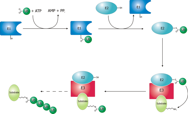
A ubiquitin ligase (also called an E3 ubiquitin ligase) is a protein that recruits an E2 ubiquitin-conjugating enzyme that has been loaded with ubiquitin, recognizes a protein substrate, and assists or directly catalyzes the transfer of ubiquitin from the E2 to the protein substrate. In simple and more general terms, the ligase enables movement of ubiquitin from a ubiquitin carrier to another thing (the substrate) by some mechanism. The ubiquitin, once it reaches its destination, ends up being attached by an isopeptide bond to a lysine residue, which is part of the target protein. E3 ligases interact with both the target protein and the E2 enzyme, and so impart substrate specificity to the E2. Commonly, E3s polyubiquitinate their substrate with Lys48-linked chains of ubiquitin, targeting the substrate for destruction by the proteasome. However, many other types of linkages are possible and alter a protein's activity, interactions, or localization. Ubiquitination by E3 ligases r ... [...More Info...] [...Related Items...] OR: [Wikipedia] [Google] [Baidu] |
Phosphorylation
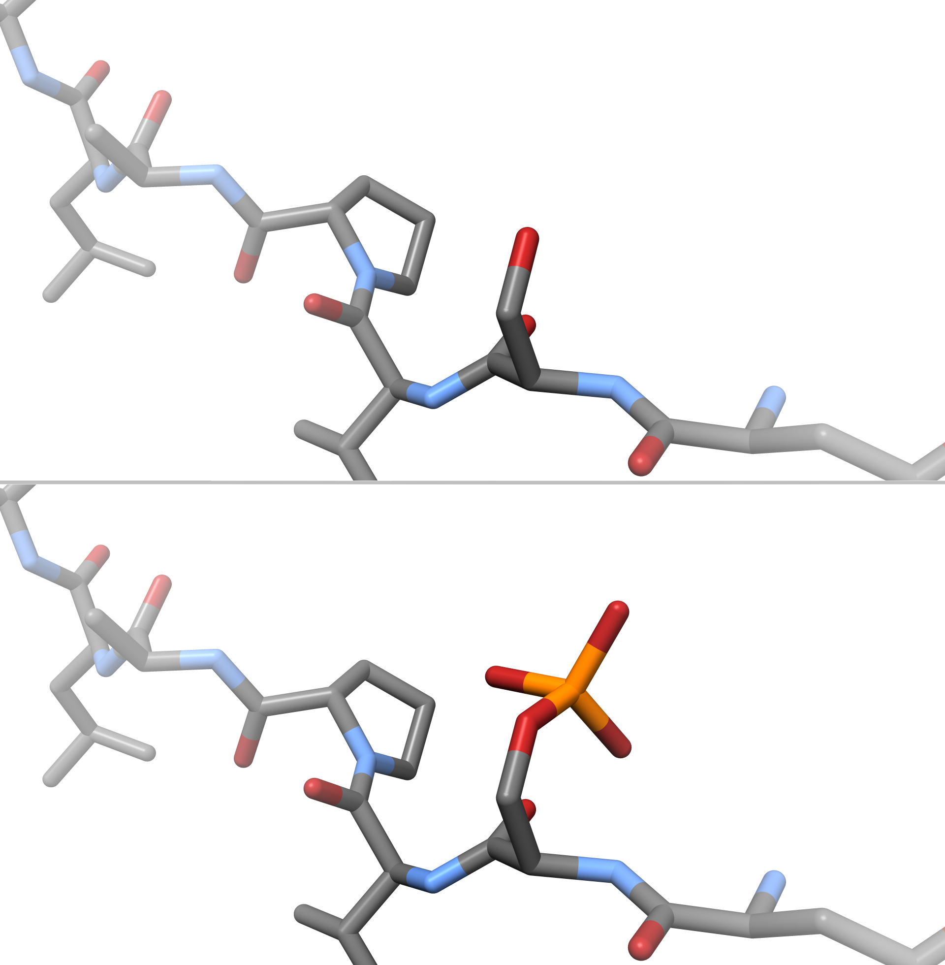
In chemistry, phosphorylation is the attachment of a phosphate group to a molecule or an ion. This process and its inverse, dephosphorylation, are common in biology and could be driven by natural selection. Text was copied from this source, which is available under a Creative Commons Attribution 4.0 International License. Protein phosphorylation often activates (or deactivates) many enzymes. Glucose Phosphorylation of sugars is often the first stage in their catabolism. Phosphorylation allows cells to accumulate sugars because the phosphate group prevents the molecules from diffusing back across their transporter. Phosphorylation of glucose is a key reaction in sugar metabolism. The chemical equation for the conversion of D-glucose to D-glucose-6-phosphate in the first step of glycolysis is given by :D-glucose + ATP → D-glucose-6-phosphate + ADP :ΔG° = −16.7 kJ/mol (° indicates measurement at standard condition) Hepatic cells are freely permeable to glucose, an ... [...More Info...] [...Related Items...] OR: [Wikipedia] [Google] [Baidu] |
BMP4
Bone morphogenetic protein 4 is a protein that in humans is encoded by ''BMP4'' gene. BMP4 is found on chromosome 14q22-q23. BMP4 is a member of the bone morphogenetic protein family which is part of the transforming growth factor-beta superfamily. The superfamily includes large families of growth and differentiation factors. BMP4 is highly conserved evolutionarily. BMP4 is found in early embryonic development in the ventral marginal zone and in the eye, heart blood and otic vesicle. Discovery Bone morphogenetic proteins were originally identified by an ability of demineralized bone extract to induce endochondral osteogenesis in vivo in an extraskeletal site. Function BMP4 is a polypeptide belonging to the TGF-β superfamily of proteins. It, like other bone morphogenetic proteins, is involved in bone and cartilage development, specifically tooth and limb development and fracture repair. This particular family member plays an important role in the onset of endochondral bone ... [...More Info...] [...Related Items...] OR: [Wikipedia] [Google] [Baidu] |


