|
Muscle Fascicles
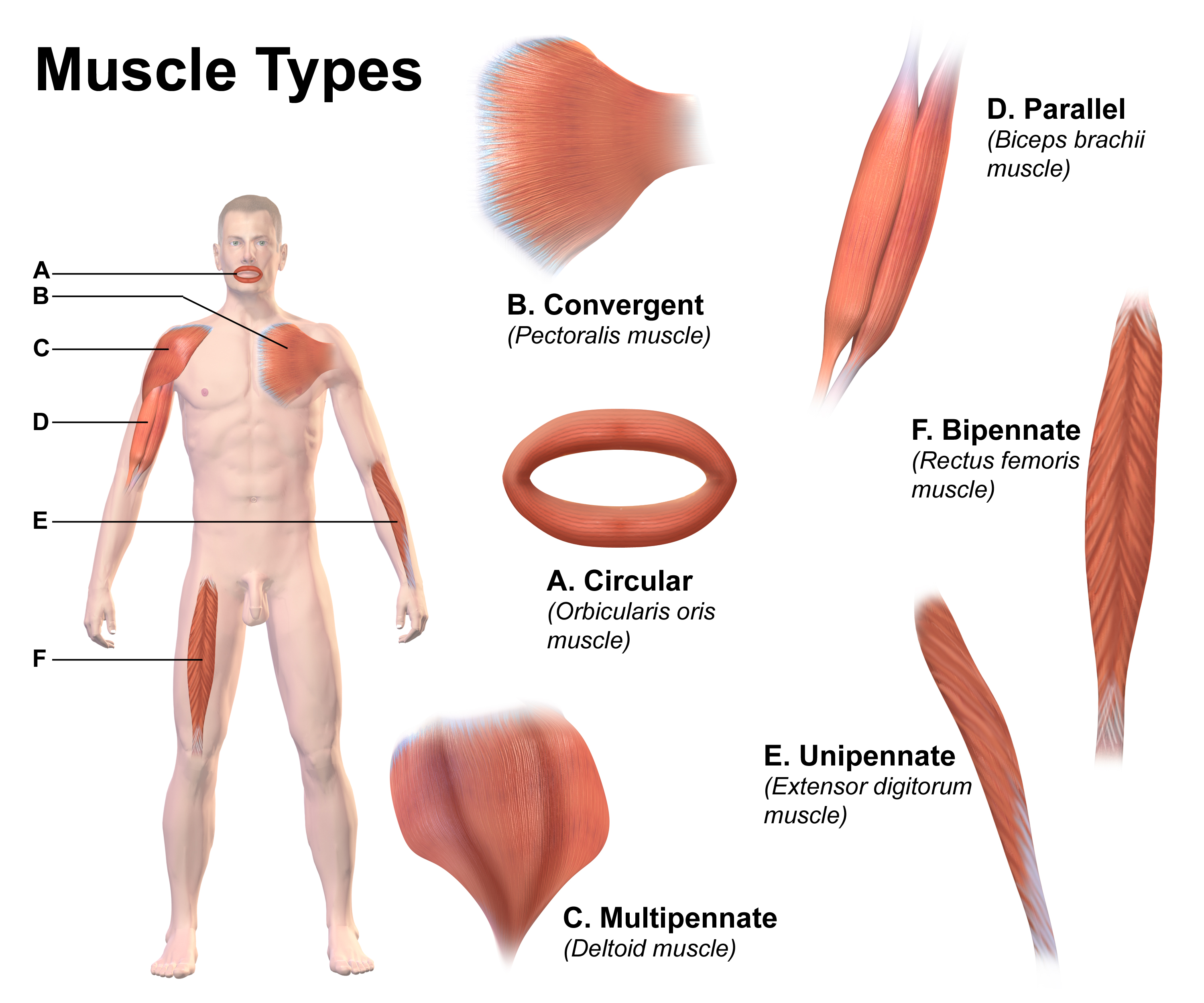
A muscle fascicle is a bundle of skeletal muscle fibers surrounded by perimysium, a type of connective tissue. Structure Muscle cells are grouped into muscle fascicles by enveloping perimysium connective tissue. Fascicles are bundled together by epimysium connective tissue. Muscle fascicles typically only contain one type of muscle cell (either type I fibres or type II fibres), but can contain a mixture of both types. Function In the heart specialized cardiac muscle cells transmit electrical impulses from the atrioventricular node (AV node) to the Purkinje fibers – fascicles, also referred to as bundle branches. These start as a single fascicle of fibers at the AV node called the bundle of His that then splits into three bundle branches: the right fascicular branch, left anterior fascicular branch, and left posterior fascicular branch. Clinical significance Myositis may cause thickening of the muscle fascicles. This may be detected with ultrasound scans. Mu ... [...More Info...] [...Related Items...] OR: [Wikipedia] [Google] [Baidu] |
Skeletal Muscle
Skeletal muscles (commonly referred to as muscles) are organs of the vertebrate muscular system and typically are attached by tendons to bones of a skeleton. The muscle cells of skeletal muscles are much longer than in the other types of muscle tissue, and are often known as muscle fibers. The muscle tissue of a skeletal muscle is striated – having a striped appearance due to the arrangement of the sarcomeres. Skeletal muscles are voluntary muscles under the control of the somatic nervous system. The other types of muscle are cardiac muscle which is also striated and smooth muscle which is non-striated; both of these types of muscle tissue are classified as involuntary, or, under the control of the autonomic nervous system. A skeletal muscle contains multiple muscle fascicle, fascicles – bundles of muscle fibers. Each individual fiber, and each muscle is surrounded by a type of connective tissue layer of fascia. Muscle fibers are formed from the cell fusion, fusion of ... [...More Info...] [...Related Items...] OR: [Wikipedia] [Google] [Baidu] |
Purkinje Fibers
The Purkinje fibers (; often incorrectly ; Purkinje tissue or subendocardial branches) are located in the inner ventricular walls of the heart, just beneath the endocardium in a space called the subendocardium. The Purkinje fibers are specialized conducting fibers composed of electrically excitable cells. They are larger than cardiomyocytes with fewer myofibrils and many mitochondria. They conduct cardiac action potentials more quickly and efficiently than any of the other cells in the heart's electrical conduction system. Purkinje fibers allow the heart's conduction system to create synchronized contractions of its ventricles, and are essential for maintaining a consistent heart rhythm. Histology Purkinje fibers are a unique cardiac end-organ. Further histologic examination reveals that these fibers are split in ventricles walls. The electrical origin of atrial Purkinje fibers arrives from the sinoatrial node. Given no aberrant channels, the Purkinje fibers are distinct ... [...More Info...] [...Related Items...] OR: [Wikipedia] [Google] [Baidu] |
Endomysium
The endomysium, meaning ''within the muscle'', is a wispy layer of areolar connective tissue that ensheaths each individual Skeletal muscle#Skeletal muscle fibers, muscle fiber, or muscle cell. It also contains capillaries and nerves. It overlies the muscle fiber's cell membrane: the sarcolemma. Endomysium is the deepest and smallest component of Muscle tissue, muscle connective tissue. This thin layer helps provide an appropriate chemical environment for the exchange of calcium, sodium, and potassium, which is essential for the excitation and subsequent contraction of a muscle fiber. Endomysium combines with perimysium and epimysium to create the collagen fibers of tendons, providing the tissue connection between muscles and bones by indirect attachment. It connects with perimysium using intermittent perimysial junction plates. Collagen is the major protein that composes connective tissues like endomysium. Endomysium has been shown to contain mainly Type I collagen, type I and Co ... [...More Info...] [...Related Items...] OR: [Wikipedia] [Google] [Baidu] |
Connective Tissue In Skeletal Muscle
Skeletal muscles (commonly referred to as muscles) are organs of the vertebrate muscular system and typically are attached by tendons to bones of a skeleton. The muscle cells of skeletal muscles are much longer than in the other types of muscle tissue, and are often known as muscle fibers. The muscle tissue of a skeletal muscle is striated – having a striped appearance due to the arrangement of the sarcomeres. Skeletal muscles are voluntary muscles under the control of the somatic nervous system. The other types of muscle are cardiac muscle which is also striated and smooth muscle which is non-striated; both of these types of muscle tissue are classified as involuntary, or, under the control of the autonomic nervous system. A skeletal muscle contains multiple fascicles – bundles of muscle fibers. Each individual fiber, and each muscle is surrounded by a type of connective tissue layer of fascia. Muscle fibers are formed from the fusion of developmental myoblasts in a proc ... [...More Info...] [...Related Items...] OR: [Wikipedia] [Google] [Baidu] |
Myokymia
Myokymia is an involuntary, spontaneous, localized quivering of a few muscles, or bundles within a muscle, but which are insufficient to move a joint. One type is superior oblique myokymia. Myokymia is commonly used to describe an involuntary eyelid muscle contraction, typically involving the Eyelid, lower eyelid or less often the Eyelid, upper eyelid. It occurs in normal individuals and typically starts and disappears spontaneously. However, it can sometimes last up to three weeks. Since the condition typically resolves itself, medical professionals do not consider it to be serious or a cause for concern. In contrast, facial myokymia is a fine rippling of muscles on one side of the face and may reflect an underlying Neoplasm, tumor in the brainstem (typically a brainstem glioma), demyelinating disease, loss of myelin in the brainstem (associated with multiple sclerosis) or in the recovery stage of Miller-Fisher syndrome, a variant of Guillain–Barré syndrome, an inflammatory pol ... [...More Info...] [...Related Items...] OR: [Wikipedia] [Google] [Baidu] |
Dermatomyositis
Dermatomyositis (DM) is a long-term inflammatory disorder which affects skin and the muscles. Its symptoms are generally a skin rash and worsening muscle weakness over time. These may occur suddenly or develop over months. Other symptoms may include weight loss, fever, lung inflammation, or light sensitivity. Complications may include calcium deposits in muscles or skin. The cause is unknown. Theories include that it is an autoimmune disease or a result of a viral infection. Dermatomyositis may develop as a paraneoplastic syndrome associated with several forms of malignancy. It is a type of inflammatory myopathy. Diagnosis is typically based on some combination of symptoms, blood tests, electromyography, and muscle biopsies. While no cure for the condition is known, treatments generally improve symptoms. Treatments may include medication, physical therapy, exercise, heat therapy, orthotics and assistive devices, and rest. Medications in the corticosteroids family are typic ... [...More Info...] [...Related Items...] OR: [Wikipedia] [Google] [Baidu] |
Medical Ultrasound
Medical ultrasound includes diagnostic techniques (mainly imaging techniques) using ultrasound, as well as therapeutic applications of ultrasound. In diagnosis, it is used to create an image of internal body structures such as tendons, muscles, joints, blood vessels, and internal organs, to measure some characteristics (e.g. distances and velocities) or to generate an informative audible sound. Its aim is usually to find a source of disease or to exclude pathology. The usage of ultrasound to produce visual images for medicine is called medical ultrasonography or simply sonography. The practice of examining pregnant women using ultrasound is called obstetric ultrasonography, and was an early development of clinical ultrasonography. Ultrasound is composed of sound waves with frequencies which are significantly higher than the range of human hearing (>20,000 Hz). Ultrasonic images, also known as sonograms, are created by sending pulses of ultrasound into tissue using a pr ... [...More Info...] [...Related Items...] OR: [Wikipedia] [Google] [Baidu] |
Inflammation
Inflammation (from la, wikt:en:inflammatio#Latin, inflammatio) is part of the complex biological response of body tissues to harmful stimuli, such as pathogens, damaged cells, or Irritation, irritants, and is a protective response involving immune cells, blood vessels, and molecular mediators. The function of inflammation is to eliminate the initial cause of cell injury, clear out necrotic cells and tissues damaged from the original insult and the inflammatory process, and initiate tissue repair. The five cardinal signs are heat, pain, redness, swelling, and Functio laesa, loss of function (Latin ''calor'', ''dolor'', ''rubor'', ''tumor'', and ''functio laesa''). Inflammation is a generic response, and therefore it is considered as a mechanism of innate immune system, innate immunity, as compared to adaptive immune system, adaptive immunity, which is specific for each pathogen. Too little inflammation could lead to progressive tissue destruction by the harmful stimulus (e.g. b ... [...More Info...] [...Related Items...] OR: [Wikipedia] [Google] [Baidu] |
Myositis
Myositis is a rare disease that involves inflammation of the muscles. This can present with a variety of symptoms such as skin involvement (i.e., rashes), muscle weakness, and other organ involvement. Systemic symptoms such as weight loss, fatigue, and low fever can also present. Causes Injury, medicines, infection, or an autoimmune disorder can lead to myositis. It can also be idiopathic. * Injury - A mild form of myositis can occur with hard exercise. A more severe form of muscle injury, called rhabdomyolysis, is also associated with myositis. This is a condition where injury to your muscles causes them to quickly break down. * Medicines - A variety of different medicines can cause myositis. One of the most common drug types that can cause myositis is statins. Statins are drugs that are used to help lower high cholesterol. One of the most common side effects of statin therapy is muscle pain. Rarely, statin therapy can lead to myositis. * Infection - The most common infectious c ... [...More Info...] [...Related Items...] OR: [Wikipedia] [Google] [Baidu] |
Bundle Of His
The bundle of His (BH) or His bundle (HB) ( "hiss"Medical Terminology for Health Professions, Spiral bound Version'. Cengage Learning; 2016. . pp. 129–.) is a collection of heart muscle cells specialized for electrical conduction. As part of the electrical conduction system of the heart, it transmits the electrical impulses from the atrioventricular node (located between the atria and the ventricles) to the point of the apex of the fascicular branches via the bundle branches. The fascicular branches then lead to the Purkinje fibers, which provide electrical conduction to the ventricles, causing the cardiac muscle of the ventricles to contract at a paced interval. Function The bundle of His is an important part of the electrical conduction system of the heart, as it transmits impulses from the atrioventricular node, located at the anterior-inferior end of the interatrial septum, to the ventricles of the heart. The bundle of His branches into the left and the right bundle br ... [...More Info...] [...Related Items...] OR: [Wikipedia] [Google] [Baidu] |
AV Node
The atrioventricular node or AV node electrically connects the heart's atria and ventricles to coordinate beating in the top of the heart; it is part of the electrical conduction system of the heart. The AV node lies at the lower back section of the interatrial septum near the opening of the coronary sinus, and conducts the normal electrical impulse from the atria to the ventricles. The AV node is quite compact (~1 x 3 x 5 mm).Full Size Picture triangle of-Koch.jpg Retrieved on 2008-12-22 Structure Location The AV node lies at the lower back section of the |
Skeletal Muscle
Skeletal muscles (commonly referred to as muscles) are organs of the vertebrate muscular system and typically are attached by tendons to bones of a skeleton. The muscle cells of skeletal muscles are much longer than in the other types of muscle tissue, and are often known as muscle fibers. The muscle tissue of a skeletal muscle is striated – having a striped appearance due to the arrangement of the sarcomeres. Skeletal muscles are voluntary muscles under the control of the somatic nervous system. The other types of muscle are cardiac muscle which is also striated and smooth muscle which is non-striated; both of these types of muscle tissue are classified as involuntary, or, under the control of the autonomic nervous system. A skeletal muscle contains multiple muscle fascicle, fascicles – bundles of muscle fibers. Each individual fiber, and each muscle is surrounded by a type of connective tissue layer of fascia. Muscle fibers are formed from the cell fusion, fusion of ... [...More Info...] [...Related Items...] OR: [Wikipedia] [Google] [Baidu] |


