|
Mullerian Anomalies
Müllerian duct anomalies are those structural anomalies caused by errors in müllerian duct development during embryonic morphogenesis. Factors that precipitate include genetics, and maternal exposure to teratogens. Genetic causes of müllerian duct anomalies are complicated and uncommon. Inheritance patterns can be autosomal dominant, autosomal recessive, and X-linked disorders. Müllerian anomalies can be part of a multiple malformation syndrome. Mullerian anomalies occur as a congenital malformation of the mullerian ducts during embryogenesis. The mullerian ducts are also referred to as paramesonephric ducts, referring to ducts next to (para) the mesonephric (Wolffian) duct during foetal development. Paramesonephric ducts are paired ducts derived from the embryo, and for females develop into the uterus, uterine tubes, cervix and upper two-thirds of the vagina. Embryogenesis of the mullerian ducts play important roles in ensuring normal development of the female reproductive tr ... [...More Info...] [...Related Items...] OR: [Wikipedia] [Google] [Baidu] |
Müllerian Duct
Paramesonephric ducts (or Müllerian ducts) are paired ducts of the embryo that run down the lateral sides of the genital ridge and terminate at the sinus tubercle in the primitive urogenital sinus. In the female, they will develop to form the fallopian tubes, uterus, cervix, and the upper one-third of the vagina. Development The female reproductive system is composed of two embryological segments: the urogenital sinus and the paramesonephric ducts. The two are conjoined at the sinus tubercle. Paramesonephric ducts are present on the embryo of both sexes. Only in females do they develop into reproductive organs. They degenerate in males of certain species, but the adjoining mesonephric ducts develop into male reproductive organs. The sex based differences in the contributions of the paramesonephric ducts to reproductive organs is based on the presence, and degree of presence, of Anti-Müllerian hormone. During the formation of the reproductive system, the paramesonephric ducts ar ... [...More Info...] [...Related Items...] OR: [Wikipedia] [Google] [Baidu] |
Magnetic Resonance Imaging
Magnetic resonance imaging (MRI) is a medical imaging technique used in radiology to form pictures of the anatomy and the physiological processes of the body. MRI scanners use strong magnetic fields, magnetic field gradients, and radio waves to generate images of the organs in the body. MRI does not involve X-rays or the use of ionizing radiation, which distinguishes it from CT and PET scans. MRI is a medical application of nuclear magnetic resonance (NMR) which can also be used for imaging in other NMR applications, such as NMR spectroscopy. MRI is widely used in hospitals and clinics for medical diagnosis, staging and follow-up of disease. Compared to CT, MRI provides better contrast in images of soft-tissues, e.g. in the brain or abdomen. However, it may be perceived as less comfortable by patients, due to the usually longer and louder measurements with the subject in a long, confining tube, though "Open" MRI designs mostly relieve this. Additionally, implants and oth ... [...More Info...] [...Related Items...] OR: [Wikipedia] [Google] [Baidu] |
Hematoma
A hematoma, also spelled haematoma, or blood suffusion is a localized bleeding outside of blood vessels, due to either disease or trauma including injury or surgery and may involve blood continuing to seep from broken capillary, capillaries. A hematoma is benign and is initially in liquid form spread among the tissues including in sacs between tissues where it may coagulate and solidify before blood is reabsorbed into blood vessels. An ecchymosis is a hematoma of the skin larger than 10 mm. They may occur among and or within many areas such as skin and other organs, connective tissues, bone, joints and muscle. A collection of blood (or even a hemorrhage) may be aggravated by anticoagulant medication (blood thinner). Blood seepage and collection of blood may occur if heparin is given via an Intramuscular injection, intramuscular route; to avoid this, heparin must be given intravenously or subcutaneous injection, subcutaneously. Signs and symptoms Some hematomas are visible ... [...More Info...] [...Related Items...] OR: [Wikipedia] [Google] [Baidu] |
Labia Minora
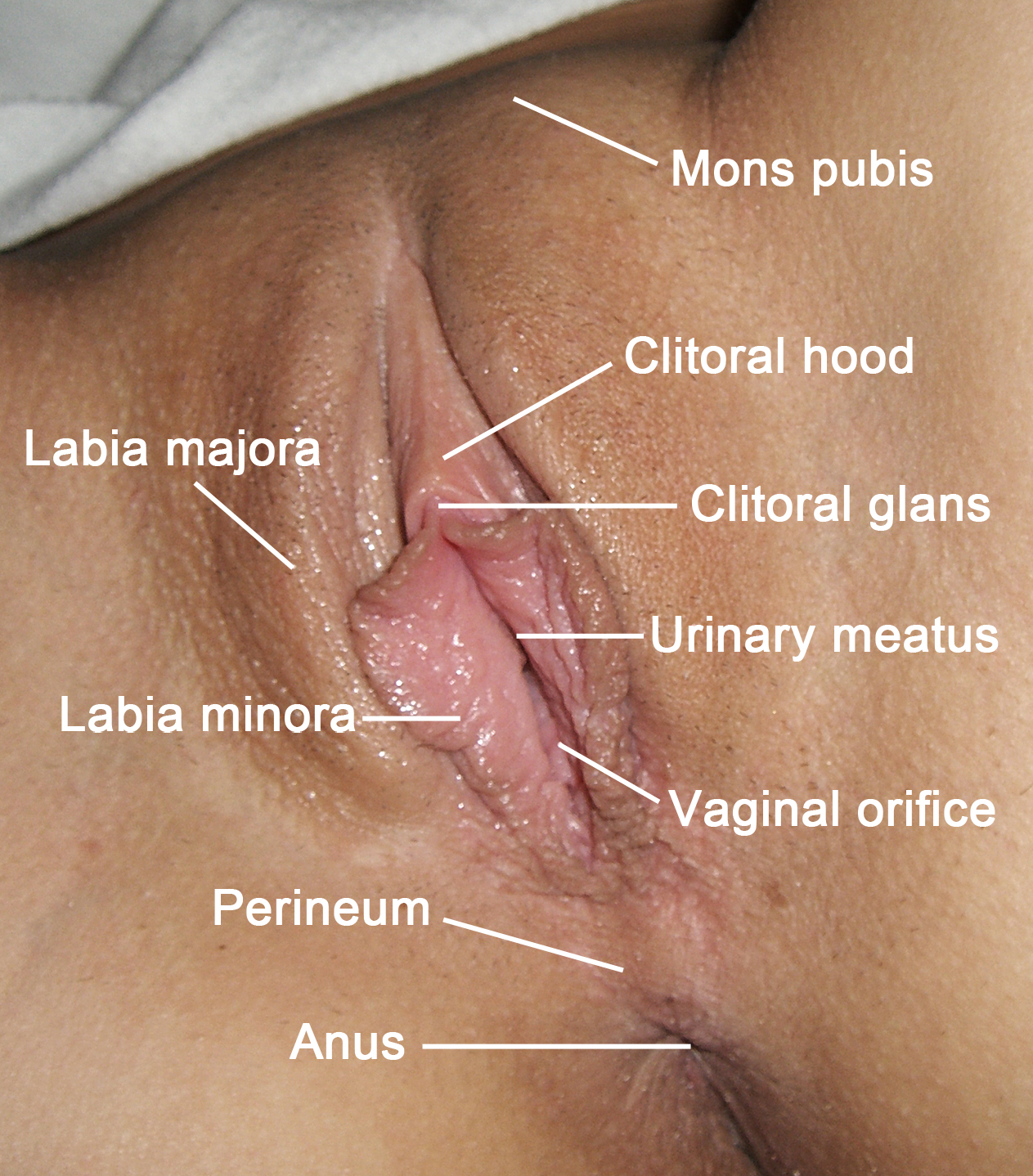
The labia minora (Latin for 'smaller lips', singular: ''labium minus'', 'smaller lip'), also known as the inner labia, inner lips, vaginal lips or nymphae are two flaps of skin on either side of the human vaginal opening in the vulva, situated between the labia majora (Latin for 'larger lips'; also called outer labia, or outer lips). The labia minora vary widely in size, color and shape from individual to individual. The labia minora are homologous to the male urethral surface of the penis. Structure and functioning The labia minora extend from the clitoris obliquely downward, laterally, and backward on either side of the vulval vestibule, ending between the bottom of the vulval vestibule and the labia majora. The posterior ends (bottom) of the labia minora are usually joined across the middle line by a flap of skin, named the frenulum of labia minora or fourchette. On the front, each lip forks dividing into two portions surrounding the clitoris. The upper part of each lip pas ... [...More Info...] [...Related Items...] OR: [Wikipedia] [Google] [Baidu] |
Neovagina
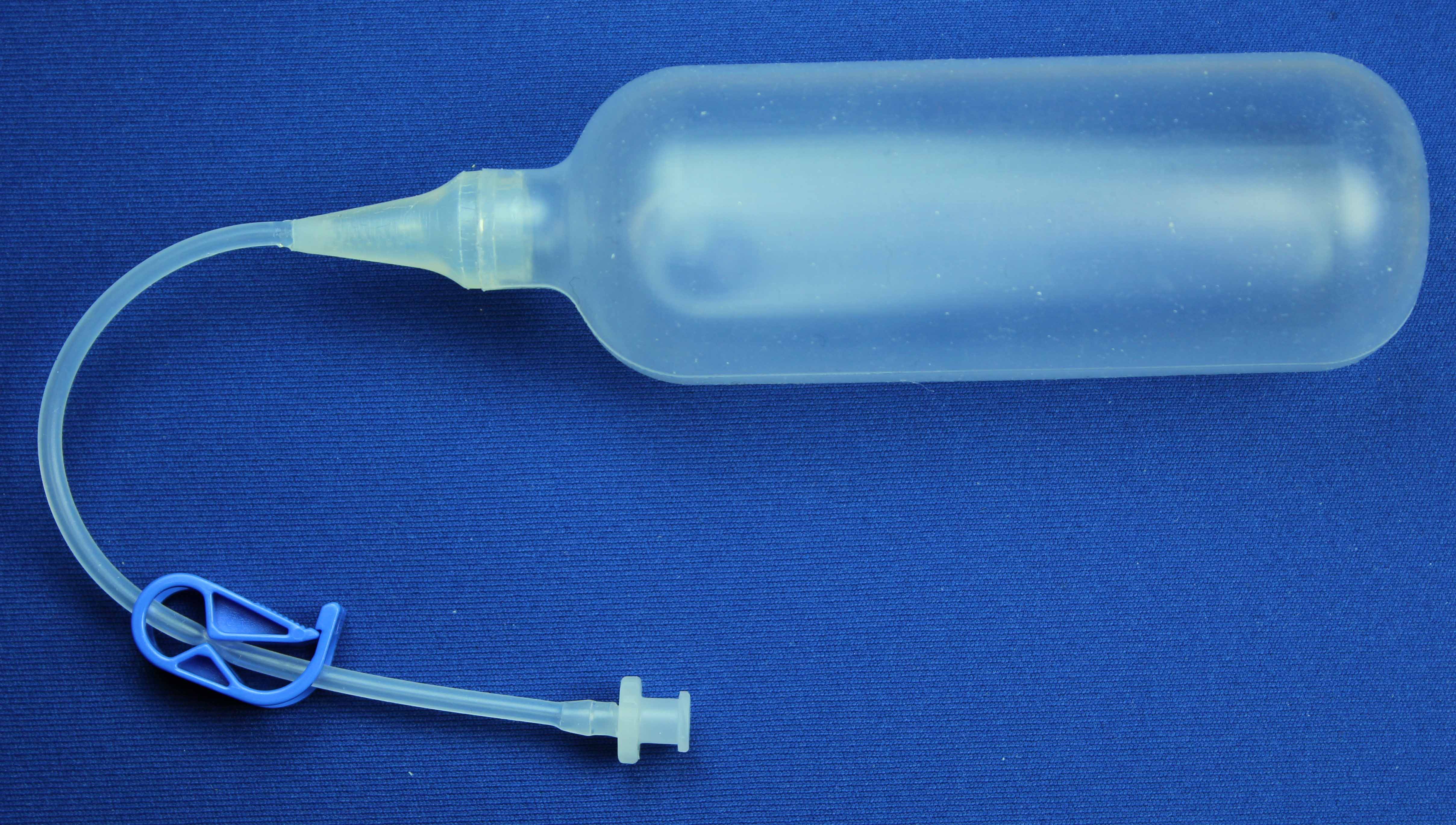
Vaginoplasty is any surgical procedure that results in the construction or reconstruction of the vagina. It is a type of genitoplasty. Pelvic organ prolapse is often treated with one or more surgeries to repair the vagina. Sometimes a vaginoplasty is needed following the treatment or removal of malignant growths or abscesses in order to restore a normal vaginal structure and function. Surgery to the vagina is done to correct congenital defects to the vagina, urethra and rectum. It will correct protrusion of the urinary bladder into the vagina (cystocele) and protrusion of the rectum (rectocele) into the vagina. Often, a vaginoplasty is performed to repair the vagina and its attached structures due to trauma or injury. Labiaplasty, which alters the appearance of the vulva, can be performed as a discrete surgery, or as a subordinate procedure within a vaginoplasty. Congenital disorders such as adrenal hyperplasia can affect the structure and function of the vagina and sometimes t ... [...More Info...] [...Related Items...] OR: [Wikipedia] [Google] [Baidu] |
HOXA13
Homeobox protein Hox-A13 is a protein that in humans is encoded by the ''HOXA13'' gene. Function In vertebrates, the genes encoding the class of transcription factors called homeobox genes are found in clusters named A, B, C, and D on four separate chromosomes. Expression of these proteins is spatially and temporally regulated during embryonic development. This gene is part of the A cluster on chromosome 7 and encodes a DNA-binding transcription factor which may regulate gene expression, morphogenesis, and Cellular differentiation, differentiation. Clinical significance Expansion of a polyalanine tract in the encoded protein can cause Hand-Foot-Genital Syndrome, hand-foot-genital syndrome, also known as hand-foot-uterus syndrome. Aberrant expression of ''HoxA13'' gene products in the esophagus, provokes Barrett’s esophagus, a form of metaplasia that is a direct precursor to esophageal cancer. See also * Homeobox References Further reading * * * * * * * ... [...More Info...] [...Related Items...] OR: [Wikipedia] [Google] [Baidu] |
HOXA11
Homeobox protein Hox-A11 is a protein that in humans is encoded by the ''HOXA11'' gene. Function In vertebrates, the genes encoding the class of transcription factors called homeobox genes are found in clusters named A, B, C, and D on four separate chromosomes. Expression of these proteins is spatially and temporally regulated during embryonic development. This gene is part of the A cluster on chromosome 7 and encodes a DNA-binding transcription factor which may regulate gene expression, morphogenesis, and differentiation. This gene is involved in the regulation of uterine development and is required for female fertility. Mutations in this gene can cause radioulnar synostosis Radioulnar synostosis is a rare condition where there is an abnormal connection between the radius and ulna bones of the forearm. This can be present at birth (congenital), when it is a result of a failure of the bones to form separately, or follo ... with amegakaryocytic thrombocytopenia. See also * ... [...More Info...] [...Related Items...] OR: [Wikipedia] [Google] [Baidu] |
HOXA10
Homeobox protein Hox-A10 is a protein that in humans is encoded by the ''HOXA10'' gene. Function In vertebrates, the genes encoding the class of transcription factors called homeobox genes are found in clusters named A, B, C, and D on four separate chromosomes. Expression of these proteins is spatially and temporally regulated during embryonic development. This gene is part of the A cluster on chromosome 7 and encodes a DNA-binding transcription factor that may regulate gene expression, morphogenesis, and differentiation. More specifically, it may function in fertility, embryo viability, and regulation of hematopoietic lineage commitment. Alternatively spliced transcript variants encoding different isoforms have been described. Downregulation of HOXA10 is observed in the human and baboon decidua after implantation and this downregulation promotes trophoblast invasion by activating STAT3 Interactions Homeobox A10 has been shown to interact with PTPN6. See also * Homeobox ... [...More Info...] [...Related Items...] OR: [Wikipedia] [Google] [Baidu] |
Spina Bifida
Spina bifida (Latin for 'split spine'; SB) is a birth defect in which there is incomplete closing of the spine and the membranes around the spinal cord during early development in pregnancy. There are three main types: spina bifida occulta, meningocele and myelomeningocele. Meningocele and myelomeningocele may be grouped as spina bifida cystica. The most common location is the lower back, but in rare cases it may be in the middle back or neck. Occulta has no or only mild signs, which may include a hairy patch, dimple, dark spot or swelling on the back at the site of the gap in the spine. Meningocele typically causes mild problems, with a sac of fluid present at the gap in the spine. Myelomeningocele, also known as open spina bifida, is the most severe form. Problems associated with this form include poor ability to walk, impaired bladder or bowel control, accumulation of fluid in the brain (hydrocephalus), a tethered spinal cord and latex allergy. Learning problems are rela ... [...More Info...] [...Related Items...] OR: [Wikipedia] [Google] [Baidu] |
Hemivertebrae
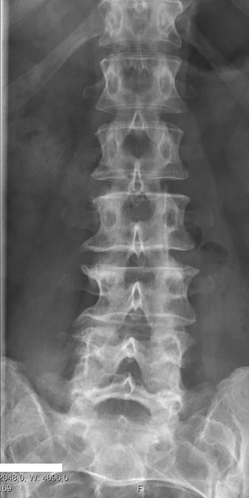
Congenital vertebral anomalies are a collection of malformations of the spine. Most, around 85%, are not clinically significant, but they can cause compression of the spinal cord by deforming the vertebral canal or causing instability. This condition occurs in the womb. Congenital vertebral anomalies include alterations of the shape and number of vertebrae. Lumbarization and sacralization ''Lumbarization'' is an anomaly in the spine. It is defined by the nonfusion of the first and second segments of the sacrum. The lumbar spine subsequently appears to have six vertebrae or segments, not five. This sixth lumbar vertebra is known as a transitional vertebra. Conversely the sacrum appears to have only four segments instead of its designated five segments. Lumbosacral transitional vertebrae consist of the process of the last lumbar vertebra fusing with the first sacral segment. While only around 10 percent of adults have a spinal abnormality due to genetics, a sixth lumbar vertebra ... [...More Info...] [...Related Items...] OR: [Wikipedia] [Google] [Baidu] |
Horseshoe Kidneys
Horseshoe kidney, also known as ''ren arcuatus'' (in Latin), renal fusion or super kidney, is a congenital disorder affecting about 1 in 500 people that is more common in men, often asymptomatic, and usually diagnosed incidentally. In this disorder, the patient's kidneys fuse to form a horseshoe-shape during development in the womb. The fused part is the isthmus of the horseshoe kidney. The abnormal anatomy can affect kidney drainage resulting in increased frequency of kidney stones and urinary tract infections as well as increase risk of certain renal cancers. Fusion abnormalities of the kidney can be categorized into two groups: horseshoe kidney and crossed fused ectopia. The 'horseshoe kidney' is the most common renal fusion anomaly. Signs and symptoms Although often asymptomatic, the most common presenting symptom of patients with a horseshoe kidney is abdominal or flank pain. However, presentation is often non-specific. Approximately a third of patients with horseshoe kidney ... [...More Info...] [...Related Items...] OR: [Wikipedia] [Google] [Baidu] |
Urological
Urology (from Greek οὖρον ''ouron'' "urine" and ''-logia'' "study of"), also known as genitourinary surgery, is the branch of medicine that focuses on surgical and medical diseases of the urinary-tract system and the reproductive organs. Organs under the domain of urology include the kidneys, adrenal glands, ureters, urinary bladder, urethra, and the male reproductive organs (testes, epididymis, vas deferens, seminal vesicles, prostate, and penis). The urinary and reproductive tracts are closely linked, and disorders of one often affect the other. Thus a major spectrum of the conditions managed in urology exists under the domain of genitourinary disorders. Urology combines the management of medical (i.e., non-surgical) conditions, such as urinary-tract infections and benign prostatic hyperplasia, with the management of surgical conditions such as bladder or prostate cancer, kidney stones, congenital abnormalities, traumatic injury, and stress incontinence. Urological t ... [...More Info...] [...Related Items...] OR: [Wikipedia] [Google] [Baidu] |


