|
Mucosal Associated Invariant T Cells
Mucosal-associated invariant T cells (MAIT cells) make up a subset of T cells in the immune system that display innate, effector-like qualities. In humans, MAIT cells are found in the blood, liver, lungs, and mucosa, defending against microbial activity and infection. The MHC class I-like protein, MR1, is responsible for presenting bacterially-produced vitamin B2 and B9 metabolites to MAIT cells. After the presentation of foreign antigen by MR1, MAIT cells secrete pro-inflammatory cytokines and are capable of lysing bacterially-infected cells. MAIT cells can also be activated through MR1-independent signaling. In addition to possessing innate-like functions, this T cell subset supports the adaptive immune response and has a memory-like phenotype. Furthermore, MAIT cells are thought to play a role in autoimmune diseases, such as multiple sclerosis, arthritis and inflammatory bowel disease, although definitive evidence is yet to be published. Molecular characteristics MAIT cells con ... [...More Info...] [...Related Items...] OR: [Wikipedia] [Google] [Baidu] |
T Cell
A T cell is a type of lymphocyte. T cells are one of the important white blood cells of the immune system and play a central role in the adaptive immune response. T cells can be distinguished from other lymphocytes by the presence of a T-cell receptor (TCR) on their cell surface. T cells are born from hematopoietic stem cells, found in the bone marrow. Developing T cells then migrate to the thymus gland to develop (or mature). T cells derive their name from the thymus. After migration to the thymus, the precursor cells mature into several distinct types of T cells. T cell differentiation also continues after they have left the thymus. Groups of specific, differentiated T cell subtypes have a variety of important functions in controlling and shaping the immune response. One of these functions is immune-mediated cell death, and it is carried out by two major subtypes: CD8+ "killer" and CD4+ "helper" T cells. (These are named for the presence of the cell surface proteins CD8 or ... [...More Info...] [...Related Items...] OR: [Wikipedia] [Google] [Baidu] |
Interleukin 18
Interleukin-18 (IL-18), also known as interferon-gamma inducing factor) is a protein which in humans is encoded by the ''IL18'' gene. The protein encoded by this gene is a proinflammatory cytokine. Many cell types, both hematopoietic cells and non-hematopoietic cells, have the potential to produce IL-18. It was first described in 1989 as a factor that induced interferon-γ (IFN-γ) production in mouse spleen cells. Originally, IL-18 production was recognized in Kupffer cells, liver-resident macrophages. However, IL-18 is constitutively expressed in non-hematopoietic cells, such as intestinal epithelial cells, keratinocytes, and endothelial cells. IL-18 can modulate both innate and adaptive immunity and its dysregulation can cause autoimmune or inflammatory diseases. Processing Cytokines usually contain the signal peptide which is necessary for their extracellular release. In this case, ''IL18'' gene, similar to other IL-1 family members, lacks this signal peptide. Furthermore, ... [...More Info...] [...Related Items...] OR: [Wikipedia] [Google] [Baidu] |
Gastrointestinal Tract
The gastrointestinal tract (GI tract, digestive tract, alimentary canal) is the tract or passageway of the digestive system that leads from the mouth to the anus. The GI tract contains all the major organ (biology), organs of the digestive system, in humans and other animals, including the esophagus, stomach, and intestines. Food taken in through the mouth is digestion, digested to extract nutrients and absorb energy, and the waste expelled at the anus as feces. ''Gastrointestinal'' is an adjective meaning of or pertaining to the stomach and intestines. Nephrozoa, Most animals have a "through-gut" or complete digestive tract. Exceptions are more primitive ones: sponges have small pores (ostium (sponges), ostia) throughout their body for digestion and a larger dorsal pore (osculum) for excretion, comb jellies have both a ventral mouth and dorsal anal pores, while cnidarians and acoels have a single pore for both digestion and excretion. The human gastrointestinal tract consists o ... [...More Info...] [...Related Items...] OR: [Wikipedia] [Google] [Baidu] |
MR1 (human Protein)
MR1 or MR-1 may refer to: * Bristol M.R.1, an experimental biplane * HAWAII MR1, a sea floor imaging system * Major histocompatibility complex, class I-related * Mercury-Redstone 1 Mercury-Redstone 1 (MR-1) was the first Mercury-Redstone uncrewed flight test in Project Mercury and the first attempt to launch a Mercury spacecraft with the Mercury-Redstone Launch Vehicle. Intended to be an uncrewed sub-orbital spaceflight, it ..., an unsuccessful unmanned American space mission * PNKD, or myofibrillogenesis regulator 1 * Shewanella oneidensis MR-1, a bacterium * Hadoop architecture Map/Reduce version 1 {{Letter-NumberCombDisambig ... [...More Info...] [...Related Items...] OR: [Wikipedia] [Google] [Baidu] |
MHC Class II
MHC Class II molecules are a class of major histocompatibility complex (MHC) molecules normally found only on professional antigen-presenting cells such as dendritic cells, mononuclear phagocytes, some endothelial cells, thymic epithelial cells, and B cells. These cells are important in initiating immune responses. The antigens presented by class II peptides are derived from extracellular proteins (not cytosolic as in MHC class I). Loading of a MHC class II molecule occurs by phagocytosis; extracellular proteins are endocytosed, digested in lysosomes, and the resulting epitopic peptide fragments are loaded onto MHC class II molecules prior to their migration to the cell surface. In humans, the MHC class II protein complex is encoded by the human leukocyte antigen gene complex (HLA). HLAs corresponding to MHC class II are HLA-DP, HLA-DM, HLA-DOA, HLA-DOB, HLA-DQ, and HLA-DR. Mutations in the HLA gene complex can lead to bare lymphocyte syndrome (BLS), which is a type of MHC ... [...More Info...] [...Related Items...] OR: [Wikipedia] [Google] [Baidu] |
Thymus
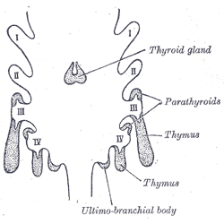
The thymus is a specialized primary lymphoid organ of the immune system. Within the thymus, thymus cell lymphocytes or ''T cells'' mature. T cells are critical to the adaptive immune system, where the body adapts to specific foreign invaders. The thymus is located in the upper front part of the chest, in the anterior superior mediastinum, behind the sternum, and in front of the heart. It is made up of two lobes, each consisting of a central medulla and an outer cortex, surrounded by a capsule. The thymus is made up of immature T cells called thymocytes, as well as lining cells called epithelial cells which help the thymocytes develop. T cells that successfully develop react appropriately with MHC immune receptors of the body (called ''positive selection'') and not against proteins of the body (called ''negative selection''). The thymus is largest and most active during the neonatal and pre-adolescent periods. By the early teens, the thymus begins to decrease in size and a ... [...More Info...] [...Related Items...] OR: [Wikipedia] [Google] [Baidu] |
L-selectin
L-selectin, also known as CD62L, is a cell adhesion molecule found on the cell surface of leukocytes, and the blastocyst. It is coded for in the human by the ''SELL'' gene. L-selectin belongs to the selectin family of proteins, which recognize sialylated carbohydrate groups containing a Sialyl LewisX (sLeX) determinant. L-selectin plays an important role in both the innate and adaptive immune responses by facilitating leukocyte-endothelial cell adhesion events. These tethering interactions are essential for the trafficking of monocytes and neutrophils into inflamed tissue as well as the homing of lymphocytes to secondary lymphoid organs. L-selectin is also expressed by lymphoid primed hematopoietic stem cells and may participate in the migration of these stem cells to the primary lymphoid organs. In addition to its function in the immune response, L-selectin is expressed on embryonic cells and facilitates the attachment of the blastocyst to the endometrial endothelium during hum ... [...More Info...] [...Related Items...] OR: [Wikipedia] [Google] [Baidu] |
C-C Chemokine Receptor Type 7
C-C chemokine receptor type 7 is a protein that in humans is encoded by the ''CCR7'' gene. Two ligands have been identified for this receptor: the chemokines (C-C motif) ligand 19 ( CCL19/ELC) and (C-C motif) ligand 21 (CCL21). CCR7 has also recently been designated CD197 (cluster of differentiation 197). Function The protein encoded by this gene is a member of the G protein-coupled receptor family. This receptor was identified as a gene induced by the Epstein–Barr virus (EBV), and is thought to be a mediator of EBV effects on B lymphocytes. This receptor is expressed in various lymphoid tissues and activates B and T lymphocytes. CCR7 has been shown to stimulate dendritic cell maturation. CCR7 is also involved in homing of T cells to various secondary lymphoid organs such as lymph nodes and the spleen as well as trafficking of T cells within the spleen. Activation of dendritic cells in peripheral tissues induces CCR7 expression on the cell's surface, which recognize CCL19 a ... [...More Info...] [...Related Items...] OR: [Wikipedia] [Google] [Baidu] |
PTPRC
Protein tyrosine phosphatase, receptor type, C also known as PTPRC is an enzyme that, in humans, is encoded by the ''PTPRC'' gene. PTPRC is also known as CD45 antigen (CD stands for cluster of differentiation), which was originally called leukocyte common antigen (LCA). Function The protein product of this gene, best known as CD45, is a member of the protein tyrosine phosphatase (PTP) family. PTPs are signaling molecules that regulate a variety of cellular processes including cell growth, differentiation, mitotic cycle, and oncogenic transformation. CD45 contains an extracellular domain, a single transmembrane segment, and two tandem intracytoplasmic catalytic domains, and thus belongs to the receptor type PTP family. CD45 is a type I transmembrane protein that is present in various isoforms on all differentiated hematopoietic cells (except erythrocytes and plasma cells). CD45 has been shown to be an essential regulator of T- and B-cell antigen receptor signaling. It function ... [...More Info...] [...Related Items...] OR: [Wikipedia] [Google] [Baidu] |
CD44
The CD44 antigen is a cell-surface glycoprotein involved in cell–cell interactions, cell adhesion and migration. In humans, the CD44 antigen is encoded by the ''CD44'' gene on chromosome 11. CD44 has been referred to as HCAM (homing cell adhesion molecule), Pgp-1 (phagocytic glycoprotein-1), Hermes antigen, lymphocyte homing receptor, ECM-III, and HUTCH-1. Tissue distribution and isoforms CD44 is expressed in a large number of mammalian cell types. The standard isoform, designated CD44s, comprising exons 1–5 and 16–20 is expressed in most cell types. CD44 splice variants containing variable exons are designated CD44v. Some epithelial cells also express a larger isoform (CD44E), which includes exons v8–10. Function CD44 participates in a wide variety of cellular functions including lymphocyte activation, recirculation and homing, hematopoiesis, and tumor metastasis. CD44 is a receptor for hyaluronic acid and can also interact with other ligands, such as osteop ... [...More Info...] [...Related Items...] OR: [Wikipedia] [Google] [Baidu] |
Memory T Cell
Memory T cells are a subset of T lymphocytes that might have some of the same functions as memory B cells. Their lineage is unclear. Function Antigen-specific memory T cells specific to viruses or other microbial molecules can be found in both central memory T cells (TCM) and effector memory T cells (TEM) subsets. Although most information is currently based on observations in the cytotoxic T cells (CD8-positive) subset, similar populations appear to exist for both the helper T cells ( CD4-positive) and the cytotoxic T cells. Primary function of memory cells is augmented immune response after reactivation of those cells by reintroduction of relevant pathogen into the body. It is important to note that this field is intensively studied and some information may not be available as of yet. * Central memory T cells (TCM): TCM lymphocytes have several attributes in common with stem cells, the most important being the ability of self-renewal, mainly because of high level of phosphoryl ... [...More Info...] [...Related Items...] OR: [Wikipedia] [Google] [Baidu] |
C-C Chemokine Receptor Type 6
Chemokine receptor 6 also known as CCR6 is a CC chemokine receptor protein which in humans is encoded by the ''CCR6'' gene. CCR6 has also recently been designated CD196 (cluster of differentiation 196). The gene is located on the long arm of Chromosome 6 (6q27) on the Watson (plus) strand. It is 139,737 bases long and encodes a protein of 374 amino acids (molecular weight 42,494 Da). Function This protein belongs to family A of G protein-coupled receptor superfamily. The gene is expressed in lymphatic and non-lymphatic tissue as spleen, lymph nodes, pancreas, colon, appendix, small intestine. CCR6 is expressed on B-cells, immature dendritic cells (DC), T-cells ( Th1, Th2, Th17, Treg), natural killer T cells ( NKT cells) and neutrophils. The ligand of this receptor is CCL20 or in the other name - macrophage inflammatory protein 3 alpha (MIP-3 alpha). This chemokine receptor is special because it binds only one chemokine ligand CCL20 in compare to other chemokine receptors. CCR ... [...More Info...] [...Related Items...] OR: [Wikipedia] [Google] [Baidu] |


