|
Middle Fossa
The middle cranial fossa, deeper than the anterior cranial fossa, is narrow medially and widens laterally to the sides of the skull. It is separated from the posterior fossa by the clivus and the petrous crest. It is bounded in front by the posterior margins of the lesser wings of the sphenoid bone, the anterior clinoid processes, and the ridge forming the anterior margin of the chiasmatic groove; behind, by the superior angles of the petrous portions of the temporal bones and the dorsum sellæ; laterally by the temporal squamæ, sphenoidal angles of the parietals, and greater wings of the sphenoid. It is traversed by the squamosal, sphenoparietal, sphenosquamosal, and sphenopetrosal sutures. It houses the temporal lobes of the brain and the pituitary gland. A middle fossa craniotomy is one means to surgically remove acoustic neuromas ( vestibular schwannoma) growing within the internal auditory canal of the temporal bone. Middle part The middle part of the fossa present ... [...More Info...] [...Related Items...] OR: [Wikipedia] [Google] [Baidu] |
Base Of Skull
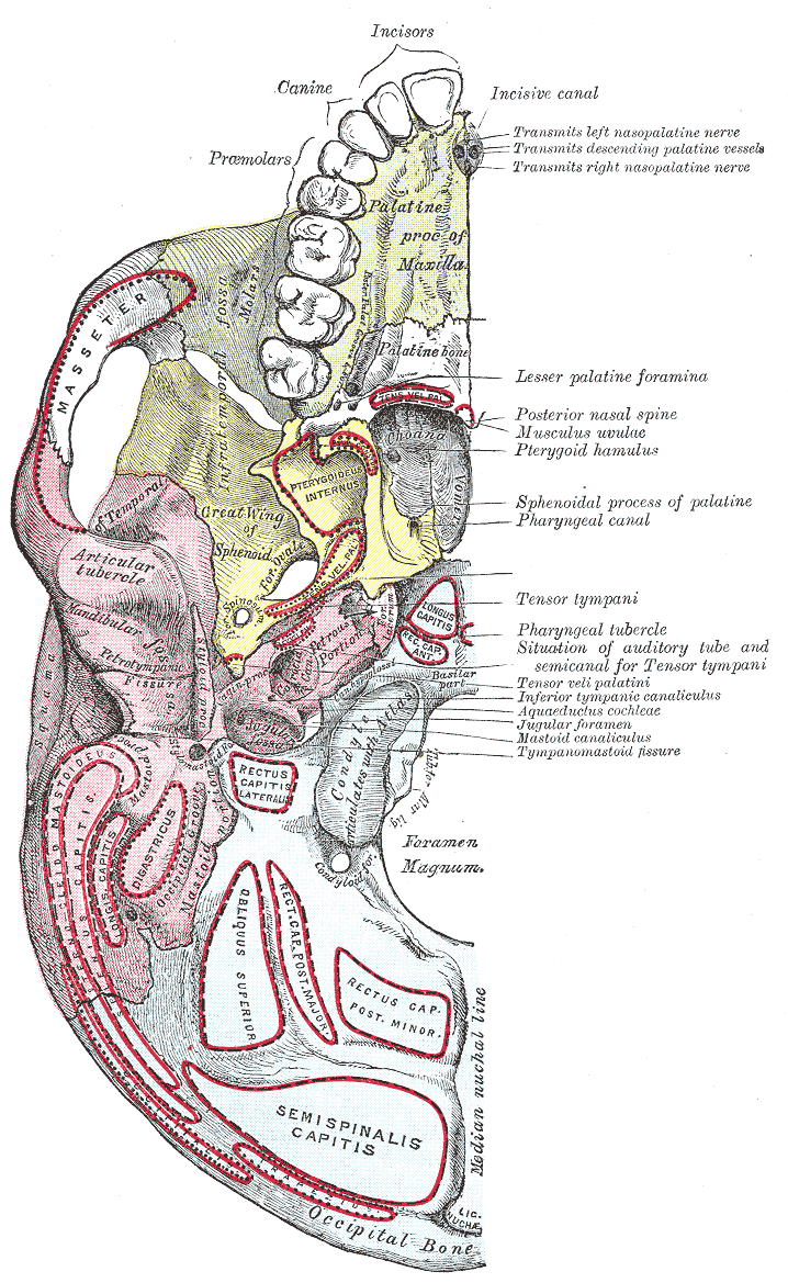
The base of skull, also known as the cranial base or the cranial floor, is the most inferior area of the skull. It is composed of the endocranium and the lower parts of the calvaria. Structure Structures found at the base of the skull are for example: Bones There are five bones that make up the base of the skull: *Ethmoid bone *Sphenoid bone *Occipital bone *Frontal bone *Temporal bone Sinuses *Occipital sinus *Superior sagittal sinus *Superior petrosal sinus Foramina of the skull * Foramen cecum *Optic foramen *Foramen lacerum *Foramen rotundum *Foramen magnum * Foramen ovale *Jugular foramen *Internal auditory meatus *Mastoid foramen *Sphenoidal emissary foramen *Foramen spinosum Sutures *Frontoethmoidal suture *Sphenofrontal suture *Sphenopetrosal suture *Sphenoethmoidal suture * Petrosquamous suture *Sphenosquamosal suture Other *Sphenoidal lingula *Subarcuate fossa *Dorsum sellae *Jugular process *Petro-occipital fissure *Condylar canal *Jugular tubercle *Tuberc ... [...More Info...] [...Related Items...] OR: [Wikipedia] [Google] [Baidu] |
Middle Cranial Fossa Surgical Anatomy - J M Kartush
Middle or The Middle may refer to: * Centre (geometry), the point equally distant from the outer limits. Places * Middle (sheading), a subdivision of the Isle of Man * Middle Bay (other) * Middle Brook (other) * Middle Creek (other) * Middle Island (other) * Middle Lake (other) * Middle Mountain, California * Middle Peninsula, Chesapeake Bay, Virginia * Middle Range, a former name of the Xueshan Range on Taiwan Island * Middle River (other) * Middle Rocks, two rocks at the eastern opening of the Straits of Singapore * Middle Sound, a bay in North Carolina * Middle Township (other) * Middle East Music * "Middle" (song), 2015 * "The Middle" (Jimmy Eat World song), 2001 * "The Middle" (Zedd, Maren Morris and Grey song), 2018 *"Middle", a song by Rocket from the Crypt from their 1995 album ''Scream, Dracula, Scream!'' *"The Middle", a song by Demi Lovato from their debut album ''Don't Forget'' *"The Middle", a song by ... [...More Info...] [...Related Items...] OR: [Wikipedia] [Google] [Baidu] |
Abducent Nerve
The abducens nerve or abducent nerve, also known as the sixth cranial nerve, cranial nerve VI, or simply CN VI, is a cranial nerve in humans and various other animals that controls the movement of the lateral rectus muscle, one of the extraocular muscles responsible for outward gaze. It is a somatic efferent nerve. Structure Nucleus The abducens nucleus is located in the pons, on the floor of the fourth ventricle, at the level of the facial colliculus. Axons from the facial nerve loop around the abducens nucleus, creating a slight bulge (the facial colliculus) that is visible on the dorsal surface of the floor of the fourth ventricle. The abducens nucleus is close to the midline, like the other motor nuclei that control eye movements (the oculomotor and trochlear nuclei). Motor axons leaving the abducens nucleus run ventrally and caudally through the pons. They pass lateral to the corticospinal tract (which runs longitudinally through the pons at this level) before exiting th ... [...More Info...] [...Related Items...] OR: [Wikipedia] [Google] [Baidu] |
Dorsum Sellae
The dorsum sellae is part of the sphenoid bone in the skull. Together with the basilar part of the occipital bone it forms the clivus. In the sphenoid bone, the anterior boundary of the sella turcica is completed by two small eminences, one on either side, called the middle clinoid processes, while the posterior boundary is formed by a square-shaped plate of bone, the dorsum sellae, ending at its superior angles in two tubercles, the posterior clinoid processes In the sphenoid bone, the anterior boundary of the sella turcica is completed by two small eminences, one on either side, called the anterior clinoid processes, while the posterior boundary is formed by a square-shaped plate of bone, the dorsum s ..., the size and form of which vary considerably in different individuals. Additional images File:Gray569.png, Tentorium cerebelli from above. File:Slide2iiii.JPG, Dorsum sellae References External links * * Bones of the head and neck {{musculoskeletal-stu ... [...More Info...] [...Related Items...] OR: [Wikipedia] [Google] [Baidu] |
Middle Clinoid Processes
The anterior boundary of the sella turcica is completed by two small eminences, one on either side, called the middle clinoid processes. It is found lateral to the sella turcica. Etymology Clinoid likely comes from the Greek root ''klinein'' or the Latin Latin (, or , ) is a classical language belonging to the Italic branch of the Indo-European languages. Latin was originally a dialect spoken in the lower Tiber area (then known as Latium) around present-day Rome, but through the power of the ... ''clinare'', both meaning "sloped" as in "inclined." References Bones of the head and neck {{musculoskeletal-stub ... [...More Info...] [...Related Items...] OR: [Wikipedia] [Google] [Baidu] |
Pituitary Gland
In vertebrate anatomy, the pituitary gland, or hypophysis, is an endocrine gland, about the size of a chickpea and weighing, on average, in humans. It is a protrusion off the bottom of the hypothalamus at the base of the brain. The hypophysis rests upon the hypophyseal fossa of the sphenoid bone in the center of the middle cranial fossa and is surrounded by a small bony cavity (sella turcica) covered by a dural fold (diaphragma sellae). The anterior pituitary (or adenohypophysis) is a lobe of the gland that regulates several physiological processes including stress, growth, reproduction, and lactation. The intermediate lobe synthesizes and secretes melanocyte-stimulating hormone. The posterior pituitary (or neurohypophysis) is a lobe of the gland that is functionally connected to the hypothalamus by the median eminence via a small tube called the pituitary stalk (also called the infundibular stalk or the infundibulum). Hormones secreted from the pituitary gland ... [...More Info...] [...Related Items...] OR: [Wikipedia] [Google] [Baidu] |
Fossa Hypophyseos
The sella turcica (Latin for 'Turkish saddle') is a saddle-shaped depression in the body of the sphenoid bone of the human skull and of the skulls of other hominids including chimpanzees, gorillas and orangutans. It serves as a cephalometric landmark. The pituitary gland or hypophysis is located within the most inferior aspect of the sella turcica, the hypophyseal fossa. Structure The sella turcica is located in the sphenoid bone behind the chiasmatic groove and the tuberculum sellae. It belongs to the middle cranial fossa. The sella turcica's most inferior portion is known as the hypophyseal fossa (the "seat of the saddle"), and contains the pituitary gland (hypophysis). In front of the hypophyseal fossa is the tuberculum sellae. Completing the formation of the saddle posteriorly is the dorsum sellae, which is continuous with the clivus, inferoposteriorly. The dorsum sellae is terminated laterally by the posterior clinoid processes. Development It is widely believed that t ... [...More Info...] [...Related Items...] OR: [Wikipedia] [Google] [Baidu] |
Sella Turcica
The sella turcica (Latin for 'Turkish saddle') is a saddle-shaped depression in the body of the sphenoid bone of the human skull and of the skulls of other hominids including chimpanzees, gorillas and orangutans. It serves as a cephalometric landmark. The pituitary gland or hypophysis is located within the most inferior aspect of the sella turcica, the hypophyseal fossa. Structure The sella turcica is located in the sphenoid bone behind the chiasmatic groove and the tuberculum sellae. It belongs to the middle cranial fossa. The sella turcica's most inferior portion is known as the hypophyseal fossa (the "seat of the saddle"), and contains the pituitary gland (hypophysis). In front of the hypophyseal fossa is the tuberculum sellae. Completing the formation of the saddle posteriorly is the dorsum sellae, which is continuous with the clivus, inferoposteriorly. The dorsum sellae is terminated laterally by the posterior clinoid processes. Development It is widely believed that t ... [...More Info...] [...Related Items...] OR: [Wikipedia] [Google] [Baidu] |
Tentorium Cerebelli
The cerebellar tentorium or tentorium cerebelli (Latin for "tent of the cerebellum") is an extension of the dura mater that separates the cerebellum from the inferior portion of the occipital lobes. Structure The cerebellar tentorium is an arched wikt:lamina, lamina, elevated in the middle, and inclining downward toward the circumference. It covers the top of the cerebellum The cerebellum (Latin for "little brain") is a major feature of the hindbrain of all vertebrates. Although usually smaller than the cerebrum, in some animals such as the mormyrid fishes it may be as large as or even larger. In humans, the cerebel ..., and supports the occipital lobes of the brain. Its anterior border is free and concave, and bounds a large oval opening, the tentorial incisure, through which pass the cerebral peduncles. It is attached, behind, by its convex border, to the transverse ridges upon the inner surface of the occipital bone, and there encloses the transverse sinuses; in front, to ... [...More Info...] [...Related Items...] OR: [Wikipedia] [Google] [Baidu] |
Anterior Clinoid Process
In the sphenoid bone, the posterior border, smooth and rounded, is received into the lateral fissure of the brain; the medial end of this border forms the anterior clinoid process, which gives attachment to the tentorium cerebelli; it is sometimes joined to the middle clinoid process by a spicule of bone, and when this occurs the termination of the groove for the internal carotid artery is converted into a foramen (carotico-clinoid). Etymology The anterior and posterior clinoid processes surround the sella turcica like the four corners of a four poster bed. Cline is Greek for bed. –oid, as usual, indicates a similarity to. The term may also come from the Greek root ''klinein'' or the Latin ''clinare'', both meaning "sloped" as in "inclined". Additional images Image:Anterior clinoid process.jpg, Anterior clinoid process Petroclinoid ligament The petroclinoid ligament is a fold of dura mater. It extends between the posterior clinoid process and anterior clinoid process and the ... [...More Info...] [...Related Items...] OR: [Wikipedia] [Google] [Baidu] |
Orbital Cavity
In anatomy, the orbit is the cavity or socket of the skull in which the eye and its appendages are situated. "Orbit" can refer to the bony socket, or it can also be used to imply the contents. In the adult human, the volume of the orbit is , of which the eye occupies . The orbital contents comprise the eye, the orbital and retrobulbar fascia, extraocular muscles, cranial nerves II, III, IV, V, and VI, blood vessels, fat, the lacrimal gland with its sac and duct, the eyelids, medial and lateral palpebral ligaments, cheek ligaments, the suspensory ligament, septum, ciliary ganglion and short ciliary nerves. Structure The orbits are conical or four-sided pyramidal cavities, which open into the midline of the face and point back into the head. Each consists of a base, an apex and four walls."eye, human."Encyclopædia Britannica from Encyclopædia Britannica 2006 Ultimate Reference Suite DVD 2009 Openings There are two important foramina, or windows, two important fis ... [...More Info...] [...Related Items...] OR: [Wikipedia] [Google] [Baidu] |
Ophthalmic Artery
The ophthalmic artery (OA) is an artery of the head. It is the first branch of the internal carotid artery distal to the cavernous sinus. Branches of the ophthalmic artery supply all the structures in the orbit around the eye, as well as some structures in the nose, face, and meninges. Occlusion of the ophthalmic artery or its branches can produce sight-threatening conditions. Structure The ophthalmic artery emerges from the internal carotid artery. This is usually just after the internal carotid artery emerges from the cavernous sinus. In some cases, the ophthalmic artery branches just before the internal carotid exits the cavernous sinus. The ophthalmic artery emerges along the medial side of the anterior clinoid process. It runs anteriorly, passing through the optic canal inferolaterally to the optic nerve. It can also pass superiorly to the optic nerve in a minority of cases. In the posterior third of the cone of the orbit, the ophthalmic artery turns sharply and medially t ... [...More Info...] [...Related Items...] OR: [Wikipedia] [Google] [Baidu] |


