|
Metarteriole
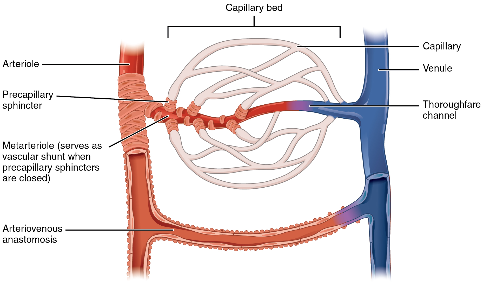
A metarteriole is a short microvessel in the microcirculation that links arterioles and capillaries. Instead of a continuous tunica media, they have individual smooth muscle cells placed a short distance apart, each forming a precapillary sphincter that encircles the entrance to that capillary bed. Constriction of these sphincters reduces or shuts off blood flow through their respective capillary beds. This allows the blood to be diverted to elsewhere in the body. Metarterioles exist in the ''mesenteric microcirculation'', and the name was originally conceived only to define the "''thoroughfare channels ''" between arterioles and venules A venule is a very small blood vessel in the microcirculation that allows blood to return from the capillary beds to drain into the larger blood vessels, the veins. Venules range from 7μm to 1mm in diameter. Veins contain approximately 70% of t .... In recent times the term has often been used instead to describe the smallest arterioles direct ... [...More Info...] [...Related Items...] OR: [Wikipedia] [Google] [Baidu] |
Precapillary Sphincter
A precapillary sphincter is a band of contractile mural cells either classified as smooth muscle or pericytes that adjusts blood flow into capillaries. They were originally described in the mesenteric microcirculation, and were thought to only reside there. At the point where each of the capillaries originates from an arteriole, contractile mural cells encircle the capillary. This is called the precapillary sphincter. The precapillary sphincter has now also been found in the brain, where it regulates blood flow to the capillary bed. The sphincter can open and close the entrance to the capillary, by which contraction causes blood flow in a capillary to change as vasomotion occurs. In some tissues, the entire capillary bed may be bypassed by blood flow through arteriovenous anastomoses or through preferential flow through metarterioles. If the sphincter is damaged or cannot contract, blood can flow into the capillary bed at high pressures. When capillary pressures are high (as per ... [...More Info...] [...Related Items...] OR: [Wikipedia] [Google] [Baidu] |
Capillary
A capillary is a small blood vessel from 5 to 10 micrometres (μm) in diameter. Capillaries are composed of only the tunica intima, consisting of a thin wall of simple squamous endothelial cells. They are the smallest blood vessels in the body: they convey blood between the arterioles and venules. These microvessels are the site of exchange of many substances with the interstitial fluid surrounding them. Substances which cross capillaries include water, oxygen, carbon dioxide, urea, glucose, uric acid, lactic acid and creatinine. Lymph capillaries connect with larger lymph vessels to drain lymphatic fluid collected in the microcirculation. During early embryonic development, new capillaries are formed through vasculogenesis, the process of blood vessel formation that occurs through a '' de novo'' production of endothelial cells that then form vascular tubes. The term '' angiogenesis'' denotes the formation of new capillaries from pre-existing blood vessels and already present ... [...More Info...] [...Related Items...] OR: [Wikipedia] [Google] [Baidu] |
Microvessel
The microcirculation is the circulation of the blood in the smallest blood vessels, the microvessels of the microvasculature present within organ tissues. The microvessels include terminal arterioles, metarterioles, capillaries, and venules. Arterioles carry oxygenated blood to the capillaries, and blood flows out of the capillaries through venules into veins. In addition to these blood vessels, the microcirculation also includes lymphatic capillaries and collecting ducts. The main functions of the microcirculation are the delivery of oxygen and nutrients and the removal of carbon dioxide (CO2). It also serves to regulate blood flow and tissue perfusion thereby affecting blood pressure and responses to inflammation which can include edema (swelling). Most vessels of the microcirculation are lined by flattened cells of the endothelium and many of them are surrounded by contractile cells called pericytes. The endothelium provides a smooth surface for the flow of blood and regu ... [...More Info...] [...Related Items...] OR: [Wikipedia] [Google] [Baidu] |
Microcirculation
The microcirculation is the circulation of the blood in the smallest blood vessels, the microvessels of the microvasculature present within organ tissues. The microvessels include terminal arterioles, metarterioles, capillaries, and venules. Arterioles carry oxygenated blood to the capillaries, and blood flows out of the capillaries through venules into veins. In addition to these blood vessels, the microcirculation also includes lymphatic capillaries and collecting ducts. The main functions of the microcirculation are the delivery of oxygen and nutrients and the removal of carbon dioxide (CO2). It also serves to regulate blood flow and tissue perfusion thereby affecting blood pressure and responses to inflammation which can include edema (swelling). Most vessels of the microcirculation are lined by flattened cells of the endothelium and many of them are surrounded by contractile cells called pericytes. The endothelium provides a smooth surface for the flow of blood and regul ... [...More Info...] [...Related Items...] OR: [Wikipedia] [Google] [Baidu] |
Arteriole
An arteriole is a small-diameter blood vessel in the microcirculation that extends and branches out from an artery and leads to capillaries. Arterioles have muscular walls (usually only one to two layers of smooth muscle cells) and are the primary site of vascular resistance. The greatest change in blood pressure and velocity of blood flow occurs at the transition of arterioles to capillaries.This function is extremely important because it prevents the thin, one-layer capillaries from exploding upon pressure. The arterioles achieve this decrease in pressure, as they are the site with the highest resistance (a large contributor to total peripheral resistance) which translates to a large decrease in the pressure. Structure Microanatomy In a healthy vascular system the endothelium lines all blood-contacting surfaces, including arteries, arterioles, veins, venules, capillaries, and heart chambers. This healthy condition is promoted by the ample production of nitric oxide by the end ... [...More Info...] [...Related Items...] OR: [Wikipedia] [Google] [Baidu] |
Arterioles
An arteriole is a small-diameter blood vessel in the microcirculation that extends and branches out from an artery and leads to capillaries. Arterioles have muscular walls (usually only one to two layers of smooth muscle cells) and are the primary site of vascular resistance. The greatest change in blood pressure and velocity of blood flow occurs at the transition of arterioles to capillaries.This function is extremely important because it prevents the thin, one-layer capillaries from exploding upon pressure. The arterioles achieve this decrease in pressure, as they are the site with the highest resistance (a large contributor to total peripheral resistance) which translates to a large decrease in the pressure. Structure Microanatomy In a healthy vascular system the endothelium lines all blood-contacting surfaces, including arteries, arterioles, veins, venules, capillaries, and heart chambers. This healthy condition is promoted by the ample production of nitric oxide by the end ... [...More Info...] [...Related Items...] OR: [Wikipedia] [Google] [Baidu] |
Tunica Media
The tunica media (New Latin "middle coat"), or media for short, is the middle tunica (layer) of an artery or vein. It lies between the tunica intima on the inside and the tunica externa on the outside. Artery Tunica media is made up of smooth muscle cells, elastic tissue and collagen. It lies between the tunica intima on the inside and the tunica externa on the outside. The middle coat (tunica media) is distinguished from the inner (tunica intima) by its color and by the transverse arrangement of its fibers. * In the ''smaller arteries'' it consists principally of smooth muscle fibers in fine bundles, arranged in lamellæ and disposed circularly around the vessel. These lamellæ vary in number according to the size of the vessel; the smallest arteries having only a single layer, and those slightly larger three or four layers - up to a maximum of six layers. It is to this coat that the thickness of the wall of the artery is mainly due. * In the ''larger arteries'', as the ilia ... [...More Info...] [...Related Items...] OR: [Wikipedia] [Google] [Baidu] |
Mesenteric
The mesentery is an organ that attaches the intestines to the posterior abdominal wall in humans and is formed by the double fold of peritoneum. It helps in storing fat and allowing blood vessels, lymphatics, and nerves to supply the intestines, among other functions. The mesocolon was thought to be a fragmented structure, with all named parts—the ascending, transverse, descending, and sigmoid mesocolons, the mesoappendix, and the mesorectum—separately terminating their insertion into the posterior abdominal wall. However, in 2012, new microscopic and electron microscopic examinations showed the mesocolon to be a single structure derived from the duodenojejunal flexure and extending to the distal mesorectal layer. Thus, the mesentery is an internal organ. Structure The mesentery of the small intestine arises from the root of the mesentery (or mesenteric root) and is the part connected with the structures in front of the vertebral column. The root is narrow, about 15 ... [...More Info...] [...Related Items...] OR: [Wikipedia] [Google] [Baidu] |
Venules
A venule is a very small blood vessel in the microcirculation that allows blood to return from the capillary beds to drain into the larger blood vessels, the veins. Venules range from 7μm to 1mm in diameter. Veins contain approximately 70% of total blood volume, while about 25% is contained in the venules. Many venules unite to form a vein. Structure Venule walls have three layers: An inner endothelium composed of squamous endothelial cells that act as a membrane, a middle layer of muscle and elastic tissue and an outer layer of fibrous connective tissue. The middle layer is poorly developed so that venules have thinner walls than arterioles. They are porous so that fluid and blood cells can move easily from the bloodstream through their walls. Short portal venules between the neural and anterior pituitary lobes provide an avenue for rapid hormonal exchange via the blood. Specifically within and between the pituitary lobes is anatomical evidence for confluent interlobe ve ... [...More Info...] [...Related Items...] OR: [Wikipedia] [Google] [Baidu] |


