|
Mesangial Cells
Mesangial cells are specialised cells in the kidney that make up the mesangium of the glomerulus. Together with the mesangial matrix, they form the vascular pole of the renal corpuscle. The mesangial cell population accounts for approximately 30-40% of the total cells in the glomerulus. Mesangial cells can be categorized as either extraglomerular mesangial cells or intraglomerular mesangial cells, based on their relative location to the glomerulus. The extraglomerular mesangial cells are found between the afferent and efferent arterioles towards the vascular pole of the glomerulus. The extraglomerular mesangial cells are adjacent to the intraglomerular mesangial cells that are located inside the glomerulus and in between the capillaries. The primary function of mesangial cells is to remove trapped residues and aggregated protein from the basement membrane thus keeping the filter free of debris. The contractile properties of mesangial cells have been shown to be insignificant ... [...More Info...] [...Related Items...] OR: [Wikipedia] [Google] [Baidu] |
Mesangium
The glomerulus (plural glomeruli) is a network of small blood vessels (capillaries) known as a ''tuft'', located at the beginning of a nephron in the kidney. Each of the two kidneys contains about one million nephrons. The tuft is structurally supported by the mesangium (the space between the blood vessels), composed of intraglomerular mesangial cells. The blood is filtered across the capillary walls of this tuft through the glomerular filtration barrier, which yields its filtrate of water and soluble substances to a cup-like sac known as Bowman's capsule. The filtrate then enters the renal tubule of the nephron. The glomerulus receives its blood supply from an afferent arteriole of the renal arterial circulation. Unlike most capillary beds, the glomerular capillaries exit into efferent arterioles rather than venules. The resistance of the efferent arterioles causes sufficient hydrostatic pressure within the glomerulus to provide the force for ultrafiltration. The glomerulus a ... [...More Info...] [...Related Items...] OR: [Wikipedia] [Google] [Baidu] |
Ultrafiltration (renal)
In renal physiology, ultrafiltration occurs at the barrier between the blood and the filtrate in the glomerular capsule (Bowman's capsule) in the kidneys. As in nonbiological examples of ultrafiltration, pressure (in this case blood pressure) and concentration gradients lead to a separation through a semipermeable membrane (provided by the podocytes). The Bowman's capsule contains a dense capillary network called the glomerulus. Blood flows into these capillaries through the afferent arterioles and leaves through the efferent arterioles. The high hydrostatic pressure forces small molecules in the tubular fluid such as water, glucose, amino acids, sodium chloride and urea through the filter, from the blood in the glomerular capsule across the basement membrane of the Bowman's capsule and into the renal tubules. This process is called ultrafiltration; the resulting fluid, virtually free of large proteins and blood cells, is referred to as glomerular filtrate, or ultrafiltrate. ... [...More Info...] [...Related Items...] OR: [Wikipedia] [Google] [Baidu] |
Podocyte
Podocytes are cells in Bowman's capsule in the kidneys that wrap around capillaries of the glomerulus. Podocytes make up the epithelial lining of Bowman's capsule, the third layer through which filtration of blood takes place. Bowman's capsule filters the blood, retaining large molecules such as proteins while smaller molecules such as water, salts, and sugars are filtered as the first step in the formation of urine. Although various viscera have epithelial layers, the name visceral epithelial cells usually refers specifically to podocytes, which are specialized epithelial cells that reside in the visceral layer of the capsule. One type of specialized epithelial cell is podocalyxin. The podocytes have long foot processes called ''pedicels'', for which the cells are named (''podo-'' + '' -cyte''). The pedicels wrap around the capillaries and leave slits between them. Blood is filtered through these slits, each known as a filtration slit, slit diaphragm, or slit pore. Severa ... [...More Info...] [...Related Items...] OR: [Wikipedia] [Google] [Baidu] |
Platelet-derived Growth Factor
Platelet-derived growth factor (PDGF) is one among numerous growth factors that regulate cell growth and division. In particular, PDGF plays a significant role in blood vessel formation, the growth of blood vessels from already-existing blood vessel tissue, mitogenesis, i.e. proliferation, of mesenchymal cells such as fibroblasts, osteoblasts, tenocytes, vascular smooth muscle cells and mesenchymal stem cells as well as chemotaxis, the directed migration, of mesenchymal cells. Platelet-derived growth factor is a dimeric glycoprotein that can be composed of two A subunits (PDGF-AA), two B subunits (PDGF-BB), or one of each (PDGF-AB). PDGF is a potent mitogen for cells of mesenchymal origin, including fibroblasts, smooth muscle cells and glial cells. In both mouse and human, the PDGF signalling network consists of five ligands, PDGF-AA through -DD (including -AB), and two receptors, PDGFRalpha and PDGFRbeta. All PDGFs function as secreted, disulphide-linked homodimers, but only ... [...More Info...] [...Related Items...] OR: [Wikipedia] [Google] [Baidu] |
Vascular Smooth Muscle
Vascular smooth muscle is the type of smooth muscle that makes up most of the walls of blood vessels. Structure Vascular smooth muscle refers to the particular type of smooth muscle found within, and composing the majority of the wall of blood vessels. Nerve supply Vascular smooth muscle is innervated primarily by the sympathetic nervous system through adrenergic receptors (adrenoceptors). The three types present are: alpha-1, alpha-2 and beta-2 adrenergic receptors, . The main endogenous agonist of these cell receptors is norepinephrine (NE). The adrenergic receptors exert opposite physiologic effects in the vascular smooth muscle under activation: * alpha-1 receptors. Under NE binding alpha-1 receptors cause vasoconstriction ( contraction of the vascular smooth muscle cells decreasing the diameter of the vessels). Thesea receptors are activated in response to shock or low blood pressure as a defensive reaction trying to restore the normal blood pressure. Antagonists of a ... [...More Info...] [...Related Items...] OR: [Wikipedia] [Google] [Baidu] |
Pericyte
Pericytes (previously known as Rouget cells) are multi-functional mural cells of the microcirculation that wrap around the endothelial cells that line the capillaries throughout the body. Pericytes are embedded in the basement membrane of blood capillaries, where they communicate with endothelial cells by means of both direct physical contact and paracrine signaling. The morphology, distribution, density and molecular fingerprints of pericytes vary between organs and vascular beds. Pericytes help to maintain homeostatic and hemostatic functions in the brain, one of the organs with higher pericyte coverage, and also sustain the blood–brain barrier. These cells are also a key component of the neurovascular unit, which includes endothelial cells, astrocytes, and neurons. Pericytes have been postulated to regulate capillary blood flow and the clearance and phagocytosis of cellular debris ''in vitro.'' Pericytes stabilize and monitor the maturation of endothelial cells by means of ... [...More Info...] [...Related Items...] OR: [Wikipedia] [Google] [Baidu] |
ACTA2
ACTA2 (actin alpha 2) is an actin protein with several aliases including alpha-actin, alpha-actin-2, aortic smooth muscle or alpha smooth muscle actin (α-SMA, SMactin, alpha-SM-actin, ASMA). Actins are a family of globular multi-functional proteins that form microfilaments. ACTA2 is one of 6 different actin isoforms and is involved in the contractile apparatus of smooth muscle. ACTA2 (as with all the actins) is extremely highly conserved and found in nearly all mammals. In humans, ACTA2 is encoded by the ''ACTA2'' gene located on 10q22-q24. Mutations in this gene cause a variety of vascular diseases, such as thoracic aortic disease, coronary artery disease, stroke, Moyamoya disease, and multisystemic smooth muscle dysfunction syndrome. ACTA2 (commonly referred to as alpha-smooth muscle actin or α-SMA) is often used as a marker of myofibroblast A myofibroblast is a cell phenotype that was first described as being in a state between a fibroblast and a smooth muscle cell. ... [...More Info...] [...Related Items...] OR: [Wikipedia] [Google] [Baidu] |
Stromal Cell
Stromal cells, or mesenchymal stromal cells, are differentiating cells found in abundance within bone marrow but can also be seen all around the body. Stromal cells can become connective tissue cells of any organ, for example in the uterine mucosa (endometrium), prostate, bone marrow, lymph node and the ovary. They are cells that support the function of the parenchymal cells of that organ. The most common stromal cells include fibroblasts and pericytes. The term ''stromal'' comes from Latin , "bed covering", and Ancient Greek , , "bed". Stromal cells are an important part of the body's immune response and modulate inflammation through multiple pathways. They also aid in differentiation of hematopoietic cells and forming necessary blood elements. The interaction between stromal cells and tumor cells is known to play a major role in cancer growth and progression. In addition, by regulating local cytokine networks (e.g. M-CSF, LIF), bone marrow stromal cells have been described to ... [...More Info...] [...Related Items...] OR: [Wikipedia] [Google] [Baidu] |
Mesenchyme
Mesenchyme () is a type of loosely organized animal embryonic connective tissue of undifferentiated cells that give rise to most tissues, such as skin, blood or bone. The interactions between mesenchyme and epithelium help to form nearly every organ in the developing embryo. Vertebrates Structure Mesenchyme is characterized morphologically by a prominent ground substance matrix containing a loose aggregate of reticular fibers and unspecialized mesenchymal stem cells. Mesenchymal cells can migrate easily (in contrast to epithelial cells, which lack mobility), are organized into closely adherent sheets, and are polarized in an apical-basal orientation. Development The mesenchyme originates from the mesoderm. From the mesoderm, the mesenchyme appears as an embryologically primitive "soup". This "soup" exists as a combination of the mesenchymal cells plus serous fluid plus the many different tissue proteins. Serous fluid is typically stocked with the many serous elements, su ... [...More Info...] [...Related Items...] OR: [Wikipedia] [Google] [Baidu] |
Proteoglycan
Proteoglycans are proteins that are heavily glycosylated. The basic proteoglycan unit consists of a "core protein" with one or more covalently attached glycosaminoglycan (GAG) chain(s). The point of attachment is a serine (Ser) residue to which the glycosaminoglycan is joined through a tetrasaccharide bridge (e.g. chondroitin sulfate-GlcA- Gal-Gal- Xyl-PROTEIN). The Ser residue is generally in the sequence -Ser- Gly-X-Gly- (where X can be any amino acid residue but proline), although not every protein with this sequence has an attached glycosaminoglycan. The chains are long, linear carbohydrate polymers that are negatively charged under physiological conditions due to the occurrence of sulfate and uronic acid groups. Proteoglycans occur in connective tissue. Types Proteoglycans are categorized by their relative size (large and small) and the nature of their glycosaminoglycan chains. Types include: Certain members are considered members of the "small leucine-rich proteogly ... [...More Info...] [...Related Items...] OR: [Wikipedia] [Google] [Baidu] |
Fibronectin
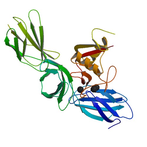
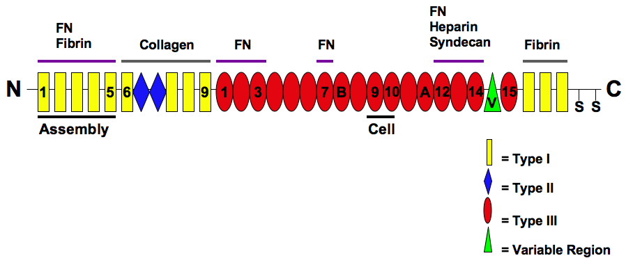
Fibronectin is a high- molecular weight (~500-~600 kDa) glycoprotein of the extracellular matrix that binds to membrane-spanning receptor proteins called integrins. Fibronectin also binds to other extracellular matrix proteins such as collagen, fibrin, and heparan sulfate proteoglycans (e.g. syndecans). Fibronectin exists as a protein dimer, consisting of two nearly identical monomers linked by a pair of disulfide bonds. The fibronectin protein is produced from a single gene, but alternative splicing of its pre-mRNA leads to the creation of several isoforms. Two types of fibronectin are present in vertebrates: * soluble plasma fibronectin (formerly called "cold-insoluble globulin", or CIg) is a major protein component of blood plasma (300 μg/ml) and is produced in the liver by hepatocytes. * insoluble cellular fibronectin is a major component of the extracellular matrix. It is secreted by various cells, primarily fibroblasts, as a soluble protein dimer and is then ass ... [...More Info...] [...Related Items...] OR: [Wikipedia] [Google] [Baidu] |
Laminin
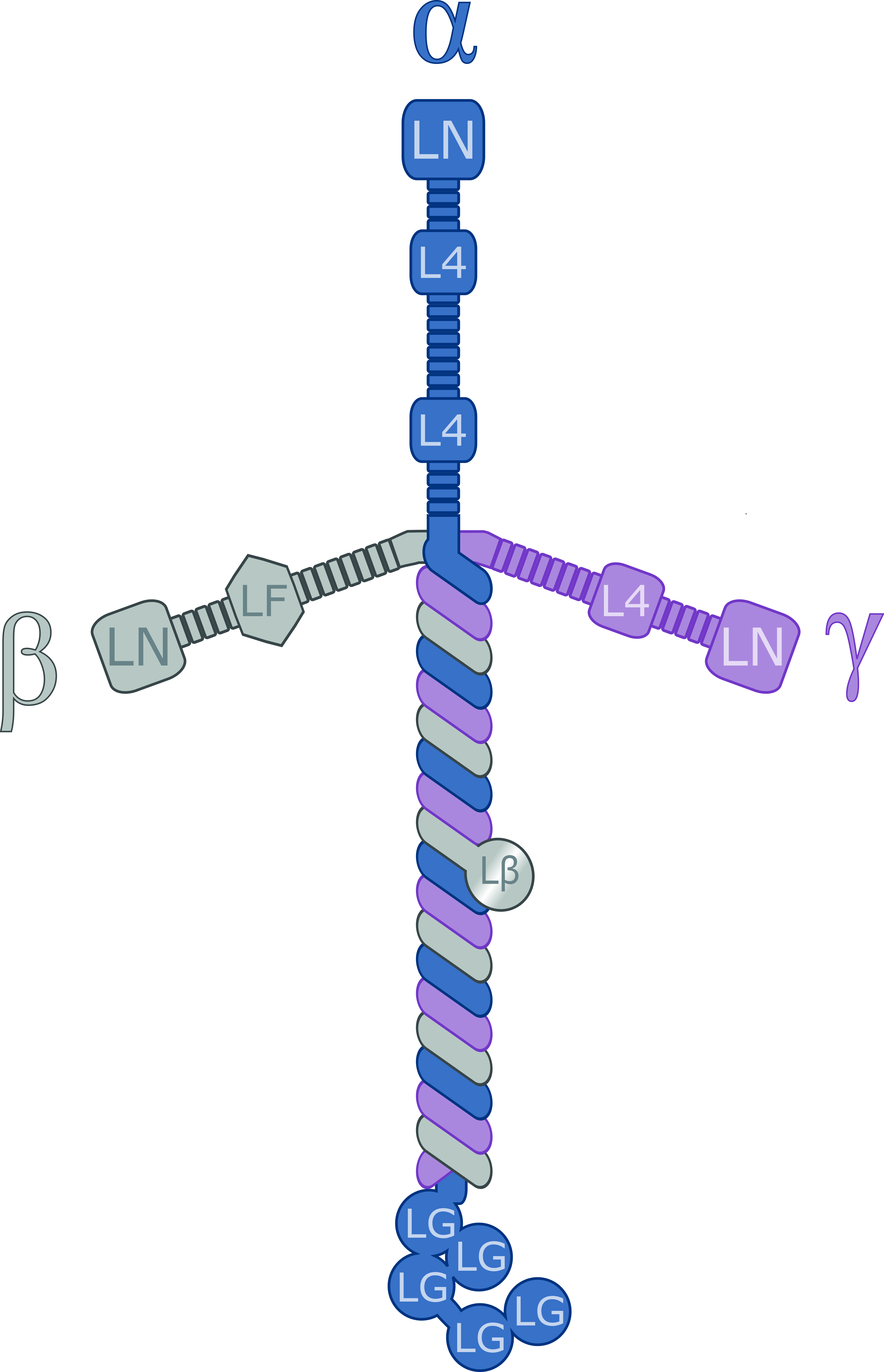
Laminins are a family of glycoproteins of the extracellular matrix of all animals. They are major components of the basal lamina (one of the layers of the basement membrane), the protein network foundation for most cells and organs. The laminins are an important and biologically active part of the basal lamina, influencing cell differentiation, migration, and adhesion. Laminins are heterotrimeric proteins with a high molecular mass (~400 to ~900 kDa). They contain three different chains (α, β and γ) encoded by five, four, and three paralogous genes in humans, respectively. The laminin molecules are named according to their chain composition. Thus, laminin-511 contains α5, β1, and γ1 chains. Fourteen other chain combinations have been identified ''in vivo''. The trimeric proteins intersect to form a cross-like structure that can bind to other cell membrane and extracellular matrix molecules. The three shorter arms are particularly good at binding to other laminin molecules, ... [...More Info...] [...Related Items...] OR: [Wikipedia] [Google] [Baidu] |


