|
Mediastinotomy
Mediastinoscopy is a procedure that enables visualization of the contents of the mediastinum, usually for the purpose of obtaining a biopsy. Mediastinoscopy is often used for staging of lymph nodes of lung cancer or for diagnosing other conditions affecting structures in the mediastinum such as sarcoidosis or lymphoma. Mediastinoscopy involves making an incision approximately 1 cm above the suprasternal notch of the sternum, or breast bone. Dissection is carried out down to the pretracheal space and down to the carina. A scope (mediastinoscope) is then advanced into the created tunnel which provides a view of the mediastinum. The scope may provide direct visualization or may be attached to a video monitor. Mediastinoscopy provides access to mediastinal lymph node levels 2, 4, and 7. Current use Historically, mediastinoscopy has been the gold standard for the staging of lung cancer. However, with advances in minimally invasive procedures and imaging, mediastinoscopy usage has ... [...More Info...] [...Related Items...] OR: [Wikipedia] [Google] [Baidu] |
Mediastinum
The mediastinum (from ) is the central compartment of the thoracic cavity. Surrounded by loose connective tissue, it is an undelineated region that contains a group of structures within the thorax, namely the heart and its vessels, the esophagus, the trachea, the phrenic nerve, phrenic and cardiac nerves, the thoracic duct, the thymus and the lymph nodes of the central chest. Anatomy The mediastinum lies within the thorax and is enclosed on the right and left by pulmonary pleurae, pleurae. It is surrounded by the chest wall in front, the lungs to the sides and the Spine (anatomy), spine at the back. It extends from the sternum in front to the vertebral column behind. It contains all the organs of the thorax except the lungs. It is continuous with the loose connective tissue of the neck. The mediastinum can be divided into an upper (or superior) and lower (or inferior) part: * The superior mediastinum starts at the superior thoracic aperture and ends at the #Thoracic plane, t ... [...More Info...] [...Related Items...] OR: [Wikipedia] [Google] [Baidu] |
Biopsy
A biopsy is a medical test commonly performed by a surgeon, interventional radiologist, or an interventional cardiologist. The process involves extraction of sample cells or tissues for examination to determine the presence or extent of a disease. The tissue is then fixed, dehydrated, embedded, sectioned, stained and mounted before it is generally examined under a microscope by a pathologist; it may also be analyzed chemically. When an entire lump or suspicious area is removed, the procedure is called an excisional biopsy. An incisional biopsy or core biopsy samples a portion of the abnormal tissue without attempting to remove the entire lesion or tumor. When a sample of tissue or fluid is removed with a needle in such a way that cells are removed without preserving the histological architecture of the tissue cells, the procedure is called a needle aspiration biopsy. Biopsies are most commonly performed for insight into possible cancerous or inflammatory conditions. History T ... [...More Info...] [...Related Items...] OR: [Wikipedia] [Google] [Baidu] |
Lymph Node
A lymph node, or lymph gland, is a kidney-shaped organ of the lymphatic system and the adaptive immune system. A large number of lymph nodes are linked throughout the body by the lymphatic vessels. They are major sites of lymphocytes that include B and T cells. Lymph nodes are important for the proper functioning of the immune system, acting as filters for foreign particles including cancer cells, but have no detoxification function. In the lymphatic system a lymph node is a secondary lymphoid organ. A lymph node is enclosed in a fibrous capsule and is made up of an outer cortex and an inner medulla. Lymph nodes become inflamed or enlarged in various diseases, which may range from trivial throat infections to life-threatening cancers. The condition of lymph nodes is very important in cancer staging, which decides the treatment to be used and determines the prognosis. Lymphadenopathy refers to glands that are enlarged or swollen. When inflamed or enlarged, lymph nodes can be ... [...More Info...] [...Related Items...] OR: [Wikipedia] [Google] [Baidu] |
Lung Cancer
Lung cancer, also known as lung carcinoma (since about 98–99% of all lung cancers are carcinomas), is a malignant lung tumor characterized by uncontrolled cell growth in tissue (biology), tissues of the lung. Lung carcinomas derive from transformed, malignant cells that originate as epithelial cells, or from tissues composed of epithelial cells. Other lung cancers, such as the rare sarcomas of the lung, are generated by the malignant transformation of connective tissues (i.e. nerve, fat, muscle, bone), which arise from mesenchymal cells. Lymphomas and melanomas (from lymphoid and melanocyte cell lineages) can also rarely result in lung cancer. In time, this uncontrolled neoplasm, growth can metastasis, metastasize (spreading beyond the lung) either by direct extension, by entering the lymphatic circulation, or via hematogenous, bloodborne spread – into nearby tissue or other, more distant parts of the body. Most cancers that originate from within the lungs, known as primary ... [...More Info...] [...Related Items...] OR: [Wikipedia] [Google] [Baidu] |
Sarcoidosis
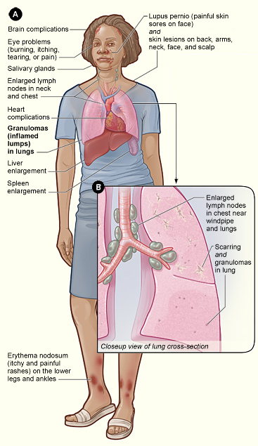
Sarcoidosis (also known as ''Besnier-Boeck-Schaumann disease'') is a disease involving abnormal collections of inflammatory cells that form lumps known as granulomata. The disease usually begins in the lungs, skin, or lymph nodes. Less commonly affected are the eyes, liver, heart, and brain. Any organ can be affected though. The signs and symptoms depend on the organ involved. Often, no, or only mild, symptoms are seen. When it affects the lungs, wheezing, coughing, shortness of breath, or chest pain may occur. Some may have Löfgren syndrome with fever, large lymph nodes, arthritis, and a rash known as erythema nodosum. The cause of sarcoidosis is unknown. Some believe it may be due to an immune reaction to a trigger such as an infection or chemicals in those who are genetically predisposed. Those with affected family members are at greater risk. Diagnosis is partly based on signs and symptoms, which may be supported by biopsy. Findings that make it likely include large lymph n ... [...More Info...] [...Related Items...] OR: [Wikipedia] [Google] [Baidu] |
Lymphoma
Lymphoma is a group of blood and lymph tumors that develop from lymphocytes (a type of white blood cell). In current usage the name usually refers to just the cancerous versions rather than all such tumours. Signs and symptoms may include enlarged lymph nodes, fever, drenching sweats, unintended weight loss, itching, and constantly feeling tired. The enlarged lymph nodes are usually painless. The sweats are most common at night. Many subtypes of lymphomas are known. The two main categories of lymphomas are the non-Hodgkin lymphoma (NHL) (90% of cases) and Hodgkin lymphoma (HL) (10%). The World Health Organization (WHO) includes two other categories as types of lymphoma – multiple myeloma and immunoproliferative diseases. Lymphomas and leukemias are a part of the broader group of tumors of the hematopoietic and lymphoid tissues. Risk factors for Hodgkin lymphoma include infection with Epstein–Barr virus and a history of the disease in the family. Risk factors for common ... [...More Info...] [...Related Items...] OR: [Wikipedia] [Google] [Baidu] |
Suprasternal Notch
The suprasternal notch, also known as the fossa jugularis sternalis, jugular notch, or Plender gap, is a large, visible dip in between the neck in humans, between the clavicles, and above the manubrium of the sternum. Structure The suprasternal notch is a visible dip in between the neck, between the clavicles, and above the manubrium of the sternum. It is at the level of the T2 and T3 vertebrae. The trachea lies just behind it, rising about 5 cm above it in adults. Clinical significance Intrathoracic pressure is measured by using a transducer held in such a way over the body that an actuator engages the soft tissue that is located above the suprasternal notch. Arcot J. Chandrasekhar, MD of Loyola University, Chicago, is the author of an evaluative test for the aorta using the suprasternal notch. - Evaluative tests using the suprasternal notch The test can help recognize the following conditions: * Aneurysm * Dissecting aneurysm * Atherosclerosis * Hypertension Hypert ... [...More Info...] [...Related Items...] OR: [Wikipedia] [Google] [Baidu] |
Human Sternum
The sternum or breastbone is a long flat bone located in the central part of the chest. It connects to the ribs via cartilage and forms the front of the rib cage, thus helping to protect the heart, lungs, and major blood vessels from injury. Shaped roughly like a necktie, it is one of the largest and longest flat bones of the body. Its three regions are the manubrium, the body, and the xiphoid process. The word "sternum" originates from the Ancient Greek στέρνον (stérnon), meaning "chest". Structure The sternum is a narrow, flat bone, forming the middle portion of the front of the chest. The top of the sternum supports the clavicles (collarbones) and its edges join with the costal cartilages of the first two pairs of ribs. The inner surface of the sternum is also the attachment of the sternopericardial ligaments. Its top is also connected to the sternocleidomastoid muscle. The sternum consists of three main parts, listed from the top: * Manubrium * Body (gladiolus) * X ... [...More Info...] [...Related Items...] OR: [Wikipedia] [Google] [Baidu] |
Carina Of Trachea
In anatomy, the carina or tracheal bifurcation is a ridge of cartilage in the trachea that occurs between the division of the two main bronchi. Structure The carina occurs at the lower end of the trachea (usually at the level of the 4th to 5th thoracic vertebra). This is in line with the sternal angle, but the carina may raise or descend up to two vertebrae higher or lower with breathing. The carina lies to the left of the midline, and runs antero-posteriorly (front to back). The bronchial arteries supply the carina and the rest of the lower trachea. The carina is around the area posterior to where the aortic arch crosses to the left of the trachea. The azygos vein crosses right to the trachea above the carina. Clinical significance Foreign bodies that fall down the trachea are more likely to enter the right bronchus. The mucous membrane of the carina is the most sensitive area of the trachea and larynx for triggering a cough reflex. Widening and distortion of the carina i ... [...More Info...] [...Related Items...] OR: [Wikipedia] [Google] [Baidu] |
Mediastinoscope
A mediastinoscope is a thin, tube-like instrument used to examine the tissues and lymph nodes in the area between the lungs (mediastinum) in a procedure known as mediastinoscopy. These tissues include the heart and its large blood vessels, trachea, esophagus, and bronchi. The mediastinoscope has a light and a lens for viewing and may also have a tool to remove tissue. It is inserted into the chest through a cut above the breastbone The sternum or breastbone is a long flat bone located in the central part of the chest. It connects to the ribs via cartilage and forms the front of the rib cage, thus helping to protect the heart, lungs, and major blood vessels from injury. Sh .... External links Mediastinoscopeentry in the public domain NCI Dictionary of Cancer Terms Medical equipment {{medical-equipment-stub ... [...More Info...] [...Related Items...] OR: [Wikipedia] [Google] [Baidu] |
Aortopulmonary Window
Aortopulmonary window (APW) refers to a congenital heart defect similar in some ways to persistent truncus arteriosus. Persistent truncus arteriosus involves a single valve; aortopulmonary window is a septal defect A congenital heart defect (CHD), also known as a congenital heart anomaly and congenital heart disease, is a defect in the structure of the heart or great vessels that is present at birth. A congenital heart defect is classed as a cardiovascular .... A large number of patients with a large APW usually die within 1 year of age. It is extremely rare to find cases of APW surviving till adult age and it is still rare to surgically treat such patients who are incidentally detected in adult age because such subsets of patients invariably have associated pulmonary vascular obstructive disease in advanced stage and thus there is therapeutic dilemma to surgically correct these patients. Although cases of uncorrected APW presenting in adulthood have been reported but literat ... [...More Info...] [...Related Items...] OR: [Wikipedia] [Google] [Baidu] |
Lymph Node Stations
Lymph (from Latin, , meaning "water") is the fluid that flows through the lymphatic system, a system composed of lymph vessels (channels) and intervening lymph nodes whose function, like the venous system, is to return fluid from the tissues to be recirculated. At the origin of the fluid-return process, interstitial fluid—the fluid between the cells in all body tissues—enters the lymph capillaries. This lymphatic fluid is then transported via progressively larger lymphatic vessels through lymph nodes, where substances are removed by tissue lymphocytes and circulating lymphocytes are added to the fluid, before emptying ultimately into the right or the left subclavian vein, where it mixes with central venous blood. Because it is derived from interstitial fluid, with which blood and surrounding cells continually exchange substances, lymph undergoes continual change in composition. It is generally similar to blood plasma, which is the fluid component of blood. Lymph returns prote ... [...More Info...] [...Related Items...] OR: [Wikipedia] [Google] [Baidu] |

.jpg)


