|
Median Nerve Palsy
Injuries to the arm, forearm or wrist area can lead to various nerve disorders. One such disorder is median nerve palsy. The median nerve controls the majority of the muscles in the forearm. It controls abduction of the thumb, flexion of hand at wrist, flexion of digital phalanx of the fingers, is the sensory nerve for the first three fingers, etc. Because of this major role of the median nerve, it is also called the eye of the hand. If the median nerve is damaged, the ability to abduct and oppose the thumb may be lost due to paralysis of the thenar muscles. Various other symptoms can occur which may be repaired through surgery and tendon transfers. Tendon transfers have been very successful in restoring motor function and improving functional outcomes in patients with median nerve palsy.http://www5.aaos.org/oko/description.cfm?topic=HAN027&referringPage=mainmenu.cfm Signs and symptoms *Lack of ability to abduct and oppose the thumb due to paralysis of the thenar muscles. This ... [...More Info...] [...Related Items...] OR: [Wikipedia] [Google] [Baidu] |
Gray's Anatomy
''Gray's Anatomy'' is a reference book of human anatomy written by Henry Gray, illustrated by Henry Vandyke Carter, and first published in London in 1858. It has gone through multiple revised editions and the current edition, the 42nd (October 2020), remains a standard reference, often considered "the doctors' bible". Earlier editions were called ''Anatomy: Descriptive and Surgical'', ''Anatomy of the Human Body'' and ''Gray's Anatomy: Descriptive and Applied'', but the book's name is commonly shortened to, and later editions are titled, ''Gray's Anatomy''. The book is widely regarded as an extremely influential work on the subject. Publication history Origins The English anatomist Henry Gray was born in 1827. He studied the development of the endocrine glands and spleen and in 1853 was appointed Lecturer on Anatomy at St George's Hospital Medical School in London. In 1855, he approached his colleague Henry Vandyke Carter with his idea to produce an inexpensive and ac ... [...More Info...] [...Related Items...] OR: [Wikipedia] [Google] [Baidu] |
Cubital Fossa
The cubital fossa, chelidon, or elbow pit, is the triangular area on the anterior side of the upper limb between the arm and forearm of a human or other hominid animals. It lies anteriorly to the elbow (Latin ) when in standard anatomical position. Boundaries * superior (proximal) boundary – an imaginary horizontal line connecting the medial epicondyle of the humerus to the lateral epicondyle of the humerus * medial (ulnar) boundary – lateral border of pronator teres muscle originating from the medial epicondyle of the humerus. * lateral (radial) boundary – medial border of brachioradialis muscle originating from the lateral supraepicondylar ridge of the humerus. * apex – it is directed inferiorly, and is formed by the meeting point of the lateral and medial boundaries * superficial boundary (roof) – skin, superficial fascia containing the median cubital vein, the lateral cutaneous nerve of the forearm and the medial cutaneous nerve of the forearm, deep fascia reinforce ... [...More Info...] [...Related Items...] OR: [Wikipedia] [Google] [Baidu] |
Phalen Maneuver
Phalen's maneuver is a diagnostic test for carpal tunnel syndrome by an American orthopedist named George S. Phalen. Technique The patient is asked to hold their Wrist, wrists in complete and forced flexion (pushing the dorsal surfaces of both hands together) for 30–60 seconds. The lumbricals attach in part to the flexor digitorum profundus tendons. As the wrists flex, the flexor digitorum profundus contracts in a anatomical terms of location#Relative directions, proximal direction, drawing the Lumbricals of the hand, lumbricals along with it. In some individuals, the lumbricals can be "dragged" into the carpal tunnel with flexor digitorum profundus contraction. As such, Phalen's maneuver can moderately increase the pressure in the carpal tunnel via this mass effect, pinching the median nerve between the anatomical terms of location#Relative directions, proximal edge of the transverse carpal ligament and the anterior border of the distal end of the radius (bone), radius. ... [...More Info...] [...Related Items...] OR: [Wikipedia] [Google] [Baidu] |
Nerve Conduction Velocity
In neuroscience, nerve conduction velocity (CV) is an important aspect of nerve conduction studies. It is the speed at which an electrochemical impulse propagates down a neural pathway. Conduction velocities are affected by a wide array of factors, which include; age, sex, and various medical conditions. Studies allow for better diagnoses of various neuropathies, especially demyelinating diseases as these conditions result in reduced or non-existent conduction velocities. Normal conduction velocities Ultimately, conduction velocities are specific to each individual and depend largely on an axon's diameter and the degree to which that axon is myelinated, but the majority of 'normal' individuals fall within defined ranges. Nerve impulses are extremely slow compared to the speed of electricity, where the electric field can propagate with a speed on the order of 50–99% of the speed of light; however, it is very fast compared to the speed of blood flow, with some myelinated neurons ... [...More Info...] [...Related Items...] OR: [Wikipedia] [Google] [Baidu] |
Carpal Tunnel Syndrome
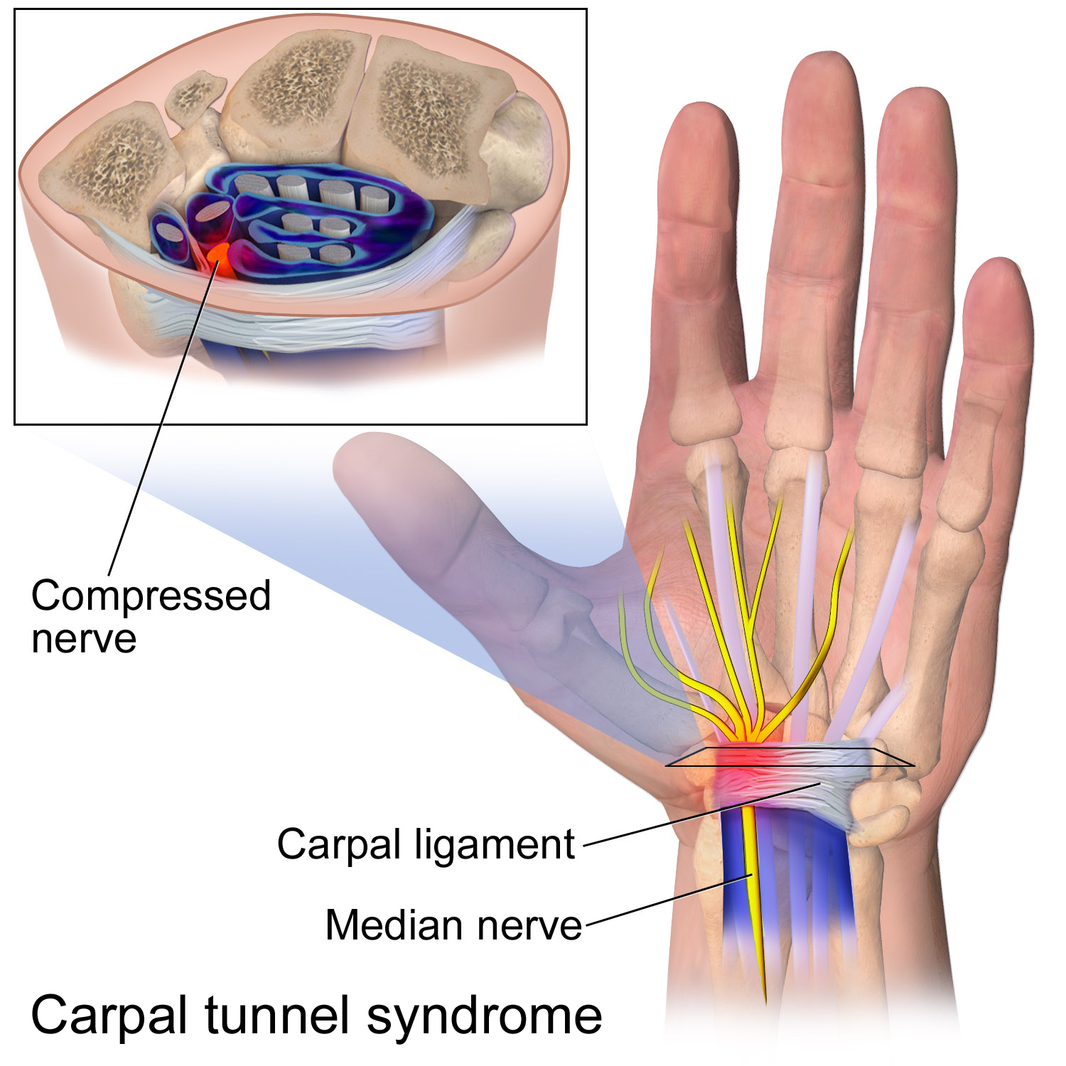
Carpal tunnel syndrome (CTS) is the collection of symptoms and signs associated with median neuropathy at the carpal tunnel. Most CTS is related to idiopathic compression of the median nerve as it travels through the wrist at the carpal tunnel (IMNCT). Idiopathic means that there is no other disease process contributing to pressure on the nerve. As with most structural issues, it occurs in both hands, and the strongest risk factor is genetics. Other conditions can cause CTS such as wrist fracture or rheumatoid arthritis. After fracture, swelling, bleeding, and deformity compress the median nerve. With rheumatoid arthritis, the enlarged synovial lining of the tendons causes compression. The main symptoms are numbness and tingling in the thumb, index finger, middle finger and the thumb side of the ring finger. People often report pain, but pain without tingling is not characteristic of IMNCT. Rather, the numbness can be so intense that it is described as painful. Symptoms are ... [...More Info...] [...Related Items...] OR: [Wikipedia] [Google] [Baidu] |
Hand Of Benediction
The hand of benediction, also known as benediction sign or preacher's hand, has been said to occur as a result of prolonged compression or injury of the median nerve at the forearm or elbow. More recently it has been shown that the clinical appearance of a high median nerve palsy is different from the classical hand of benediction or preacher's hand posture pointing finger. In this article "High Median Nerve Paralysis: Is the Hand of Benediction or Preacher's Hand A Correct Sign?" it shows that the hand of benediction or preacher’s hand is incorrectly associated with a high median nerve lesion. https://pubmed.ncbi.nlm.nih.gov/36320624/ Cause The term "hand of benediction" has been used to refer to damage of the median nerve. However, the name is misleading as the patients with this median nerve problem usually can flex all fingers except for the index finger. The index finger is still extended at the metacarpophalangeal joint (MCP joint) when the ulnar nerve innervated muscle ... [...More Info...] [...Related Items...] OR: [Wikipedia] [Google] [Baidu] |
MCP Joint
The metacarpophalangeal joints (MCP) are situated between the metacarpal bones and the proximal phalanges of the fingers. These joints are of the condyloid kind, formed by the reception of the rounded heads of the metacarpal bones into shallow cavities on the proximal ends of the proximal phalanges. Being condyloid, they allow the movements of flexion, extension, abduction, adduction and circumduction at the joint. Structure Ligaments Each joint has: * palmar ligaments of metacarpophalangeal articulations * collateral ligaments of metacarpophalangeal articulations Dorsal surfaces The dorsal surfaces of these joints are covered by the expansions of the Extensor tendons, together with some loose areolar tissue which connects the deep surfaces of the tendons to the bones. Function The movements which occur in these joints are flexion, extension, adduction, abduction, and circumduction; the movements of abduction and adduction are very limited, and cannot be performed while the ... [...More Info...] [...Related Items...] OR: [Wikipedia] [Google] [Baidu] |
Lateral Epicondylitis
Tennis elbow, also known as lateral epicondylitis or enthesopathy of the extensor carpi radialis origin, is a condition in which the outer part of the elbow becomes painful and tender. The pain may also extend into the back of the forearm. Onset of symptoms is generally gradual although they can seem sudden and be misinterpreted as an injury. Golfer's elbow is a similar condition that affects the inside of the elbow. Enthesopathies are idiopathic, meaning science has not yet determined the cause. Enthesopathies are most common in middle age (ages 35 to 60). It is often stated that the condition is caused by excessive use of the muscles of the back of the forearm, but this is not supported by experimental evidence and is a common misinterpretation or unhelpful thought about symptoms. It may be associated with work or sports, classically racquet sports, but most people with the condition are not exposed to these activities. The diagnosis is based on the symptoms and examination ... [...More Info...] [...Related Items...] OR: [Wikipedia] [Google] [Baidu] |
Radiography
Radiography is an imaging technique using X-rays, gamma rays, or similar ionizing radiation and non-ionizing radiation to view the internal form of an object. Applications of radiography include medical radiography ("diagnostic" and "therapeutic") and industrial radiography. Similar techniques are used in airport security (where "body scanners" generally use backscatter X-ray). To create an image in conventional radiography, a beam of X-rays is produced by an X-ray generator and is projected toward the object. A certain amount of the X-rays or other radiation is absorbed by the object, dependent on the object's density and structural composition. The X-rays that pass through the object are captured behind the object by a detector (either photographic film or a digital detector). The generation of flat two dimensional images by this technique is called projectional radiography. In computed tomography (CT scanning) an X-ray source and its associated detectors rotate around the su ... [...More Info...] [...Related Items...] OR: [Wikipedia] [Google] [Baidu] |
Thenar Eminence
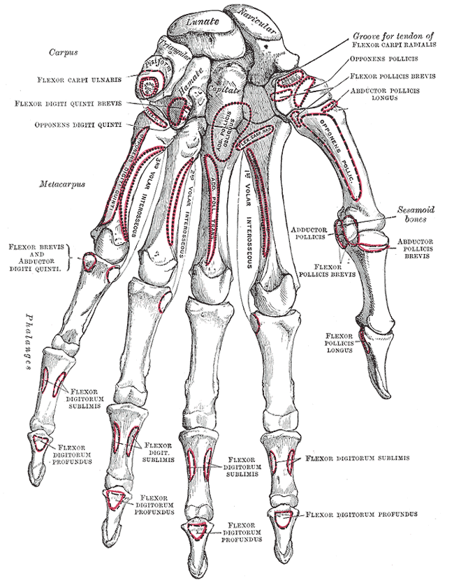
The thenar eminence is the mound formed at the base of the thumb on the palm of the hand by the intrinsic group of muscles of the thumb. The skin overlying this region is the area stimulated when trying to elicit a palmomental reflex. The word thenar comes . Structure The following three muscles are considered part of the thenar eminence: * Abductor pollicis brevis abducts the thumb. This muscle is the most superficial of the thenar group. * Flexor pollicis brevis, which lies next to the abductor, will flex the thumb, curling it up in the palm. (The Flexor pollicis longus, which is inserted into the distal phalanx of the thumb, is not considered part of the thenar eminence.) * Opponens pollicis lies deep to abductor pollicis brevis. As its name suggests it opposes the thumb, bringing it against the fingers. This is a very important movement, as most of human hand dexterity comes from this action. Another muscle that controls movement of the thumb is adductor pollicis. It lies ... [...More Info...] [...Related Items...] OR: [Wikipedia] [Google] [Baidu] |
Carpal Tunnel
In the human body, the carpal tunnel or carpal canal is the passageway on the palmar side of the wrist that connects the forearm to the hand. The tunnel is bounded by the bones of the wrist and flexor retinaculum from connective tissue. Normally several tendons from the flexor group of forearm muscles and the median nerve pass through it. There are described cases of variable median artery occurrence. When any of the nine long flexor tendons passing through the narrow carpal canal swell or degenerate, the narrowing of the canal may result in the median nerve becoming entrapped or compressed, a common medical condition known as carpal tunnel syndrome (CTS). Structure The carpal bones that make up the wrist form an arch which is convex on the dorsal side of the hand and concave on the palmar side. The groove on the palmar side, the ''sulcus carpi'', is covered by the flexor retinaculum, a sheath of tough connective tissue, thus forming the carpal tunnel. On the side of the ... [...More Info...] [...Related Items...] OR: [Wikipedia] [Google] [Baidu] |
Palmar Cutaneous Branch
The palmar branch of the median nerve is a branch of the median nerve which arises at the distal part of the forearm. Branches It pierces the palmar carpal ligament, and divides into a lateral and a medial branch; * The ''lateral branch'' supplies the skin over the ball of the thumb, and communicates with the volar branch of the lateral antebrachial cutaneous nerve. * The ''medial branch'' supplies the skin of the Hand, palm and communicates with the palmar cutaneous branch of the ulnar. Clinical significance Unlike most of the median nerve innervation of the hand, the palmar branch travels superficial to the Flexor retinaculum of the hand. Therefore, this portion of the median nerve usually remains functioning during carpal tunnel syndrome. Additional images File:Gray812and814.PNG, Diagram of segmental distribution of the cutaneous nerves of the right upper extremity. References * http://nervesurgery.wustl.edu/NerveImages/Anatomy%20and%20Physiology/AP-Median-Nerve---I ... [...More Info...] [...Related Items...] OR: [Wikipedia] [Google] [Baidu] |


