|
Medial Cord
The medial cord is the part of the brachial plexus formed by of the anterior division of the lower trunk (C8-T1). Its name comes from it being medial to the axillary artery as it passes through the axilla. The other cords of the brachial plexus are the posterior cord and lateral cord. The medial cord gives rise to the following nerves from proximal to distal: *medial pectoral nerve (C8-T1) *medial brachial cutaneous nerve (T1) *medial antebrachial cutaneous nerve (C8-T1) *medial head of median nerve (C8-T1) [other part of median nerve comes from lateral cord The lateral cord is the part of the brachial plexus formed by the anterior divisions of the upper (C5-C6) and middle trunks (C7). Its name comes from it being lateral to the axillary artery as it passes through the axilla. The other cords of the br ...] *ulnar nerve (C8-T1, occasionally C7) Additional images File:PLEXUS BRACHIALIS.jpg, Brachial plexus References Nerves of the upper limb {{Neuroanatomy-stub ... [...More Info...] [...Related Items...] OR: [Wikipedia] [Google] [Baidu] |
Brachial Plexus
The brachial plexus is a network () of nerves formed by the anterior rami of the lower four cervical nerves and first thoracic nerve ( C5, C6, C7, C8, and T1). This plexus extends from the spinal cord, through the cervicoaxillary canal in the neck, over the first rib, and into the armpit, it supplies afferent and efferent nerve fibers the to chest, shoulder, arm, forearm, and hand. Structure The brachial plexus is divided into five ''roots'', three ''trunks'', six ''divisions'' (three anterior and three posterior), three ''cords'', and five ''branches''. There are five "terminal" branches and numerous other "pre-terminal" or "collateral" branches, such as the subscapular nerve, the thoracodorsal nerve, and the long thoracic nerve, that leave the plexus at various points along its length. A common structure used to identify part of the brachial plexus in cadaver dissections is the M or W shape made by the musculocutaneous nerve, lateral cord, median nerve, medial cord, and ... [...More Info...] [...Related Items...] OR: [Wikipedia] [Google] [Baidu] |
Medial Pectoral Nerve
The medial pectoral nerve (also known as the medial anterior thoracic nerve) arises from the medial cord (sometimes directly from the anterior division of the inferior trunk) of the brachial plexus, and through it from the eighth cervical and first thoracic roots (C8/T1). It passes behind the first part of the axillary artery, curves forward between the axillary artery and vein, and unites in front of the artery with a filament from the lateral nerve. It then enters the deep surface of the pectoralis minor muscle, where it divides into a number of branches, which supply the muscle. Two or three branches pierce the muscle and end in the sternocostal head of the pectoralis major muscle. The medial pectoral nerve pierces both the pectoralis minor and the sternocostal head of the pectoralis major. The lateral pectoral nerve pierces only the clavicular head of the pectoralis major. Clinical relevance The medial pectoral nerve can be used as a donor nerve when reconstructing a ... [...More Info...] [...Related Items...] OR: [Wikipedia] [Google] [Baidu] |
Medial Brachial Cutaneous Nerve
The medial brachial cutaneous nerve (lesser internal cutaneous nerve; medial cutaneous nerve of arm) is distributed to the skin on the medial brachial side of the arm. Anatomy It is the smallest branch of the brachial plexus, and arising from the medial cord receives its fibers from the eighth cervical and first thoracic nerves. It passes through the axilla, at first lying behind, and then medial to the axillary vein, and communicates with the intercostobrachial nerve. It descends along the medial side of the brachial artery to the middle of the arm, where it pierces the deep fascia, and is distributed to the skin of the back of the lower third of the arm, extending as far as the elbow, where some filaments are lost in the skin in front of the medial epicondyle, and others over the olecranon. It communicates with the ulnar branch of the medial antebrachial cutaneous nerve. Eponym The term nerve of Wrisberg (after Heinrich August Wrisberg) has been used to describe this nerve. ... [...More Info...] [...Related Items...] OR: [Wikipedia] [Google] [Baidu] |
Medial Antebrachial Cutaneous Nerve
The medial cutaneous nerve of the forearm (medial antebrachial cutaneous nerve) branches from the medial cord of the brachial plexus. It contains axons from the ventral rami of the eighth cervical (C8) and first thoracic (T1) nerves. It gives off a branch near the axilla, which pierces the fascia and supplies the skin covering the biceps brachii, nearly as far as the elbow. The nerve then runs down the ulnar side of the arm medial to the brachial artery, pierces the deep fascia with the basilic vein, about the middle of the arm, and divides into a volar and an ulnar branch. Volar branch The ''volar branch'' (ramus volaris; anterior branch), the larger, passes usually in front of, but occasionally behind, the vena mediana cubiti (median basilic vein). It then descends on the front of the ulnar side of the forearm, distributing filaments to the skin as far as the wrist, and communicating with the palmar cutaneous branch of the ulnar nerve. Ulnar branch The ''ulnar branch'' ... [...More Info...] [...Related Items...] OR: [Wikipedia] [Google] [Baidu] |
Median Nerve
The median nerve is a nerve in humans and other animals in the upper limb. It is one of the five main nerves originating from the brachial plexus. The median nerve originates from the lateral and medial cords of the brachial plexus, and has contributions from ventral roots of C5-C7 (lateral cord) and C8 and T1 (medial cord). The median nerve is the only nerve that passes through the carpal tunnel. Carpal tunnel syndrome is the disability that results from the median nerve being pressed in the carpal tunnel. Structure The median nerve arises from the branches from lateral and medial cords of the brachial plexus, courses through the anterior part of arm, forearm, and hand, and terminates by supplying the muscles of the hand. Arm After receiving inputs from both the lateral and medial cords of the brachial plexus, the median nerve enters the arm from the axilla at the inferior margin of the teres major muscle. It then passes vertically down and courses lateral to the brachial ar ... [...More Info...] [...Related Items...] OR: [Wikipedia] [Google] [Baidu] |
Ulnar Nerve
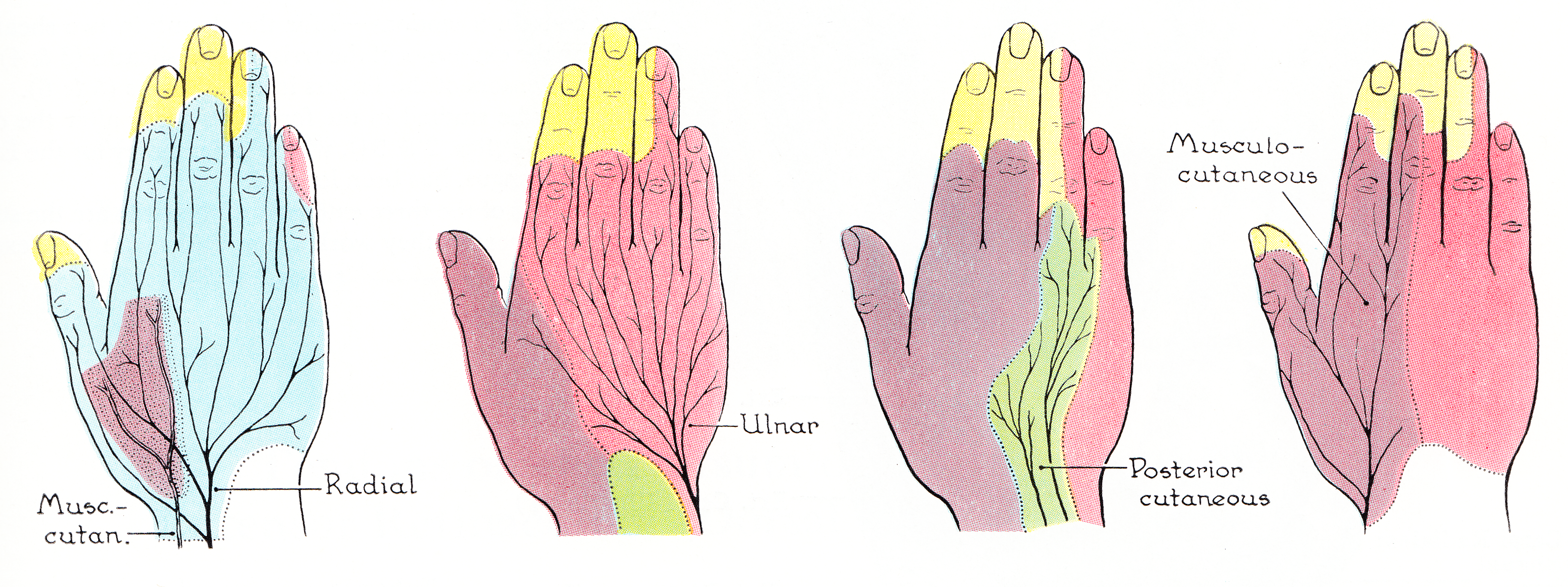
In human anatomy, the ulnar nerve is a nerve that runs near the ulna bone. The ulnar collateral ligament of elbow joint is in relation with the ulnar nerve. The nerve is the largest in the human body unprotected by muscle or bone, so injury is common. This nerve is directly connected to the little finger, and the adjacent half of the ring finger, innervating the palmar aspect of these fingers, including both front and back of the tips, perhaps as far back as the fingernail beds. This nerve can cause an electric shock-like sensation by striking the medial epicondyle of the humerus posteriorly, or inferiorly with the elbow flexed. The ulnar nerve is trapped between the bone and the overlying skin at this point. This is commonly referred to as bumping one's "funny bone". This name is thought to be a pun, based on the sound resemblance between the name of the bone of the upper arm, the humerus, and the word "humorous". Alternatively, according to the Oxford English Dictionary, i ... [...More Info...] [...Related Items...] OR: [Wikipedia] [Google] [Baidu] |
Axillary Artery
In human anatomy, the axillary artery is a large blood vessel that conveys oxygenated blood to the lateral aspect of the thorax, the axilla (armpit) and the upper limb. Its origin is at the lateral margin of the first rib, before which it is called the subclavian artery. After passing the lower margin of teres major it becomes the brachial artery. Structure The axillary artery is often referred to as having three parts, with these divisions based on its location relative to the Pectoralis minor muscle, which is superficial to the artery. * First part – the part of the artery superior to the pectoralis minor * Second part – the part of the artery posterior to the pectoralis minor * Third part – the part of the artery inferior to the pectoralis minor. Relations The axillary artery is accompanied by the axillary vein, which lies medial to the artery, along its length. In the axilla, the axillary artery is surrounded by the brachial plexus. The second part of the axi ... [...More Info...] [...Related Items...] OR: [Wikipedia] [Google] [Baidu] |
Axilla
The axilla (also, armpit, underarm or oxter) is the area on the human body directly under the shoulder joint. It includes the axillary space, an anatomical space within the shoulder girdle between the arm and the thoracic cage, bounded superiorly by the imaginary plane between the superior borders of the first rib, clavicle and scapula (above which are considered part of the neck), medially by the serratus anterior muscle and thoracolumbar fascia, anteriorly by the pectoral muscles and posteriorly by the subscapularis, teres major and latissimus dorsi muscle. The soft skin covering the lateral axilla contains many hair and sweat glands. In humans, the formation of body odor happens mostly in the axilla. These odorant substances have been suggested by some to serve as pheromones, which play a role related to mate selection, although this is a controversial topic within the scientific community. The underarms seem more important than the pubic area for emitting body odor, whi ... [...More Info...] [...Related Items...] OR: [Wikipedia] [Google] [Baidu] |
Posterior Cord
The posterior cord is a part of the brachial plexus The brachial plexus is a network () of nerves formed by the anterior rami of the lower four cervical nerves and first thoracic nerve ( C5, C6, C7, C8, and T1). This plexus extends from the spinal cord, through the cervicoaxillary canal in th .... It consists of contributions from all of the roots of the brachial plexus. The posterior cord gives rise to the following nerves: Additional images File:PLEXUS BRACHIALIS.jpg, Brachial plexus File:Slide12OOO.JPG, Posterior cord File:Slide1SSS.JPG, Posterior cord File:Slide1cord.JPG, Brachial plexus.Deep dissection. File:Slide1ecc.JPG, Brachial plexus.Deep dissection.Anterolateral view References MBBS resources http://mbbsbasic.googlepages.com/ External links * - "Axilla, dissection, anterior view" Nerves of the upper limb {{neuroscience-stub ... [...More Info...] [...Related Items...] OR: [Wikipedia] [Google] [Baidu] |
Lateral Cord
The lateral cord is the part of the brachial plexus formed by the anterior divisions of the upper (C5-C6) and middle trunks (C7). Its name comes from it being lateral to the axillary artery as it passes through the axilla. The other cords of the brachial plexus are the posterior cord and medial cord. The lateral cord gives rise to the following nerves from proximal to distal: *lateral pectoral nerve (C5-C7) *musculocutaneous nerve (C5-C7) *lateral head of median nerve (C5-C7) [other part of median nerve comes from medial cord The medial cord is the part of the brachial plexus formed by of the anterior division of the lower trunk (C8-T1). Its name comes from it being medial to the axillary artery as it passes through the axilla. The other cords of the brachial plexus ar ...] Additional images File:Slide10EEEE.JPG, Lateral cord File:Slide1cord.JPG, Brachial plexus.Deep dissection. Nerves of the upper limb {{neuroanatomy-stub ... [...More Info...] [...Related Items...] OR: [Wikipedia] [Google] [Baidu] |


