|
Medial Calcaneal Branches Of The Tibial Nerve
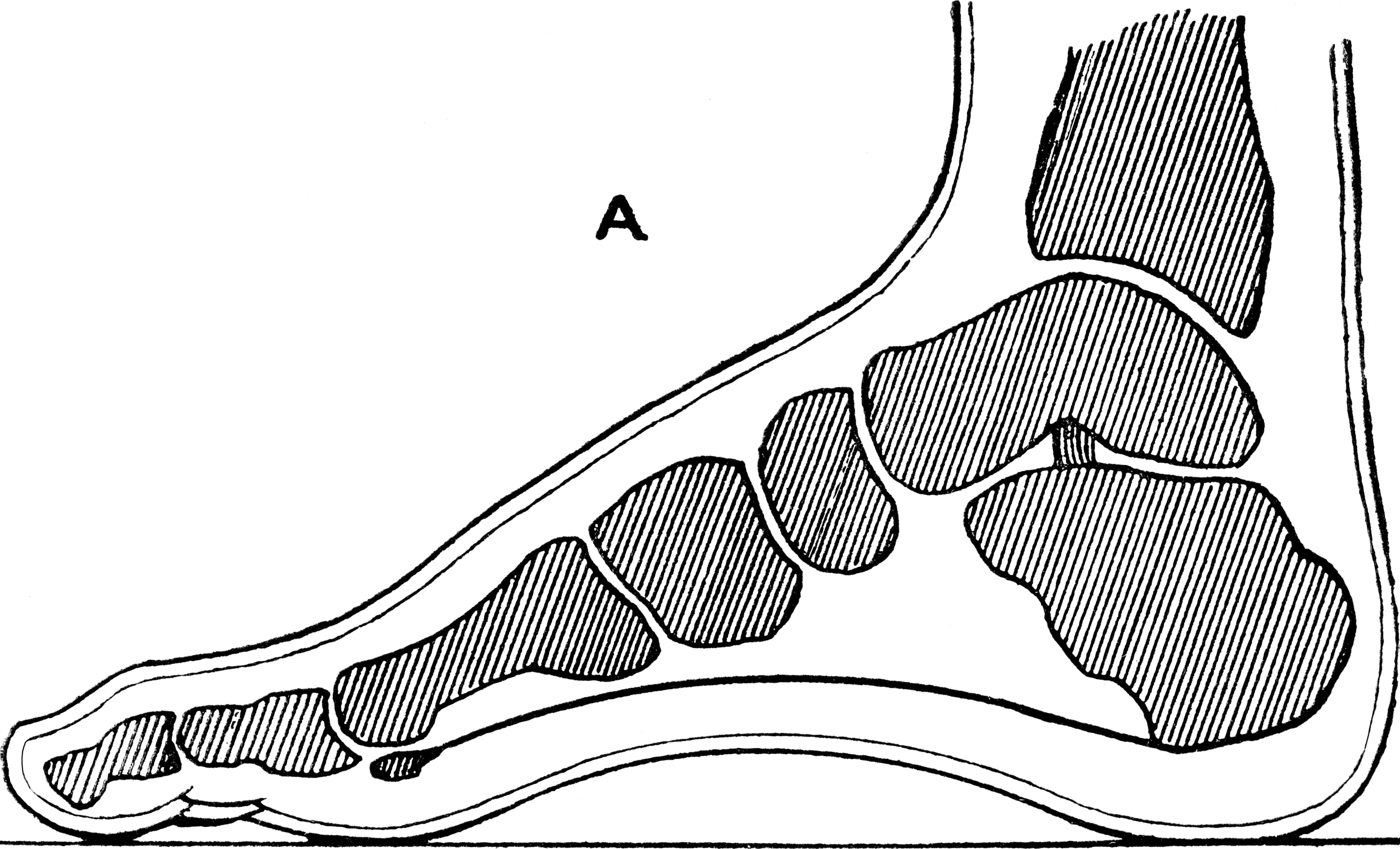
The medial calcaneal branches of the tibial nerve (internal calcaneal branches) perforate the laciniate ligament, and supply the skin of the heel and medial side of the sole of the foot. Structure The medial calcaneal nerve originates either from the tibial nerve or the lateral plantar nerve. It splits into two cutaneous branches. Function The medial calcaneal nerve provides sensory innervation to the medial side of the heel. See also * Cutaneous innervation of the lower limbs Cutaneous innervation refers to the area of the skin which is supplied by a specific nerve. Modern texts are in agreement about which areas of the skin are served by which nerves, but there are minor variations in some of the details. The borde ... References Nerves of the lower limb and lower torso {{Neuroanatomy-stub ... [...More Info...] [...Related Items...] OR: [Wikipedia] [Google] [Baidu] |
Tibial Nerve
The tibial nerve is a branch of the sciatic nerve. The tibial nerve passes through the popliteal fossa to pass below the arch of soleus. Structure Popliteal fossa The tibial nerve is the larger terminal branch of the sciatic nerve with root values of L4, L5, S1, S2, and S3. It lies superficial (or posterior) to the popliteal vessels, extending from the superior angle to the inferior angle of the popliteal fossa, crossing the popliteal vessels from lateral to medial side. It gives off branches as shown below: * Muscular branches - Muscular branches arise from the distal part of the popliteal fossa. It supplies the medial and lateral heads of gastrocnemius, soleus, plantaris and popliteus muscles. Nerve to popliteus crosses the popliteus muscle, runs downwards and laterally, winds around the lower border of the popliteus to supply the deep (or anterior) surface of the popliteus. This nerve also supplies the tibialis posterior muscle, superior tibiofibular joint, tibia bone, intero ... [...More Info...] [...Related Items...] OR: [Wikipedia] [Google] [Baidu] |
Laciniate Ligament
The flexor retinaculum of foot (laciniate ligament, internal annular ligament) is a strong fibrous band in the foot. Structure The flexor retinaculum of the foot extends from the medial malleolus above, to the calcaneus below. This converts a series of bony grooves into canals for the passage of the tendons of the flexor muscles and the posterior tibial vessels and tibial nerve into the sole of the foot, known as the tarsal tunnel. It is continuous by its upper border with the deep fascia of the leg, and by its lower border with the plantar aponeurosis and the fibers of origin of the abductor hallucis muscle. Enumerated from the medial side, the four canals which it forms transmit the tendons of the tibialis posterior and flexor digitorum longus muscles; the posterior tibial artery and tibial nerve, which run through a broad space beneath the ligament; and lastly, in a canal formed partly by the talus bone, talus, the tendon of the flexor hallucis longus. Clinical significanc ... [...More Info...] [...Related Items...] OR: [Wikipedia] [Google] [Baidu] |
Heel
The heel is the prominence at the posterior end of the foot. It is based on the projection of one bone, the calcaneus or heel bone, behind the articulation of the bones of the lower Human leg, leg. Structure To distribute the compressive forces exerted on the heel during gait, and especially the stance phase when the heel contacts the ground, the sole (foot), sole of the foot is covered by a layer of subcutaneous connective tissue up to 2 cm thick (under the heel). This tissue has a system of pressure chambers that both acts as a shock absorber and stabilises the sole. Each of these chambers contains fibrofatty tissue covered by a layer of tough connective tissue made of collagen fibers. These septum, septa ("walls") are firmly attached both to the plantar aponeurosis above and the sole's Dermis, skin below. The sole of the foot is one of the most highly vascularized regions of the body surface, and the dense system of blood vessels further stabilize the septa. The Ach ... [...More Info...] [...Related Items...] OR: [Wikipedia] [Google] [Baidu] |
Sole Of The Foot
The sole is the bottom of the foot. In humans the sole of the foot is anatomically referred to as the plantar aspect. Structure The glabrous skin on the sole of the foot lacks the hair and pigmentation found elsewhere on the body, and it has a high concentration of sweat pores. The sole contains the thickest layers of skin on the body due to the weight that is continually placed on it. It is crossed by a set of creases that form during the early stages of embryonic development. Like those of the palm, the sweat pores of the sole lack sebaceous glands. The sole is a sensory organ by which we can perceive the ground while standing and walking. The subcutaneous tissue in the sole has adapted to deal with the high local compressive forces on the heel and the ball (between the toes and the arch) by developing a system of "pressure chambers." Each chamber is composed of internal fibrofatty tissue covered by external collagen connective tissue. The septa (internal walls) of th ... [...More Info...] [...Related Items...] OR: [Wikipedia] [Google] [Baidu] |
Lateral Plantar Nerve
The lateral plantar nerve (external plantar nerve) is a branch of the tibial nerve, in turn a branch of the sciatic nerve and supplies the skin of the fifth toe and lateral half of the fourth, as well as most of the deep muscles, its distribution being similar to that of the ulnar nerve in the hand. It passes obliquely forward with the lateral plantar artery to the lateral side of the foot, lying between the flexor digitorum brevis and quadratus plantae and, in the interval between the flexor muscle and the abductor digiti minimi, divides into a superficial and a deep branch. Before its division, it supplies the quadratus plantae and abductor digiti minimi. It divides into deep and superficial branches. Additional images File:Gray357.png, Coronal section through right talocrural and talocalcaneal joint In human anatomy, the subtalar joint, also known as the talocalcaneal joint, is a joint of the foot. It occurs at the meeting point of the talus and the calcaneus. The joi ... [...More Info...] [...Related Items...] OR: [Wikipedia] [Google] [Baidu] |
ScienceDirect
ScienceDirect is a website which provides access to a large bibliographic database of scientific and medical publications of the Dutch publisher Elsevier. It hosts over 18 million pieces of content from more than 4,000 academic journals and 30,000 e-books of this publisher. The access to the full-text requires subscription, while the bibliographic metadata is free to read. ScienceDirect is operated by Elsevier. It was launched in March 1997. Usage The journals are grouped into four main sections: ''Physical Sciences and Engineering'', ''Life Sciences'', ''Health Sciences'', and ''Social Sciences and Humanities''. Article abstracts are freely available, and access to their full texts (in PDF and, for newer publications, also HTML) generally requires a subscription or pay-per-view purchase unless the content is freely available in open access. Subscriptions to the overall offering hosted on ScienceDirect, rather than to specific titles it carries, are usually acquired through a ... [...More Info...] [...Related Items...] OR: [Wikipedia] [Google] [Baidu] |
Cutaneous Innervation Of The Lower Limbs
Cutaneous innervation refers to the area of the skin which is supplied by a specific nerve. Modern texts are in agreement about which areas of the skin are served by which nerves, but there are minor variations in some of the details. The borders designated by the diagrams in the 1918 edition of '' Gray's Anatomy'', provided below, are similar but not identical to those generally accepted today. Pelvis and buttocks * Lateral cutaneous nerve of thigh - labeled as "lateral femoral cutaneous" (pink) * Lumboinguinal nerve (green) and Ilioinguinal nerve (purple). In modern texts, these two regions are often considered to be innervated by the genitofemoral nerve. * Medial cluneal nerves (pink) - labeled as "post. division of sacral" * Inferior cluneal nerves (pink region, not designated with its own section) * Perforating cutaneous nerve (pink region, not designated with its own section) * Superior cluneal nerves (yellow) - labeled as "post. division of lumbar" * Iliohypogastric n ... [...More Info...] [...Related Items...] OR: [Wikipedia] [Google] [Baidu] |

