|
Mdc1
Mediator of DNA damage checkpoint protein 1 is a 2080 amino acid long protein that in humans is encoded by the ''MDC1'' gene located on the short arm (p) of chromosome 6. MDC1 protein is a regulator of the Intra-S phase and the G2/M cell cycle checkpoints and recruits repair proteins to the site of DNA damage. It is involved in determining cell survival fate in association with tumor suppressor protein p53. This protein also goes by the name Nuclear Factor with BRCT Domain 1 (NFBD1). Function Role in DNA damage response The ''MDC1'' gene encodes the MDC1 nuclear protein which is part of the DNA damage response (DDR) pathway, the mechanism through which eukaryotic cells respond to damaged DNA, specifically DNA double-strand breaks (DSB) that are caused by ionizing radiation or chemical clastogens. The DDR of mammalian cells is made up of kinases, and mediator/adaptors factors. In mammalian cells the DDR is a network of pathways made up of proteins that function as either ... [...More Info...] [...Related Items...] OR: [Wikipedia] [Google] [Baidu] |
Amino Acid
Amino acids are organic compounds that contain both amino and carboxylic acid functional groups. Although over 500 amino acids exist in nature, by far the most important are the 22 α-amino acids incorporated into proteins. Only these 22 appear in the genetic code of life. Amino acids can be classified according to the locations of the core structural functional groups ( alpha- , beta- , gamma- amino acids, etc.); other categories relate to polarity, ionization, and side-chain group type ( aliphatic, acyclic, aromatic, polar, etc.). In the form of proteins, amino-acid '' residues'' form the second-largest component (water being the largest) of human muscles and other tissues. Beyond their role as residues in proteins, amino acids participate in a number of processes such as neurotransmitter transport and biosynthesis. It is thought that they played a key role in enabling life on Earth and its emergence. Amino acids are formally named by the IUPAC- IUBMB Joint Commi ... [...More Info...] [...Related Items...] OR: [Wikipedia] [Google] [Baidu] |
Phosphorylation
In biochemistry, phosphorylation is described as the "transfer of a phosphate group" from a donor to an acceptor. A common phosphorylating agent (phosphate donor) is ATP and a common family of acceptor are alcohols: : This equation can be written in several ways that are nearly equivalent that describe the behaviors of various protonated states of ATP, ADP, and the phosphorylated product. As is clear from the equation, a phosphate group per se is not transferred, but a phosphoryl group (PO3-). Phosphoryl is an electrophile. This process and its inverse, dephosphorylation, are common in biology. Text was copied from this source, which is available under a Creative Commons Attribution 4.0 International License. Protein phosphorylation often activates (or deactivates) many enzymes. During respiration Phosphorylation is essential to the processes of both anaerobic and aerobic respiration, which involve the production of adenosine triphosphate (ATP), the "high-energy" exc ... [...More Info...] [...Related Items...] OR: [Wikipedia] [Google] [Baidu] |
KIAA1524
Protein CIP2A also known as cancerous inhibitor of PP2A (CIP2A) is a protein that in humans is encoded by the ''KIAA1524'' gene. CIP2A is a regulatory protein involved in the inhibition of the serine-threonine phosphatase activity of Protein phosphatase 2A (PP2A). PP2A is a trimeric enzyme composed of a catalytic C-subunit, a scaffolding A-subunit, and various regulatory B-subunits, which collectively dephosphorylate a vast majority of cellular serine/threonine phosphorylated proteins, including many involved in cancer progression. CIP2A has been shown to regulate phosphorylation and activity of numerous oncoproteins, promoting malignant cell growth and tumorigenesis in various human cancers. High expression levels of CIP2A have been observed in multiple cancer types, such as head and neck cancer, colon cancer, gastric cancer, breast cancer, prostate cancer, and lung cancer, correlating with poor patient prognosis and disease aggressivity. Additionally, CIP2A is implicated in res ... [...More Info...] [...Related Items...] OR: [Wikipedia] [Google] [Baidu] |
Microtubule
Microtubules are polymers of tubulin that form part of the cytoskeleton and provide structure and shape to eukaryotic cells. Microtubules can be as long as 50 micrometres, as wide as 23 to 27 nanometer, nm and have an inner diameter between 11 and 15 nm. They are formed by the polymerization of a Protein dimer, dimer of two globular proteins, Tubulin#Eukaryotic, alpha and beta tubulin into #Structure, protofilaments that can then associate laterally to form a hollow tube, the microtubule. The most common form of a microtubule consists of 13 protofilaments in the tubular arrangement. Microtubules play an important role in a number of cellular processes. They are involved in maintaining the structure of the cell and, together with microfilaments and intermediate filaments, they form the cytoskeleton. They also make up the internal structure of cilia and flagella. They provide platforms for intracellular transport and are involved in a variety of cellular processes, in ... [...More Info...] [...Related Items...] OR: [Wikipedia] [Google] [Baidu] |
DNA Damage (naturally Occurring)
Natural DNA damage is an alteration in the chemical structure of DNA, such as a break in a strand of DNA, a nucleobase missing from the backbone of DNA, or a chemically changed base such as 8-OHdG. DNA damage can occur naturally or via environmental factors, but is distinctly different from mutation, although both are types of error in DNA. DNA damage is an abnormal chemical structure in DNA, while a mutation is a change in the sequence of base pairs. DNA damages cause changes in the structure of the genetic material and prevents the replication mechanism from functioning and performing properly. The DNA damage response (DDR) is a complex signal transduction pathway which recognizes when DNA is damaged and initiates the cellular response to the damage. DNA damage and mutation have different biological consequences. While most DNA damages can undergo DNA repair, such repair is not 100% efficient. Un-repaired DNA damages accumulate in non-replicating cells, such as cells in the brai ... [...More Info...] [...Related Items...] OR: [Wikipedia] [Google] [Baidu] |
Oocyte
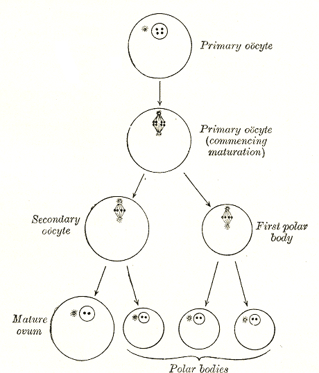
An oocyte (, oöcyte, or ovocyte) is a female gametocyte or germ cell involved in reproduction. In other words, it is an immature ovum, or egg cell. An oocyte is produced in a female fetus in the ovary during female gametogenesis. The female germ cells produce a primordial germ cell (PGC), which then undergoes mitosis, forming oogonia. During oogenesis, the oogonia become primary oocytes. An oocyte is a form of genetic material that can be collected for cryoconservation. Formation The formation of an oocyte is called oocytogenesis, which is a part of oogenesis. Oogenesis results in the formation of both primary oocytes during fetal period, and of secondary oocytes after it as part of ovulation. Characteristics Cytoplasm Oocytes are rich in cytoplasm, which contains yolk granules to nourish the cell early in development. Nucleus During the primary oocyte stage of oogenesis, the nucleus is called a germinal vesicle. The only normal human type of secondary oocyte has the ... [...More Info...] [...Related Items...] OR: [Wikipedia] [Google] [Baidu] |
Prophase
Prophase () is the first stage of cell division in both mitosis and meiosis. Beginning after interphase, DNA has already been replicated when the cell enters prophase. The main occurrences in prophase are the condensation of the chromatin reticulum and the disappearance of the nucleolus. Staining and microscopy Microscopy can be used to visualize condensed chromosomes as they move through meiosis and mitosis. Various DNA stains are used to treat cells such that condensing chromosomes can be visualized as the move through prophase. The giemsa G-banding technique is commonly used to identify mammalian chromosomes, but utilizing the technology on plant cells was originally difficult due to the high degree of chromosome compaction in plant cells. G-banding was fully realized for plant chromosomes in 1990. During both meiotic and mitotic prophase, giemsa staining can be applied to cells to elicit G-banding in chromosomes. Silver staining, a more modern technology, in conj ... [...More Info...] [...Related Items...] OR: [Wikipedia] [Google] [Baidu] |
Meiosis
Meiosis () is a special type of cell division of germ cells in sexually-reproducing organisms that produces the gametes, the sperm or egg cells. It involves two rounds of division that ultimately result in four cells, each with only one copy of each chromosome (haploid). Additionally, prior to the division, genetic material from the paternal and maternal copies of each chromosome is crossed over, creating new combinations of code on each chromosome. Later on, during fertilisation, the haploid cells produced by meiosis from a male and a female will fuse to create a zygote, a cell with two copies of each chromosome. Errors in meiosis resulting in aneuploidy (an abnormal number of chromosomes) are the leading known cause of miscarriage and the most frequent genetic cause of developmental disabilities. In meiosis, DNA replication is followed by two rounds of cell division to produce four daughter cells, each with half the number of chromosomes as the original parent cell. ... [...More Info...] [...Related Items...] OR: [Wikipedia] [Google] [Baidu] |
Genotoxic
Genotoxicity is the property of chemical agents that damage the genetic information within a cell causing mutations, which may lead to cancer. While genotoxicity is often confused with mutagenicity, all mutagens are genotoxic, but some genotoxic substances are not mutagenic. The alteration can have direct or indirect effects on the DNA: the induction of mutations, mistimed event activation, and direct DNA damage leading to mutations. The permanent, heritable changes can affect either somatic cells of the organism or germ cells to be passed on to future generations. Cells prevent expression of the genotoxic mutation by either DNA repair or apoptosis; however, the damage may not always be fixed leading to mutagenesis. To assay for genotoxic molecules, researchers assay for DNA damage in cells exposed to the toxic substrates. This DNA damage can be in the form of single- and double-strand breaks, loss of excision repair, cross-linking, alkali-labile sites, point mutations, and stru ... [...More Info...] [...Related Items...] OR: [Wikipedia] [Google] [Baidu] |
A549 Cell
A549 cells are adenocarcinoma, adenocarcinomic human Pulmonary alveolus, alveolar basal lamina, basal epithelial cell (biology), cells, and constitute a cell line that was first developed in 1972 by D. J. Giard, et al. through the removal and cell culture, culturing of cancerous lung tissue in the explanted tumor of a 58-year-old caucasian male. The cells are used as models for the study of lung cancer and the development of drug therapies against it. Characteristics A549 cells, as found in the lung tissue of their origin, are squamous and responsible for the diffusion of some substances, such as water and electrolytes, across Pulmonary alveolus, alveoli. If A549 cells are cultured ''in vitro'', they grow as a monolayer; adherent or attaching to the culture flask. The cells are able to synthesize lecithin and contain high levels of unsaturated fatty acids, which are important to maintain phospholipid bilayer, membrane phospholipids. A549 cells are widely used as a type II pulmonar ... [...More Info...] [...Related Items...] OR: [Wikipedia] [Google] [Baidu] |
Chk2
CHEK2 (Checkpoint kinase 2) is a tumor suppressor gene that encodes the protein CHK2, a serine-threonine kinase. CHK2 is involved in DNA repair, cell cycle arrest or apoptosis in response to DNA damage. Mutations to the CHEK2 gene have been linked to a wide range of cancers. Gene location The CHEK2 gene is located on the long (q) arm of chromosome 22 at position 12.1. Its location on chromosome 22 stretches from base pair 28,687,742 to base pair 28,741,904. Protein structure The CHEK2 protein encoded by the CHEK2 gene is a serine threonine kinase. The protein consists of 543 amino acids and the following domains: * N-terminal SQ/TQ cluster domain (SCD) * Central forkhead-associated (FHA) domain * C-terminal serine/threonine kinase domain (KD) The SCD domain contains multiple SQ/TQ motifs that serve as sites for phosphorylation in response to DNA damage. The most notable and frequently phosphorylated site being Thr68. CHK2 appears as a monomer in its inactive state. ... [...More Info...] [...Related Items...] OR: [Wikipedia] [Google] [Baidu] |


