|
Matrix Gla Protein
Matrix Gla protein (MGP) is member of a family of vitamin K2 dependent, Gla-containing proteins. MGP has a high affinity binding to calcium ions, similar to other Gla-containing proteins. The protein acts as an inhibitor of vascular mineralization and plays a role in bone organization. MGP is found in a number of body tissues in mammals, birds, and fish. Its mRNA is present in bone, cartilage, heart, and kidney. It is present in bone together with the related vitamin K2-dependent protein osteocalcin. In bone, its production is increased by vitamin D. Genetics The ''MGP'' was linked to the short arm of chromosome 12 in 1990. Its mRNA sequence length is 585 bases long in humans. Physiology MGP and osteocalcin are both calcium-binding proteins that may participate in the organisation of bone tissue. Both have glutamate residues that are post-translationally carboxylated by the enzyme gamma-glutamyl carboxylase in a reaction that requires Vitamin K hydroquinone. Role in ... [...More Info...] [...Related Items...] OR: [Wikipedia] [Google] [Baidu] |
Vitamin K2
Vitamin K2 or menaquinone (MK) () is one of three types of vitamin K, the other two being vitamin K1 (phylloquinone) and K3 (menadione). K2 is both a tissue and bacterial product (derived from vitamin K1 in both cases) and is usually found in animal products or fermented foods. The number ''n'' of isoprenyl units in their side chain differs and ranges from 4 to 13, hence Vitamin K2 consists of various forms. It is indicated as a suffix (-n), e. g. MK-7 or MK-9. The most common in the human diet is the short-chain, water-soluble menatetrenone (MK-4), which is usually produced by tissue and/or bacterial conversion of vitamin K1, and is commonly found in animal products. It is known that production of MK-4 from dietary plant vitamin K1 can be accomplished by animal tissues alone, as it proceeds in germ-free rodents. However, at least one published study concluded that "MK-4 present in food does not contribute to the vitamin K status as measured by serum vitamin K levels. MK-7, howe ... [...More Info...] [...Related Items...] OR: [Wikipedia] [Google] [Baidu] |
Post-translational Modification
Post-translational modification (PTM) is the covalent and generally enzymatic modification of proteins following protein biosynthesis. This process occurs in the endoplasmic reticulum and the golgi apparatus. Proteins are synthesized by ribosomes translating mRNA into polypeptide chains, which may then undergo PTM to form the mature protein product. PTMs are important components in cell signaling, as for example when prohormones are converted to hormones. Post-translational modifications can occur on the amino acid side chains or at the protein's C- or N- termini. They can extend the chemical repertoire of the 20 standard amino acids by modifying an existing functional group or introducing a new one such as phosphate. Phosphorylation is a highly effective mechanism for regulating the activity of enzymes and is the most common post-translational modification. Many eukaryotic and prokaryotic proteins also have carbohydrate molecules attached to them in a process called glycosyla ... [...More Info...] [...Related Items...] OR: [Wikipedia] [Google] [Baidu] |
Extracellular Matrix Proteins
In biology, the extracellular matrix (ECM), also called intercellular matrix, is a three-dimensional network consisting of extracellular macromolecules and minerals, such as collagen, enzymes, glycoproteins and hydroxyapatite that provide structural and biochemical support to surrounding cells. Because multicellularity evolved independently in different multicellular lineages, the composition of ECM varies between multicellular structures; however, cell adhesion, cell-to-cell communication and differentiation are common functions of the ECM. The animal extracellular matrix includes the interstitial matrix and the basement membrane. Interstitial matrix is present between various animal cells (i.e., in the intercellular spaces). Gels of polysaccharides and fibrous proteins fill the interstitial space and act as a compression buffer against the stress placed on the ECM. Basement membranes are sheet-like depositions of ECM on which various epithelial cells rest. Each type of connecti ... [...More Info...] [...Related Items...] OR: [Wikipedia] [Google] [Baidu] |
Arterial
An artery (plural arteries) () is a blood vessel in humans and most animals that takes blood away from the heart to one or more parts of the body (tissues, lungs, brain etc.). Most arteries carry oxygenated blood; the two exceptions are the pulmonary and the umbilical arteries, which carry deoxygenated blood to the organs that oxygenate it (lungs and placenta, respectively). The effective arterial blood volume is that extracellular fluid which fills the arterial system. The arteries are part of the circulatory system, that is responsible for the delivery of oxygen and nutrients to all cells, as well as the removal of carbon dioxide and waste products, the maintenance of optimum blood pH, and the circulation of proteins and cells of the immune system. Arteries contrast with veins, which carry blood back towards the heart. Structure The anatomy of arteries can be separated into gross anatomy, at the macroscopic level, and microanatomy, which must be studied with a microscop ... [...More Info...] [...Related Items...] OR: [Wikipedia] [Google] [Baidu] |
Hypoplasia
Hypoplasia (from Ancient Greek ὑπo- ''hypo-'' 'under' + πλάσις ''plasis'' 'formation'; adjective form ''hypoplastic'') is underdevelopment or incomplete development of a tissue or organ. Dictionary of Cell and Molecular Biology (11 March 2008) Although the term is not always used precisely, it properly refers to an inadequate or below-normal number of cells.Hypoplasia Stedman's Medical Dictionary. lww.com Hypoplasia is similar to |
Pulmonary Artery
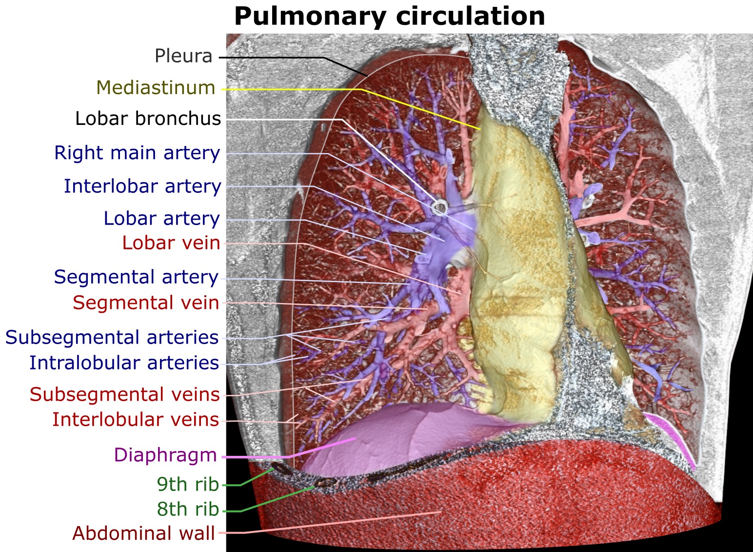
A pulmonary artery is an artery in the pulmonary circulation that carries deoxygenated blood from the right side of the heart to the lungs. The largest pulmonary artery is the ''main pulmonary artery'' or ''pulmonary trunk'' from the heart, and the smallest ones are the arterioles, which lead to the capillaries that surround the pulmonary alveoli. Structure The pulmonary arteries are blood vessels that carry systemic venous blood from the right ventricle of the heart to the microcirculation of the lungs. Unlike in other organs where arteries supply oxygenated blood, the blood carried by the pulmonary arteries is deoxygenated, as it is venous blood returning to the heart. The main pulmonary arteries emerge from the right side of the heart, and then split into smaller arteries that progressively divide and become arterioles, eventually narrowing into the capillary microcirculation of the lungs where gas exchange occurs. Pulmonary trunk In order of blood flow, the pulmonary art ... [...More Info...] [...Related Items...] OR: [Wikipedia] [Google] [Baidu] |
Stenosis
A stenosis (from Ancient Greek στενός, "narrow") is an abnormal narrowing in a blood vessel or other tubular organ or structure such as foramina and canals. It is also sometimes called a stricture (as in urethral stricture). ''Stricture'' as a term is usually used when narrowing is caused by contraction of smooth muscle (e.g. achalasia, prinzmetal angina); ''stenosis'' is usually used when narrowing is caused by lesion that reduces the space of lumen (e.g. atherosclerosis). The term coarctation is another synonym, but is commonly used only in the context of aortic coarctation. Restenosis is the recurrence of stenosis after a procedure. Types The resulting syndrome depends on the structure affected. Examples of vascular stenotic lesions include: * Intermittent claudication (peripheral artery stenosis) * Angina ( coronary artery stenosis) * Carotid artery stenosis which predispose to (strokes and transient ischaemic episodes) * Renal artery stenosis The types of sten ... [...More Info...] [...Related Items...] OR: [Wikipedia] [Google] [Baidu] |
Peripheral
A peripheral or peripheral device is an auxiliary device used to put information into and get information out of a computer. The term ''peripheral device'' refers to all hardware components that are attached to a computer and are controlled by the computer system, but they are not the core components of the computer, such as the CPU or power supply unit. In other words, peripherals can also be defined as devices that can be easily removed and plugged into a computer system. Several categories of peripheral devices may be identified, based on their relationship with the computer: *An ''input device'' sends data or instructions to the computer, such as a mouse, keyboard, graphics tablet, image scanner, barcode reader, game controller, light pen, light gun, microphone and webcam; *An ''output device'' provides output data from the computer, such as a computer monitor, projector, printer, headphones and computer speaker; *An ''input/output device'' performs both input and output fun ... [...More Info...] [...Related Items...] OR: [Wikipedia] [Google] [Baidu] |
Cartilage
Cartilage is a resilient and smooth type of connective tissue. In tetrapods, it covers and protects the ends of long bones at the joints as articular cartilage, and is a structural component of many body parts including the rib cage, the neck and the bronchial tubes, and the intervertebral discs. In other taxa, such as chondrichthyans, but also in cyclostomes, it may constitute a much greater proportion of the skeleton. It is not as hard and rigid as bone, but it is much stiffer and much less flexible than muscle. The matrix of cartilage is made up of glycosaminoglycans, proteoglycans, collagen fibers and, sometimes, elastin. Because of its rigidity, cartilage often serves the purpose of holding tubes open in the body. Examples include the rings of the trachea, such as the cricoid cartilage and carina. Cartilage is composed of specialized cells called chondrocytes that produce a large amount of collagenous extracellular matrix, abundant ground substance that is rich in pro ... [...More Info...] [...Related Items...] OR: [Wikipedia] [Google] [Baidu] |
Keutel Syndrome
Keutel syndrome (KS) is a rare autosomal recessive genetic disorder characterized by abnormal diffuse cartilage calcification, hypoplasia of the mid-face, peripheral pulmonary stenosis, hearing loss, short distal phalanges (tips) of the fingers and mild mental retardation. Individuals with KS often present with peripheral pulmonary stenosis, brachytelephalangism, sloping forehead, midface hypoplasia, and receding chin. It is associated with abnormalities in the gene coding for matrix gla protein, ''MGP''. Being an autosomal recessive disorder, it may be inherited from two unaffected, abnormal MGP-carrying parents. Thus, people who inherit two affected ''MGP'' alleles will likely inherit KS. It was first identified in 1972 as a novel rare genetic disorder sharing similar symptoms with chondrodysplasia punctata. Multiple forms of chondrodysplasia punctata share symptoms consistent with KS including abnormal cartilage calcification, forceful respiration, brachytelephalangism, hypoto ... [...More Info...] [...Related Items...] OR: [Wikipedia] [Google] [Baidu] |
Vitamin K
Vitamin K refers to structurally similar, fat-soluble vitamers found in foods and marketed as dietary supplements. The human body requires vitamin K for post-synthesis modification of certain proteins that are required for blood coagulation (K from ''Koagulation'', German for "coagulation") or for controlling binding of calcium in bones and other tissues. The complete synthesis involves final modification of these so-called "Gla proteins" by the enzyme gamma-glutamyl carboxylase that uses vitamin K as a cofactor. Vitamin K is used in the liver as the intermediate VKH2 to deprotonate a glutamate residue and then is reprocessed into vitamin K through a vitamin K oxide intermediate. The presence of uncarboxylated proteins indicates a vitamin K deficiency. Carboxylation allows them to bind (chelate) calcium ions, which they cannot do otherwise. Without vitamin K, blood coagulation is seriously impaired, and uncontrolled bleeding occurs. Research suggests that deficiency of v ... [...More Info...] [...Related Items...] OR: [Wikipedia] [Google] [Baidu] |
Gamma-glutamyl Carboxylase
Gamma-glutamyl carboxylase is an enzyme that in humans is encoded by the ''GGCX'' gene, located on chromosome 2 at 2p12. Function Gamma-glutamyl carboxylase is an enzyme that catalyzes the posttranslational modification of vitamin K-dependent proteins. Many of these vitamin K-dependent proteins are involved in coagulation so the function of the encoded enzyme is essential for hemostasis. Most gla domain-containing proteins depend on this carboxylation reaction for posttranslational modification. In humans, the gamma-glutamyl carboxylase enzyme is most highly expressed in the liver. Catalytic reaction Gamma-glutamyl carboxylase oxidizes Vitamin K hydroquinone to Vitamin K 2,3 epoxide, while simultaneously adding CO2 to protein-bound glutamic acid (abbreviation = Glu) to form gamma-carboxyglutamic acid (also called gamma-carboxyglutamate, abbreviation = Gla). Presence of two carboxylate groups causes chelation of Ca2+ , resulting in change in tertiary structure of protein and its ... [...More Info...] [...Related Items...] OR: [Wikipedia] [Google] [Baidu] |


