|
Mastoid Cells
The mastoid cells (also called air cells of Lenoir or mastoid cells of Lenoir) are air-filled cavities within the mastoid process of the temporal bone of the cranium The skull is a bone protective cavity for the brain. The skull is composed of four types of bone i.e., cranial bones, facial bones, ear ossicles and hyoid bone. However two parts are more prominent: the cranium and the mandible. In humans, .... The mastoid cells are a form of skeletal pneumaticity. Infection in these cells is called mastoiditis. The term "cells" refers to enclosed spaces, not cells as living, biological units. Anatomy A section of the mastoid process will show it to be hollowed out into a number of spaces which exhibit great variety in their size and number. At the upper and front part of the process they are large and irregular and contain air, but toward the lower part they diminish in size, while those at the apex of the process are frequently quite small and contain marrow. Occasio ... [...More Info...] [...Related Items...] OR: [Wikipedia] [Google] [Baidu] |
Temporal Bone
The temporal bones are situated at the sides and base of the skull, and lateral to the temporal lobes of the cerebral cortex. The temporal bones are overlaid by the sides of the head known as the temples, and house the structures of the ears. The lower seven cranial nerves and the major vessels to and from the brain traverse the temporal bone. Structure The temporal bone consists of four parts— the squamous, mastoid, petrous and tympanic parts. The squamous part is the largest and most superiorly positioned relative to the rest of the bone. The zygomatic process is a long, arched process projecting from the lower region of the squamous part and it articulates with the zygomatic bone. Posteroinferior to the squamous is the mastoid part. Fused with the squamous and mastoid parts and between the sphenoid and occipital bones lies the petrous part, which is shaped like a pyramid. The tympanic part is relatively small and lies inferior to the squamous part, anterior to t ... [...More Info...] [...Related Items...] OR: [Wikipedia] [Google] [Baidu] |
Stylomastoid Artery
The stylomastoid artery enters the stylomastoid foramen and supplies the tympanic cavity, the tympanic antrum and mastoid cells, and the semicircular canals. It is a branch of the posterior auricular artery, and thus part of the external carotid arterial system. In the young subject a branch from this vessel forms, with the anterior tympanic artery from the internal maxillary, a vascular circle, which surrounds the tympanic membrane, and from which delicate vessels ramify on that membrane. It anastomoses with the superficial petrosal branch of the middle meningeal artery by a twig which enters the hiatus canalis facialis. References External links ArcLab Arteries of the head and neck {{circulatory-stub ... [...More Info...] [...Related Items...] OR: [Wikipedia] [Google] [Baidu] |
Mastoid Process
The mastoid part of the temporal bone is the posterior (back) part of the temporal bone, one of the bones of the skull. Its rough surface gives attachment to various muscles (via tendons) and it has openings for blood vessels. From its borders, the mastoid part articulates with two other bones. Etymology The word "mastoid" is derived from the Greek word for "breast", a reference to the shape of this bone. Surfaces Outer surface Its outer surface is rough and gives attachment to the occipitalis and posterior auricular muscles. It is perforated by numerous foramina (holes); for example, the mastoid foramen is situated near the posterior border and transmits a vein to the transverse sinus and a small branch of the occipital artery to the dura mater. The position and size of this foramen are very variable; it is not always present; sometimes it is situated in the occipital bone, or in the suture between the temporal and the occipital. Mastoid process The mastoid proce ... [...More Info...] [...Related Items...] OR: [Wikipedia] [Google] [Baidu] |
Cranium
The skull is a bone protective cavity for the brain. The skull is composed of four types of bone i.e., cranial bones, facial bones, ear ossicles and hyoid bone. However two parts are more prominent: the cranium and the mandible. In humans, these two parts are the neurocranium and the viscerocranium (facial skeleton) that includes the mandible as its largest bone. The skull forms the anterior-most portion of the skeleton and is a product of cephalisation—housing the brain, and several sensory structures such as the eyes, ears, nose, and mouth. In humans these sensory structures are part of the facial skeleton. Functions of the skull include protection of the brain, fixing the distance between the eyes to allow stereoscopic vision, and fixing the position of the ears to enable sound localisation of the direction and distance of sounds. In some animals, such as horned ungulates (mammals with hooves), the skull also has a defensive function by providing the mount (on the ... [...More Info...] [...Related Items...] OR: [Wikipedia] [Google] [Baidu] |
Skeletal Pneumaticity
Skeletal pneumaticity is the presence of air spaces within bones. It is generally produced during development by excavation of bone by pneumatic diverticula (air sacs) from an air-filled space, such as the lungs or nasal cavity. Pneumatization is highly variable between individuals, and bones not normally pneumatized can become pneumatized in pathological development. Cranial pneumaticity Pneumatization occurs in the skulls of mammals, crocodilians and birds among extant tetrapods. Pneumatization has been documented in extinct archosaurs including dinosaurs and pterosaurs. Pneumatic spaces include the paranasal sinuses and some of the mastoid cells. Postcranial pneumaticity Postcranial pneumaticity is found largely in certain archosaur groups, namely dinosaurs, pterosaurs, and birds. Vertebral pneumatization is widespread among saurischian dinosaurs, and some theropods have quite widespread pneumatization, for example ''Aerosteon riocoloradensis'' has pneumatization of the il ... [...More Info...] [...Related Items...] OR: [Wikipedia] [Google] [Baidu] |
Mastoiditis
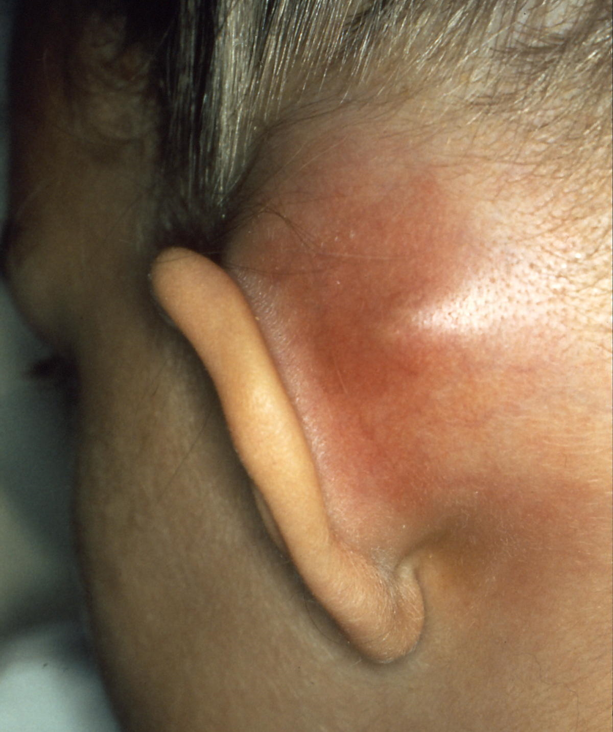
Mastoiditis is the result of an infection that extends to the air cells of the skull behind the ear. Specifically, it is an inflammation of the mucosal lining of the mastoid antrum and mastoid air cell system inside the mastoid process. The mastoid process is the portion of the temporal bone of the skull that is behind the ear. The mastoid process contains open, air-containing spaces. Mastoiditis is usually caused by untreated acute otitis media (middle ear infection) and used to be a leading cause of child mortality. With the development of antibiotics, however, mastoiditis has become quite rare in developed countries where surgical treatment is now much less frequent and more conservative, unlike former times. There is no evidence that the drop in antibiotic prescribing for otitis media has increased the incidence of mastoiditis, raising the possibility that the drop in reported cases is due to a confounding factor such as childhood immunizations against ''Haemophilus'' and ' ... [...More Info...] [...Related Items...] OR: [Wikipedia] [Google] [Baidu] |
Cell (biology)
The cell is the basic structural and functional unit of life forms. Every cell consists of a cytoplasm enclosed within a membrane, and contains many biomolecules such as proteins, DNA and RNA, as well as many small molecules of nutrients and metabolites.Cell Movements and the Shaping of the Vertebrate Body in Chapter 21 of Molecular Biology of the Cell '' fourth edition, edited by Bruce Alberts (2002) published by Garland Science. The Alberts text discusses how the "cellular building blocks" move to shape developing s. It is also common ... [...More Info...] [...Related Items...] OR: [Wikipedia] [Google] [Baidu] |
Mastoid Cells Of Lenoir
The mastoid part of the temporal bone is the posterior (back) part of the temporal bone, one of the bones of the skull. Its rough surface gives attachment to various muscles (via tendons) and it has openings for blood vessels. From its borders, the mastoid part articulates with two other bones. Etymology The word "mastoid" is derived from the Greek word for "breast", a reference to the shape of this bone. Surfaces Outer surface Its outer surface is rough and gives attachment to the occipitalis and posterior auricular muscles. It is perforated by numerous foramina (holes); for example, the mastoid foramen is situated near the posterior border and transmits a vein to the transverse sinus and a small branch of the occipital artery to the dura mater. The position and size of this foramen are very variable; it is not always present; sometimes it is situated in the occipital bone, or in the suture between the temporal and the occipital. Mastoid process The mastoid process is ... [...More Info...] [...Related Items...] OR: [Wikipedia] [Google] [Baidu] |
Bone Marrow
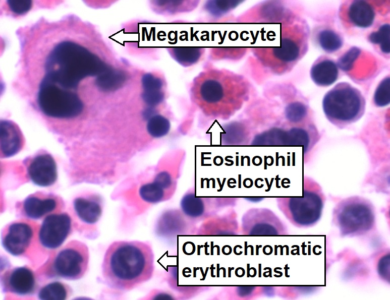
Bone marrow is a semi-solid tissue found within the spongy (also known as cancellous) portions of bones. In birds and mammals, bone marrow is the primary site of new blood cell production (or haematopoiesis). It is composed of hematopoietic cells, marrow adipose tissue, and supportive stromal cells. In adult humans, bone marrow is primarily located in the ribs, vertebrae, sternum, and bones of the pelvis. Bone marrow comprises approximately 5% of total body mass in healthy adult humans, such that a man weighing 73 kg (161 lbs) will have around 3.7 kg (8 lbs) of bone marrow. Human marrow produces approximately 500 billion blood cells per day, which join the systemic circulation via permeable vasculature sinusoids within the medullary cavity. All types of hematopoietic cells, including both myeloid and lymphoid lineages, are created in bone marrow; however, lymphoid cells must migrate to other lymphoid organs (e.g. thymus) in order to complete ... [...More Info...] [...Related Items...] OR: [Wikipedia] [Google] [Baidu] |
Otitis Media
Otitis media is a group of inflammatory diseases of the middle ear. One of the two main types is acute otitis media (AOM), an infection of rapid onset that usually presents with ear pain. In young children this may result in pulling at the ear, increased crying, and poor sleep. Decreased eating and a fever may also be present. The other main type is otitis media with effusion (OME), typically not associated with symptoms, although occasionally a feeling of fullness is described; it is defined as the presence of non-infectious fluid in the middle ear which may persist for weeks or months often after an episode of acute otitis media. Chronic suppurative otitis media (CSOM) is middle ear inflammation that results in a perforated tympanic membrane with discharge from the ear for more than six weeks. It may be a complication of acute otitis media. Pain is rarely present. All three types of otitis media may be associated with hearing loss. If children with hearing loss due to OME do ... [...More Info...] [...Related Items...] OR: [Wikipedia] [Google] [Baidu] |
Aditus Ad Antrum
The aditus to mastoid antrum (otomastoid foramen or entrance or aperture to the mastoid antrum) is a large irregular cavity that leads backward from the epitympanic recess into a considerable air space named the tympanic or mastoid antrum. The antrum communicates behind and below with the mastoid air cells, which vary considerably in number, size, and form; the antrum and mastoid air cells are lined by mucous membrane, continuous with that lining the tympanic cavity. On the medial wall of the entrance to the antrum is a rounded eminence, situated above and behind the prominence of the facial canal; it corresponds with the position of the ampullated ends of the superior and lateral semicircular canals. See also * Aditus * Mastoid antrum * Epitympanic recess The epitympanic recess is a hollow located on the superior/roof aspect of the middle ear. Clinical significance This recess is a possible route of spread of infection to the mastoid air cells located in the mastoid pro ... [...More Info...] [...Related Items...] OR: [Wikipedia] [Google] [Baidu] |
Mastoid Antrum
The mastoid antrum (tympanic antrum, antrum mastoideum, Valsalva's antrum) is an air space in the petrous portion of the temporal bone, communicating posteriorly with the mastoid cells and anteriorly with the epitympanic recess of the middle ear via the aditus to mastoid antrum (entrance to the mastoid antrum). These air spaces function as sound receptors, provide voice resonance, act as acoustic insulation and dissipation, provide protection from physical damage and reduce the mass of the cranium. The roof is formed by the tegmen antri which is a continuation of the tegmen tympani and separates it from the middle cranial fossa. The lateral wall of the antrum is formed by a plate of bone which is an average of 1.5 cm in adults. The mastoid air cell system is a major contributor to middle ear inflammatory diseases. Additional images File:Gray1209.png, Left temporal bone showing surface markings for the tympanic antrum (red), transverse sinus (blue), and facial nerve T ... [...More Info...] [...Related Items...] OR: [Wikipedia] [Google] [Baidu] |


