|
Malpighian
Malpighian is an attribute to several anatomical structures discovered by, described by or attributed to Marcello Malpighi: * Malpighian corpuscle (other) ** Renal corpuscle, the initial filtering component of nephrons in the kidneys ** Splenic lymphoid nodules or white nodules, follicles in the white pulp of the spleen * Malpighian layer of the skin, a term with various definitions * Malpighian tubule system, an excretory and osmoregulatory system found in some insects, myriapods, arachnids and tardigrades See also * Malpighiales The Malpighiales comprise one of the largest orders of flowering plants, containing about 36 families and more than species, about 7.8% of the eudicots. The order is very diverse, containing plants as different as the willow, violet, poinsett ..., an order of flowering plants ** '' Malpighia'', a genus in the order {{disambiguation ... [...More Info...] [...Related Items...] OR: [Wikipedia] [Google] [Baidu] |
Malpighian Tubule System
The Malpighian tubule system is a type of excretory and osmoregulatory system found in some insects, myriapods, arachnids and tardigrades. The system consists of branching tubules extending from the alimentary canal that absorbs solutes, water, and wastes from the surrounding hemolymph. The wastes then are released from the organism in the form of solid nitrogenous compounds and calcium oxalate. The system is named after Marcello Malpighi, a seventeenth-century anatomist. Structure Malpighian tubules are slender tubes normally found in the posterior regions of arthropod alimentary canals. Each tubule consists of a single layer of cells that is closed off at the distal end with the proximal end joining the alimentary canal at the junction between the midgut and hindgut. Most tubules are normally highly convoluted. The number of tubules varies between species although most occur in multiples of two. Tubules are usually bathed in hemolymph and are in proximity to fat body tissue. ... [...More Info...] [...Related Items...] OR: [Wikipedia] [Google] [Baidu] |
Marcello Malpighi
Marcello Malpighi (10 March 1628 – 30 November 1694) was an Italian biologist and physician, who is referred to as the "Founder of microscopical anatomy, histology & Father of physiology and embryology". Malpighi's name is borne by several physiological features related to the biological excretory system, such as the Malpighian corpuscles and Malpighian pyramids of the kidneys and the Malpighian tubule system of insects. The splenic lymphoid nodules are often called the "Malpighian bodies of the spleen" or Malpighian corpuscles. The botanical family Malpighiaceae is also named after him. He was the first person to see capillaries in animals, and he discovered the link between arteries and veins that had eluded William Harvey. Malpighi was one of the earliest people to observe red blood cells under a microscope, after Jan Swammerdam. His treatise ''De polypo cordis'' (1666) was important for understanding blood composition, as well as how blood clots. In it, Malpighi describ ... [...More Info...] [...Related Items...] OR: [Wikipedia] [Google] [Baidu] |
Malpighian Layer
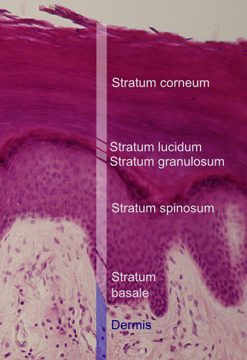
The Malpighian layer (''stratum mucosum'' or ''stratum malpighii'') of the epidermis, the outermost layer of the skin, is generally defined as both the stratum basale (basal layer) and the thicker stratum spinosum (spinous layer/prickle cell layer) immediately above it as a single unit,McGrath, J.A.; Eady, R.A.; Pope, F.M. (2004). ''Rook's Textbook of Dermatology'' (Seventh Edition). Blackwell Publishing. Pages 3.1-3.6. . although it is occasionally defined as the stratum basale specifically, or the stratum spinosum specifically. It is named after the Italian biologist and physician Marcello Malpighi. Basal cell carcinoma originates from the basal layer of the stratum malpighii. This layer is where almost all of the mitotic activity in the epidermis occurs. The activity of these cells is increased by IL-1 (interleukin-1) and epidermal growth factor. The activity is decreased by transforming growth factor beta Transforming growth factor beta (TGF-β) is a multifunctional cyto ... [...More Info...] [...Related Items...] OR: [Wikipedia] [Google] [Baidu] |
Renal Corpuscle
A renal corpuscle (also called malpighian body) is the blood-filtering component of the nephron of the kidney. It consists of a glomerulus - a tuft of capillaries composed of endothelial cells, and a glomerular capsule known as Bowman's capsule. Structure The renal corpuscle is composed of two structures, the glomerulus and the Bowman's capsule. The glomerulus is a small tuft of capillaries containing two cell types. Endothelial cells, which have large fenestrae, are not covered by diaphragms. Mesangial cells are modified smooth muscle cells that lie between the capillaries. They regulate blood flow by their contractile activity and secrete extracellular matrix, prostaglandins, and cytokines. Mesangial cells also have phagocytic activity, removing proteins and other molecules trapped in the glomerular basement membrane or filtration barrier. The Bowman's capsule has an outer parietal layer composed of simple squamous epithelium. The visceral layer, composed of modified ... [...More Info...] [...Related Items...] OR: [Wikipedia] [Google] [Baidu] |
Malpighian Corpuscle (other) .
{{disambiguation ...
There are at least two anatomical structures called a Malpighian corpuscle. They are also known as: * Renal corpuscles — the initial filtering component of nephrons in the kidneys * White pulp, splenic lymphoid nodules, or white nodules — follicles in the white pulp of the spleen, containing many lymphocytes These structures are named after Marcello Malpighi (1628–1694), an Italian physician and biologist regarded as the father of microscopical anatomy and histology Histology, also known as microscopic anatomy or microanatomy, is the branch of biology which studies the microscopic anatomy of biological tissues. Histology is the microscopic counterpart to gross anatomy, which looks at larger structures vis ... [...More Info...] [...Related Items...] OR: [Wikipedia] [Google] [Baidu] |
Splenic Lymphoid Nodules
White pulp is a histological designation for regions of the spleen (named because it appears whiter than the surrounding red pulp on gross section), that encompasses approximately 25% of splenic tissue. White pulp consists entirely of lymphoid tissue. Specifically, the white pulp encompasses several areas with distinct functions: * The periarteriolar lymphoid sheaths (PALS) are typically associated with the arteriole supply of the spleen; they contain T lymphocytes. * Lymph follicles with dividing B lymphocytes are located between the PALS and the marginal zone bordering on the red pulp. IgM and IgG2 are produced in this zone. These molecules play a role in opsonization of extracellular organisms, encapsulated bacteria in particular. * The marginal zone exists between the white pulp and red pulp. It is located farther away from the central arteriole, in proximity to the red pulp. It contains antigen-presenting cells (APCs), such as dendritic cells and macrophages. Some of the w ... [...More Info...] [...Related Items...] OR: [Wikipedia] [Google] [Baidu] |
Malpighiales
The Malpighiales comprise one of the largest orders of flowering plants, containing about 36 families and more than species, about 7.8% of the eudicots. The order is very diverse, containing plants as different as the willow, violet, poinsettia, manchineel, rafflesia and coca plant, and are hard to recognize except with molecular phylogenetic evidence. It is not part of any of the classification systems based only on plant morphology. Molecular clock calculations estimate the origin of stem group Malpighiales at around 100 million years ago ( Mya) and the origin of crown group Malpighiales at about 90 Mya. The Malpighiales are divided into 32 to 42 families, depending upon which clades in the order are given the taxonomic rank of family. In the APG III system, 35 families were recognized. Medusagynaceae, Quiinaceae, Peraceae, Malesherbiaceae, Turneraceae, Samydaceae, and Scyphostegiaceae were consolidated into other families. The largest family, by far, is the Euphorbiaceae, ... [...More Info...] [...Related Items...] OR: [Wikipedia] [Google] [Baidu] |


