|
Magnuviator
''Magnuviator'' is a genus of extinct iguanomorph lizard from the Late Cretaceous of Montana, US. It contains one species, ''M. ovimonsensis'', described in 2017 by DeMar ''et al.'' from two specimens that were discovered in the Egg Mountain nesting site. ''Magnuviator'' is closest related to the Asian ''Saichangurvel'' and ''Temujinia'', which form the group Temujiniidae. Unlike other members of the Iguanomorpha, however, ''Magnuviator'' bears a distinct articulating notch on its tibia for the ankle bones (astragalus and calcaneum), which has traditionally been considered a characteristic of non-iguanomorph lizards. The morphology of its teeth suggests that its diet would have mainly consisted of wasps, like the modern phyrnosomatid iguanians ''Callisaurus'' and ''Urosaurus'', although it also shows some adaptations to herbivory. Description For an iguanomorph, ''Magnuviator'' was large, measuring long without the tail. Both of the known specimens were adults, judging by the ... [...More Info...] [...Related Items...] OR: [Wikipedia] [Google] [Baidu] |
Egg Mountain
The Two Medicine Formation is a geological formation, or rock body, in northwestern Montana and southern Alberta that was deposited between and (million years ago), during Campanian (Late Cretaceous) time. It crops out to the east of the Rocky Mountain Overthrust Belt, and the western portion (about thick) of this formation is folded and faulted while the eastern part, which thins out into the Sweetgrass Arch, is mostly undeformed plains. Below the formation are the nearshore (beach and tidal zone) deposits of the Virgelle Sandstone, and above it is the marine Bearpaw Shale. Throughout the Campanian, the Two Medicine Formation was deposited between the western shoreline of the Late Cretaceous Interior Seaway and the eastward advancing margin of the Cordilleran Overthrust Belt. The Two Medicine Formation is mostly sandstone, deposited by rivers and deltas. History of research In 1913, a US Geological Survey crew headed by Eugene Stebinger and a US National Museum crew headed ... [...More Info...] [...Related Items...] OR: [Wikipedia] [Google] [Baidu] |
Campanian
The Campanian is the fifth of six ages of the Late Cretaceous Epoch on the geologic timescale of the International Commission on Stratigraphy (ICS). In chronostratigraphy, it is the fifth of six stages in the Upper Cretaceous Series. Campanian spans the time from 83.6 (± 0.2) to 72.1 (± 0.2) million years ago. It is preceded by the Santonian and it is followed by the Maastrichtian. The Campanian was an age when a worldwide sea level rise covered many coastal areas. The morphology of some of these areas has been preserved: it is an unconformity beneath a cover of marine sedimentary rocks. Etymology The Campanian was introduced in scientific literature by Henri Coquand in 1857. It is named after the French village of Champagne in the department of Charente-Maritime. The original type locality was a series of outcrop near the village of Aubeterre-sur-Dronne in the same region. Definition The base of the Campanian Stage is defined as a place in the stratigraphic column wher ... [...More Info...] [...Related Items...] OR: [Wikipedia] [Google] [Baidu] |
Ilium (bone)
The ilium () (plural ilia) is the uppermost and largest part of the hip bone, and appears in most vertebrates including mammals and birds, but not bony fish. All reptiles have an ilium except snakes, although some snake species have a tiny bone which is considered to be an ilium. The ilium of the human is divisible into two parts, the body and the wing; the separation is indicated on the top surface by a curved line, the arcuate line, and on the external surface by the margin of the acetabulum. The name comes from the Latin (''ile'', ''ilis''), meaning "groin" or "flank". Structure The ilium consists of the body and wing. Together with the ischium and pubis, to which the ilium is connected, these form the pelvic bone, with only a faint line indicating the place of union. The body ( la, corpus) forms less than two-fifths of the acetabulum; and also forms part of the acetabular fossa. The internal surface of the body is part of the wall of the lesser pelvis and gives ... [...More Info...] [...Related Items...] OR: [Wikipedia] [Google] [Baidu] |
Postfrontal Bone
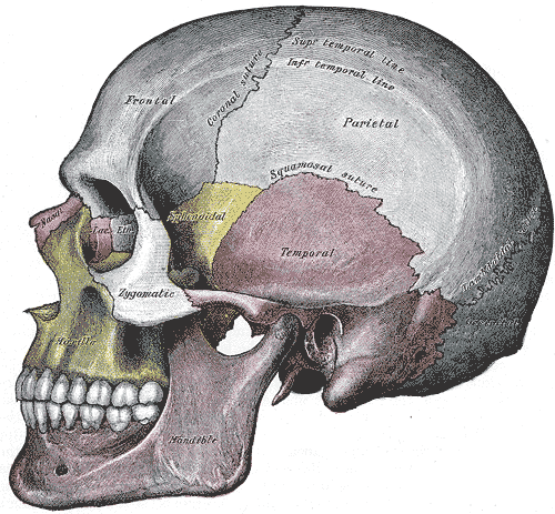
The skull is a bone protective cavity for the brain. The skull is composed of four types of bone i.e., cranial bones, facial bones, ear ossicles and hyoid bone. However two parts are more prominent: the cranium and the mandible. In humans, these two parts are the neurocranium and the viscerocranium (facial skeleton) that includes the mandible as its largest bone. The skull forms the anterior-most portion of the skeleton and is a product of cephalisation—housing the brain, and several sensory structures such as the eyes, ears, nose, and mouth. In humans these sensory structures are part of the facial skeleton. Functions of the skull include protection of the brain, fixing the distance between the eyes to allow stereoscopic vision, and fixing the position of the ears to enable sound localisation of the direction and distance of sounds. In some animals, such as horned ungulates (mammals with hooves), the skull also has a defensive function by providing the mount (on the fronta ... [...More Info...] [...Related Items...] OR: [Wikipedia] [Google] [Baidu] |
Squamosal Bone
The squamosal is a skull bone found in most reptiles, amphibians, and birds. In fishes, it is also called the pterotic bone. In most tetrapods, the squamosal and quadratojugal bones form the cheek series of the skull. The bone forms an ancestral component of the dermal roof and is typically thin compared to other skull bones. The squamosal bone lies ventral to the temporal series and otic notch, and is bordered anteriorly by the postorbital. Posteriorly, the squamosal articulates with the quadrate and pterygoid bones. The squamosal is bordered anteroventrally by the jugal and ventrally by the quadratojugal. Function in reptiles In reptiles, the quadrate and articular bones of the skull articulate to form the jaw joint. The squamosal bone lies anterior to the quadrate bone. Anatomy in synapsids Non-mammalian synapsids In non-mammalian synapsids, the jaw is composed of four bony elements and referred to as a quadro-articular jaw because the joint is between the articular an ... [...More Info...] [...Related Items...] OR: [Wikipedia] [Google] [Baidu] |
Jugal Bone
The jugal is a skull bone found in most reptiles, amphibians and birds. In mammals, the jugal is often called the malar or zygomatic. It is connected to the quadratojugal and maxilla, as well as other bones, which may vary by species. Anatomy The jugal bone is located on either side of the skull in the circumorbital region. It is the origin of several masticatory muscles in the skull. The jugal and lacrimal bones are the only two remaining from the ancestral circumorbital series: the prefrontal, postfrontal, postorbital, jugal, and lacrimal bones. During development, the jugal bone originates from dermal bone. In dinosaurs This bone is considered key in the determination of general traits in cases in which the entire skull has not been found intact (for instance, as with dinosaurs in paleontology). In some dinosaur genera the jugal also forms part of the lower margin of either the antorbital fenestra or the infratemporal fenestra, or both. Most commonly, this bone articu ... [...More Info...] [...Related Items...] OR: [Wikipedia] [Google] [Baidu] |
Skull
The skull is a bone protective cavity for the brain. The skull is composed of four types of bone i.e., cranial bones, facial bones, ear ossicles and hyoid bone. However two parts are more prominent: the cranium and the mandible. In humans, these two parts are the neurocranium and the viscerocranium ( facial skeleton) that includes the mandible as its largest bone. The skull forms the anterior-most portion of the skeleton and is a product of cephalisation—housing the brain, and several sensory structures such as the eyes, ears, nose, and mouth. In humans these sensory structures are part of the facial skeleton. Functions of the skull include protection of the brain, fixing the distance between the eyes to allow stereoscopic vision, and fixing the position of the ears to enable sound localisation of the direction and distance of sounds. In some animals, such as horned ungulates (mammals with hooves), the skull also has a defensive function by providing the mount (on the front ... [...More Info...] [...Related Items...] OR: [Wikipedia] [Google] [Baidu] |
Postorbital Bone
The ''postorbital'' is one of the bones in vertebrate skulls which forms a portion of the dermal skull roof and, sometimes, a ring about the orbit. Generally, it is located behind the postfrontal and posteriorly to the orbital fenestra. In some vertebrates, the postorbital is fused with the postfrontal to create a postorbitofrontal. Birds have a separate postorbital as an embryo, but the bone fuses with the frontal Front may refer to: Arts, entertainment, and media Films * ''The Front'' (1943 film), a 1943 Soviet drama film * ''The Front'', 1976 film Music * The Front (band), an American rock band signed to Columbia Records and active in the 1980s and e ... before it hatches. References * Roemer, A. S. 1956. ''Osteology of the Reptiles''. University of Chicago Press. 772 pp. Skull {{Vertebrate anatomy-stub ... [...More Info...] [...Related Items...] OR: [Wikipedia] [Google] [Baidu] |
Parietal Eye
A parietal eye, also known as a third eye or pineal eye, is a part of the epithalamus present in some vertebrates. The eye is located at the top of the head, is photoreceptive and is associated with the pineal gland, regulating circadian rhythmicity and hormone production for thermoregulation. The hole in the head which contains the eye is known as a pineal foramen or parietal foramen, since it is often enclosed by the parietal bones. Presence in various animals The parietal eye is found in the tuatara, most lizards, frogs, salamanders, certain bony fish, sharks, and lampreys. It is absent in mammals, but was present in their closest extinct relatives, the therapsids, suggesting it was lost during the course of the mammalian evolution due to it being useless in endothermic animals. It is also absent in the ancestrally endothermic ("warm-blooded") archosaurs such as birds. The parietal eye is also lost in ectothermic ("cold-blooded") archosaurs like crocodilians, and in turtles, ... [...More Info...] [...Related Items...] OR: [Wikipedia] [Google] [Baidu] |
Parietal Bone
The parietal bones () are two bones in the Human skull, skull which, when joined at a fibrous joint, form the sides and roof of the Human skull, cranium. In humans, each bone is roughly quadrilateral in form, and has two surfaces, four borders, and four angles. It is named from the Latin ''paries'' (''-ietis''), wall. Surfaces External The external surface [Fig. 1] is convex, smooth, and marked near the center by an eminence, the parietal eminence (''tuber parietale''), which indicates the point where ossification commenced. Crossing the middle of the bone in an arched direction are two curved lines, the superior and inferior temporal lines; the former gives attachment to the temporal fascia, and the latter indicates the upper limit of the muscular origin of the temporal muscle. Above these lines the bone is covered by a tough layer of fibrous tissue – the epicranial aponeurosis; below them it forms part of the temporal fossa, and affords attachment to the temporal muscle. ... [...More Info...] [...Related Items...] OR: [Wikipedia] [Google] [Baidu] |
Prefrontal Bone
The prefrontal bone is a bone separating the lacrimal and frontal bones in many tetrapod skulls. It first evolved in the sarcopterygian clade Rhipidistia, which includes lungfish and the Tetrapodomorpha. The prefrontal is found in most modern and extinct lungfish, amphibians and reptiles. The prefrontal is lost in early mammaliaforms and so is not present in modern mammals either. In dinosaurs The prefrontal bone is a very small bone near the top of the skull, which is lost in many groups of coelurosaurian theropod dinosaurs and is completely absent in their modern descendants, the birds. Conversely, a well developed prefrontal is considered to be a primitive feature in dinosaurs. The prefrontal makes contact with several other bones in the skull. The anterior part of the bone articulates with the nasal bone and the lacrimal bone. The posterior part of the bone articulates with the frontal bone and more rarely the palpebral bone The palpebral bone is a small dermal bone found ... [...More Info...] [...Related Items...] OR: [Wikipedia] [Google] [Baidu] |
Suture (anatomy)
In anatomy, a suture is a fairly rigid joint between two or more hard elements of an organism, with or without significant overlap of the elements. Sutures are found in the skeletons or exoskeletons of a wide range of animals, in both invertebrates and vertebrates. Sutures are found in animals with hard parts from the Cambrian period to the present day. Sutures were and are formed by several different methods, and they exist between hard parts that are made from several different materials. Vertebrate skeletons The skeletons of vertebrate animals (fish, amphibians, reptiles, birds, and mammals) are made of bone, in which the main rigid ingredient is calcium phosphate. Cranial sutures The skulls of most vertebrates consist of sets of bony plates held together by cranial sutures. These sutures are held together mainly by Sharpey's fibers which grow from each bone into the adjoining one. Sutures in the ankles of land vertebrates In the type of crurotarsal ankle which is found ... [...More Info...] [...Related Items...] OR: [Wikipedia] [Google] [Baidu] |


.jpg)


