|
Macroplata BW
''Macroplata'' (meaning "big plate") is an extinct genus of Early Jurassic rhomaleosaurid plesiosaur which grew up to in length and weighed up to . Like other plesiosaurs, ''Macroplata'' probably lived on a diet of fish, using its sharp needle-like teeth to catch prey. Its shoulder bones were fairly large, indicating a powerful forward stroke for fast swimming. ''Macroplata'' also had a relatively long neck, twice the length of the skull, in contrast to pliosaurs. A different species, ''Macroplata longirostris'' (previously called ''Plesiosaurus longirostris''), which lived somewhat later, during the Toarcian stage, was also included in the genus; however, in 2011, Benson ''et al.'' reclassified it as a pliosaurid in the genus ''Hauffiosaurus'', ''H. longirostris''. Description ''Macroplata'' bore an elongated skull, with more than half of its cranial length taken up by a roughly triangular snout. The premaxillae (front upper jaw bones) bear six teeth each, with the first being ... [...More Info...] [...Related Items...] OR: [Wikipedia] [Google] [Baidu] |
Early Jurassic
The Early Jurassic Epoch (geology), Epoch (in chronostratigraphy corresponding to the Lower Jurassic series (stratigraphy), Series) is the earliest of three epochs of the Jurassic Period. The Early Jurassic starts immediately after the Triassic-Jurassic extinction event, 201.3 Ma (million years ago), and ends at the start of the Middle Jurassic 174.1 Ma. Certain rocks of marine origin of this age in Europe are called "Lias Group, Lias" and that name was used for the period, as well, in 19th-century geology. In southern Germany rocks of this age are called Black Jurassic. Origin of the name Lias There are two possible origins for the name Lias: the first reason is it was taken by a geologist from an England, English quarryman's dialect pronunciation of the word "layers"; secondly, sloops from north Cornwall, Cornish ports such as Bude would sail across the Bristol Channel to the Vale of Glamorgan to load up with rock from coastal limestone quarries (lias limestone from S ... [...More Info...] [...Related Items...] OR: [Wikipedia] [Google] [Baidu] |
Dental Alveolus
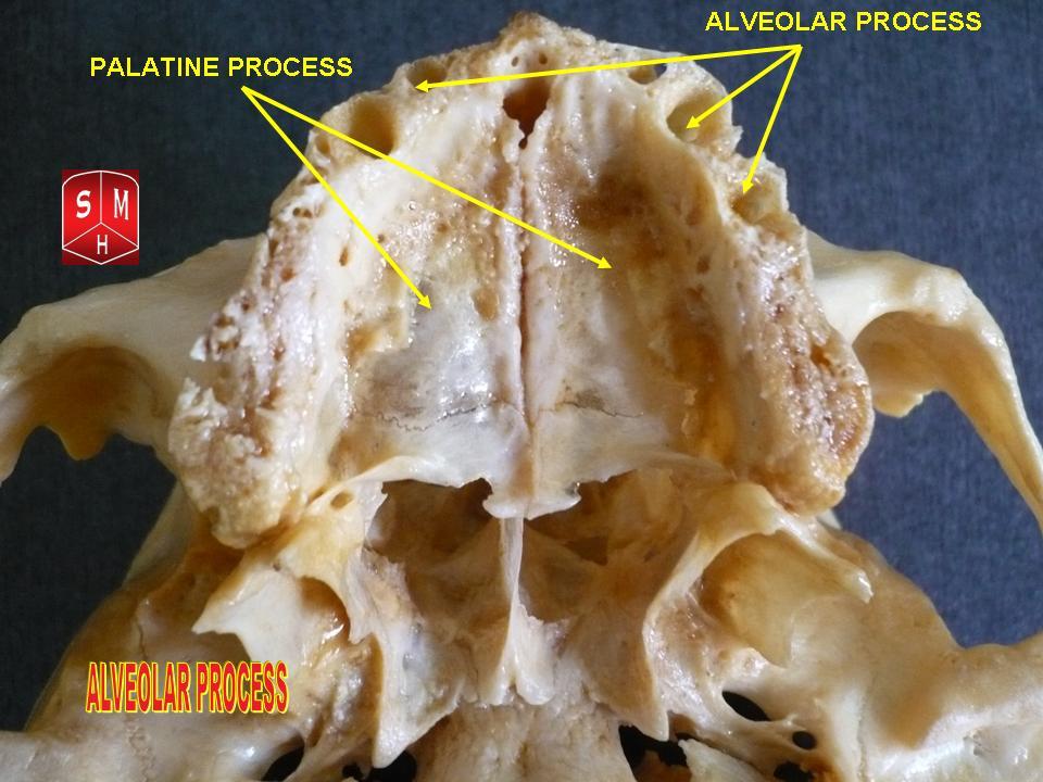
Dental alveoli (singular ''alveolus'') are sockets in the jaws in which the roots of teeth are held in the alveolar process with the periodontal ligament. The lay term for dental alveoli is tooth sockets. A joint that connects the roots of the teeth and the alveolus is called ''gomphosis'' (plural ''gomphoses''). Alveolar bone is the bone that surrounds the roots of the teeth forming bone sockets. In mammals, tooth sockets are found in the maxilla, the premaxilla, and the mandible. Etymology 1706, "a hollow," especially "the socket of a tooth," from Latin alveolus "a tray, trough, basin; bed of a small river; small hollow or cavity," diminutive of alvus "belly, stomach, paunch, bowels; hold of a ship," from PIE root *aulo- "hole, cavity" (source also of Greek aulos "flute, tube, pipe;" Serbo-Croatian, Polish, Russian ulica "street," originally "narrow opening;" Old Church Slavonic uliji, Lithuanian aulys "beehive" (hollow trunk), Armenian yli "pregnant"). The word was extended in ... [...More Info...] [...Related Items...] OR: [Wikipedia] [Google] [Baidu] |
Braincase
In human anatomy, the neurocranium, also known as the braincase, brainpan, or brain-pan is the upper and back part of the skull, which forms a protective case around the brain. In the human skull, the neurocranium includes the calvaria or skullcap. The remainder of the skull is the facial skeleton. In comparative anatomy, neurocranium is sometimes used synonymously with endocranium or chondrocranium. Structure The neurocranium is divided into two portions: * the membranous part, consisting of flat bones, which surround the brain; and * the cartilaginous part, or chondrocranium, which forms bones of the base of the skull. In humans, the neurocranium is usually considered to include the following eight bones: * 1 ethmoid bone * 1 frontal bone * 1 occipital bone * 2 parietal bones * 1 sphenoid bone * 2 temporal bones The ossicles (three on each side) are usually not included as bones of the neurocranium. There may variably also be extra sutural bones present. Below the ne ... [...More Info...] [...Related Items...] OR: [Wikipedia] [Google] [Baidu] |
Basioccipital
The basilar part of the occipital bone (also basioccipital) extends forward and upward from the foramen magnum, and presents in front an area more or less quadrilateral in outline. In the young skull this area is rough and uneven, and is joined to the body of the sphenoid by a plate of cartilage. By the twenty-fifth year this cartilaginous plate is ossified, and the occipital and sphenoid form a continuous bone. Surfaces On its ''lower surface'', about 1 cm. in front of the foramen magnum, is the pharyngeal tubercle which gives attachment to the fibrous raphe of the pharynx. On either side of the middle line the longus capitis and rectus capitis anterior are inserted, and immediately in front of the foramen magnum the anterior atlantooccipital membrane is attached. The ''upper surface'', which constitutes the lower half of the clivus, presents a broad, shallow groove which inclines upward and forward from the foramen magnum; it supports the medulla oblongata, and nea ... [...More Info...] [...Related Items...] OR: [Wikipedia] [Google] [Baidu] |
Autapomorphy
In phylogenetics, an autapomorphy is a distinctive feature, known as a derived trait, that is unique to a given taxon. That is, it is found only in one taxon, but not found in any others or outgroup taxa, not even those most closely related to the focal taxon (which may be a species, family or in general any clade). It can therefore be considered an apomorphy in relation to a single taxon. The word ''autapomorphy'', first introduced in 1950 by German entomologist Willi Hennig, is derived from the Greek words αὐτός, ''autos'' "self"; ἀπό, ''apo'' "away from"; and μορφή, ''morphḗ'' = "shape". Discussion Because autapomorphies are only present in a single taxon, they do not convey information about relationship. Therefore, autapomorphies are not useful to infer phylogenetic relationships. However, autapomorphy, like synapomorphy and plesiomorphy is a relative concept depending on the taxon in question. An autapomorphy at a given level may well be a synapomorphy at ... [...More Info...] [...Related Items...] OR: [Wikipedia] [Google] [Baidu] |
Pterygoid Bone
The pterygoid is a paired bone forming part of the palate of many vertebrates, behind the palatine bone In anatomy, the palatine bones () are two irregular bones of the facial skeleton in many animal species, located above the uvula in the throat. Together with the maxillae, they comprise the hard palate. (''Palate'' is derived from the Latin ''pa ...s. It is a flat and thin lamina, united to the medial side of the pterygoid process of the sphenoid bone, and to the perpendicular lamina of the palatine bone. Bones of the head and neck {{musculoskeletal-stub ... [...More Info...] [...Related Items...] OR: [Wikipedia] [Google] [Baidu] |
Palate
The palate () is the roof of the mouth in humans and other mammals. It separates the oral cavity from the nasal cavity. A similar structure is found in crocodilians, but in most other tetrapods, the oral and nasal cavities are not truly separated. The palate is divided into two parts, the anterior, bony hard palate and the posterior, fleshy soft palate (or velum). Structure Innervation The maxillary nerve branch of the trigeminal nerve supplies sensory innervation to the palate. Development The hard palate forms before birth. Variation If the fusion is incomplete, a cleft palate results. Function When functioning in conjunction with other parts of the mouth, the palate produces certain sounds, particularly velar, palatal, palatalized, postalveolar, alveolopalatal, and uvular consonants. History Etymology The English synonyms palate and palatum, and also the related adjective palatine (as in palatine bone), are all from the Latin ''palatum'' via Old French ''palat ... [...More Info...] [...Related Items...] OR: [Wikipedia] [Google] [Baidu] |
Parasphenoid
The parasphenoid is a bone which can be found in the cranium of many vertebrates. It is an unpaired dermal bone which lies at the midline of the roof of the mouth. In many reptiles (including birds), it fuses to the endochondral (cartilage-derived) basisphenoid bone of the lower braincase, forming a bone known as the parabasisphenoid. Early mammals have a small parasphenoid, but for the most part its function has been replaced by the vomer bone. The parasphenoid has been lost in placental mammals and caecilian amphibians. See also *Ossification of frontal bone *Terms for anatomical location Standard anatomical terms of location are used to unambiguously describe the anatomy of animals, including humans. The terms, typically derived from Latin or Greek roots, describe something in its standard anatomical position. This position prov ... References Bones of the head and neck {{musculoskeletal-stub ... [...More Info...] [...Related Items...] OR: [Wikipedia] [Google] [Baidu] |
Pineal Foramen
A parietal eye, also known as a third eye or pineal eye, is a part of the epithalamus present in some vertebrates. The eye is located at the top of the head, is photoreceptive and is associated with the pineal gland, regulating circadian rhythmicity and hormone production for thermoregulation. The hole in the head which contains the eye is known as a pineal foramen or parietal foramen, since it is often enclosed by the parietal bones. Presence in various animals The parietal eye is found in the tuatara, most lizards, frogs, salamanders, certain bony fish, sharks, and lampreys. It is absent in mammals, but was present in their closest extinct relatives, the therapsids, suggesting it was lost during the course of the mammalian evolution due to it being useless in endothermic animals. It is also absent in the ancestrally endothermic ("warm-blooded") archosaurs such as birds. The parietal eye is also lost in ectothermic ("cold-blooded") archosaurs like crocodilians, and in turtles, ... [...More Info...] [...Related Items...] OR: [Wikipedia] [Google] [Baidu] |
Postfrontal
The skull is a bone protective cavity for the brain. The skull is composed of four types of bone i.e., cranial bones, facial bones, ear ossicles and hyoid bone. However two parts are more prominent: the cranium and the mandible. In humans, these two parts are the neurocranium and the viscerocranium (facial skeleton) that includes the mandible as its largest bone. The skull forms the anterior-most portion of the skeleton and is a product of cephalisation—housing the brain, and several sensory structures such as the eyes, ears, nose, and mouth. In humans these sensory structures are part of the facial skeleton. Functions of the skull include protection of the brain, fixing the distance between the eyes to allow stereoscopic vision, and fixing the position of the ears to enable sound localisation of the direction and distance of sounds. In some animals, such as horned ungulates (mammals with hooves), the skull also has a defensive function by providing the mount (on the fronta ... [...More Info...] [...Related Items...] OR: [Wikipedia] [Google] [Baidu] |
Prefrontal Bone
The prefrontal bone is a bone separating the lacrimal and frontal bones in many tetrapod skulls. It first evolved in the sarcopterygian clade Rhipidistia, which includes lungfish and the Tetrapodomorpha. The prefrontal is found in most modern and extinct lungfish, amphibians and reptiles. The prefrontal is lost in early mammaliaforms and so is not present in modern mammals either. In dinosaurs The prefrontal bone is a very small bone near the top of the skull, which is lost in many groups of coelurosaurian theropod dinosaurs and is completely absent in their modern descendants, the birds. Conversely, a well developed prefrontal is considered to be a primitive feature in dinosaurs. The prefrontal makes contact with several other bones in the skull. The anterior part of the bone articulates with the nasal bone and the lacrimal bone. The posterior part of the bone articulates with the frontal bone and more rarely the palpebral bone The palpebral bone is a small dermal bone found ... [...More Info...] [...Related Items...] OR: [Wikipedia] [Google] [Baidu] |
Parietal Bone
The parietal bones () are two bones in the Human skull, skull which, when joined at a fibrous joint, form the sides and roof of the Human skull, cranium. In humans, each bone is roughly quadrilateral in form, and has two surfaces, four borders, and four angles. It is named from the Latin ''paries'' (''-ietis''), wall. Surfaces External The external surface [Fig. 1] is convex, smooth, and marked near the center by an eminence, the parietal eminence (''tuber parietale''), which indicates the point where ossification commenced. Crossing the middle of the bone in an arched direction are two curved lines, the superior and inferior temporal lines; the former gives attachment to the temporal fascia, and the latter indicates the upper limit of the muscular origin of the temporal muscle. Above these lines the bone is covered by a tough layer of fibrous tissue – the epicranial aponeurosis; below them it forms part of the temporal fossa, and affords attachment to the temporal muscle. ... [...More Info...] [...Related Items...] OR: [Wikipedia] [Google] [Baidu] |


