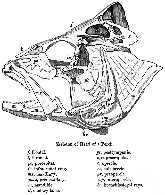
|
Braincase
In human anatomy, the neurocranium, also known as the braincase, brainpan, or brain-pan is the upper and back part of the skull, which forms a protective case around the brain. In the human skull, the neurocranium includes the calvaria or skullcap. The remainder of the skull is the facial skeleton. In comparative anatomy, neurocranium is sometimes used synonymously with endocranium or chondrocranium. Structure The neurocranium is divided into two portions: * the membranous part, consisting of flat bones, which surround the brain; and * the cartilaginous part, or chondrocranium, which forms bones of the base of the skull. In humans, the neurocranium is usually considered to include the following eight bones: * 1 ethmoid bone * 1 frontal bone * 1 occipital bone * 2 parietal bones * 1 sphenoid bone * 2 temporal bones The ossicles (three on each side) are usually not included as bones of the neurocranium. There may variably also be extra sutural bones present. Below the ne ... [...More Info...] [...Related Items...] OR: [Wikipedia] [Google] [Baidu] |
Sphenoid Bone
The sphenoid bone is an unpaired bone of the neurocranium. It is situated in the middle of the skull towards the front, in front of the basilar part of occipital bone, basilar part of the occipital bone. The sphenoid bone is one of the seven bones that articulate to form the orbit (anatomy), orbit. Its shape somewhat resembles that of a butterfly or bat with its wings extended. Structure It is divided into the following parts: * a median portion, known as the body of sphenoid bone, containing the sella turcica, which houses the pituitary gland as well as the paired paranasal sinuses, the sphenoidal sinuses * two Greater wing of sphenoid bone, greater wings on the lateral side of the body and two Lesser wing of sphenoid bone, lesser wings from the anterior side. * Pterygoid processes of the sphenoides, directed downwards from the junction of the body and the greater wings. Two sphenoidal conchae are situated at the anterior and inferior part of the body. Intrinsic ligaments of ... [...More Info...] [...Related Items...] OR: [Wikipedia] [Google] [Baidu] |
Facial Bones
The facial skeleton comprises the ''facial bones'' that may attach to build a portion of the skull. The remainder of the skull is the braincase. In human anatomy and development, the facial skeleton is sometimes called the ''membranous viscerocranium'', which comprises the human mandible, mandible and dermatocranium, dermatocranial elements that are not part of the braincase. Structure In the human skull, the facial skeleton consists of fourteen bones in the face: * Inferior turbinal (2) * Lacrimal bones (2) * Human mandible, Mandible * Maxilla (2) * Nasal bones (2) * Palatine bones (2) * Vomer * Zygomatic bones (2) Variations Elements of the ''cartilaginous viscerocranium'' (i.e., splanchnocranium, splanchnocranial elements), such as the hyoid bone, are sometimes considered part of the facial skeleton. The ethmoid bone (or a part of it) and also the sphenoid bone are sometimes included, but otherwise considered part of the neurocranium. Because the maxillary bones are fused, th ... [...More Info...] [...Related Items...] OR: [Wikipedia] [Google] [Baidu] |
Facial Skeleton
The facial skeleton comprises the ''facial bones'' that may attach to build a portion of the skull. The remainder of the skull is the braincase. In human anatomy and development, the facial skeleton is sometimes called the ''membranous viscerocranium'', which comprises the mandible and dermatocranial elements that are not part of the braincase. Structure In the human skull, the facial skeleton consists of fourteen bones in the face: * Inferior turbinal (2) * Lacrimal bones (2) * Mandible * Maxilla (2) * Nasal bones (2) * Palatine bones (2) * Vomer * Zygomatic bones (2) Variations Elements of the ''cartilaginous viscerocranium'' (i.e., splanchnocranial elements), such as the hyoid bone, are sometimes considered part of the facial skeleton. The ethmoid bone (or a part of it) and also the sphenoid bone are sometimes included, but otherwise considered part of the neurocranium. Because the maxillary bones are fused, they are often collectively listed as only one bone. The mandibl ... [...More Info...] [...Related Items...] OR: [Wikipedia] [Google] [Baidu] |
Endocranium
The endocranium in comparative anatomy is a part of the skull base in vertebrates and it represents the basal, inner part of the cranium. The term is also applied to the outer layer of the dura mater in human anatomy. Structure Structurally, the endocranium consists of a boxlike shape, open at the top. The posterior margin exhibit the '' foramen magnum'', an opening for the spinal cord. The floor of the endocranium has several paired openings for the cranial nerves, and the anterior margin holds a spongy construction, allowing for the external nasal nerves to pass through. Romer, A.S. & T.S. Parsons. 1977. ''The Vertebrate Body.'' 5th ed. Saunders, Philadelphia. (6th ed. 1985) All bones of the structure derive from the cranial neural crest during fetal development. Endocranial elements in humans In humans and other mammals, the endocranium forms during fetal development as a cartilaginous neurocranium, that ossifies from several centers. Several of these bones merge, and in t ... [...More Info...] [...Related Items...] OR: [Wikipedia] [Google] [Baidu] |
Skull
The skull is a bone protective cavity for the brain. The skull is composed of four types of bone i.e., cranial bones, facial bones, ear ossicles and hyoid bone. However two parts are more prominent: the cranium and the mandible. In humans, these two parts are the neurocranium and the viscerocranium ( facial skeleton) that includes the mandible as its largest bone. The skull forms the anterior-most portion of the skeleton and is a product of cephalisation—housing the brain, and several sensory structures such as the eyes, ears, nose, and mouth. In humans these sensory structures are part of the facial skeleton. Functions of the skull include protection of the brain, fixing the distance between the eyes to allow stereoscopic vision, and fixing the position of the ears to enable sound localisation of the direction and distance of sounds. In some animals, such as horned ungulates (mammals with hooves), the skull also has a defensive function by providing the mount (on the front ... [...More Info...] [...Related Items...] OR: [Wikipedia] [Google] [Baidu] |
Temporal Bone
The temporal bones are situated at the sides and base of the skull, and lateral to the temporal lobes of the cerebral cortex. The temporal bones are overlaid by the sides of the head known as the temples, and house the structures of the ears. The lower seven cranial nerves and the major vessels to and from the brain traverse the temporal bone. Structure The temporal bone consists of four parts— the squamous, mastoid, petrous and tympanic parts. The squamous part is the largest and most superiorly positioned relative to the rest of the bone. The zygomatic process is a long, arched process projecting from the lower region of the squamous part and it articulates with the zygomatic bone. Posteroinferior to the squamous is the mastoid part. Fused with the squamous and mastoid parts and between the sphenoid and occipital bones lies the petrous part, which is shaped like a pyramid. The tympanic part is relatively small and lies inferior to the squamous part, anterior to the mast ... [...More Info...] [...Related Items...] OR: [Wikipedia] [Google] [Baidu] |
Ossicles
The ossicles (also called auditory ossicles) are three bones in either middle ear that are among the smallest bones in the human body. They serve to transmit sounds from the air to the fluid-filled labyrinth (cochlea). The absence of the auditory ossicles would constitute a moderate-to-severe hearing loss. The term "ossicle" literally means "tiny bone". Though the term may refer to any small bone throughout the body, it typically refers to the malleus, incus, and stapes (hammer, anvil, and stirrup) of the middle ear. Structure The ossicles are, in order from the eardrum to the inner ear (from superficial to deep): the malleus, incus, and stapes, terms that in Latin are translated as "the hammer, anvil, and stirrup". * The malleus ( la, "hammer") articulates with the incus through the incudomalleolar joint and is attached to the tympanic membrane (eardrum), from which vibrational sound pressure motion is passed. * The incus ( la, "anvil") is connected to both the other bones. * ... [...More Info...] [...Related Items...] OR: [Wikipedia] [Google] [Baidu] |
Sutural Bones
Wormian bones, also known as intrasutural bones or sutural bones, are extra bone pieces that can occur within a suture (joint) in the skull. These are irregular isolated bones that can appear in addition to the usual centres of ossification of the skull and, although unusual, are not rare. They occur most frequently in the course of the lambdoid suture, which is more tortuous than other sutures. They are also occasionally seen within the sagittal and coronal sutures. A large wormian bone at lambda is often called an Inca bone (''os incae''), due to the relatively high frequency of occurrence in Peruvian mummies. Another specific Wormian bone, the pterion ossicle, sometimes exists between the sphenoidal angle of the parietal bone and the great wing of the sphenoid bone. They tend to vary in size and can be found on either side of the skull. Usually, not more than several are found in a single individual, but more than one hundred have been once found in the skull of a hydrocephali ... [...More Info...] [...Related Items...] OR: [Wikipedia] [Google] [Baidu] |
Foramen Magnum
The foramen magnum ( la, great hole) is a large, oval-shaped opening in the occipital bone of the skull. It is one of the several oval or circular openings (foramina) in the base of the skull. The spinal cord, an extension of the medulla oblongata, passes through the foramen magnum as it exits the cranial cavity. Apart from the transmission of the medulla oblongata and its membranes, the foramen magnum transmits the vertebral arteries, the anterior and posterior spinal arteries, the tectorial membranes and alar ligaments. It also transmits the accessory nerve into the skull. The foramen magnum is a very important feature in bipedal mammals. One of the attributes of a biped's foramen magnum is a forward shift of the anterior border of the cerebellar tentorium; this is caused by the shortening of the cranial base. Studies on the foramen magnum position have shown a connection to the functional influences of both posture and locomotion. The forward shift of the foramen magnum i ... [...More Info...] [...Related Items...] OR: [Wikipedia] [Google] [Baidu] |
Foramen
In anatomy and osteology, a foramen (; in Merriam-Webster Online Dictionary '. plural foramina, or foramens ) is an open hole that is present in extant or extinct s. Foramina inside the of typically allow , |
Occipital Bone
The occipital bone () is a neurocranium, cranial dermal bone and the main bone of the occiput (back and lower part of the skull). It is trapezoidal in shape and curved on itself like a shallow dish. The occipital bone overlies the occipital lobes of the cerebrum. At the base of skull in the occipital bone, there is a large oval opening called the foramen magnum, which allows the passage of the spinal cord. Like the other cranial bones, it is classed as a flat bone. Due to its many attachments and features, the occipital bone is described in terms of separate parts. From its front to the back is the basilar part of occipital bone, basilar part, also called the basioccipital, at the sides of the foramen magnum are the lateral parts of occipital bone, lateral parts, also called the exoccipitals, and the back is named as the squamous part of occipital bone, squamous part. The basilar part is a thick, somewhat quadrilateral piece in front of the foramen magnum and directed towards the ... [...More Info...] [...Related Items...] OR: [Wikipedia] [Google] [Baidu] |


