|
M. Malmoense
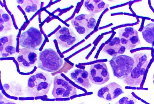
''Mycobacterium malmoense'' is a Gram-positive bacterium from the genus ''Mycobacterium''. Etymology From the city of Malmö, Sweden where the strain used for the description was isolated from patients. Description Gram-positive, nonmotile, acid-fast and coccus, coccoid to short rods. *Environmental reservoir: soil and water. Colony characteristics *Smooth and nonpigmented colonies, growth below the surface of semisolid agar medium after deep inoculation (as seen with Mycobacterium bovis, M. bovis), 0.9 - 1.7mm in diameter. Physiology *Growth on inspissated egg medium and oleic acid-albumin agar at a temperature range of 22 °C-37 °C requires over 1 week. *Susceptible to ethambutol, ethionamide, kanamycin and cycloserine. Differential characteristics *Antigenic structure: seroagglutination demonstrates a single serovar distinct from that of other species. Pathogenesis *Usually infects young children with cervical lymphadenitis or adults with chronic pulmonary dise ... [...More Info...] [...Related Items...] OR: [Wikipedia] [Google] [Baidu] |
Gram-positive
In bacteriology, gram-positive bacteria are bacteria that give a positive result in the Gram stain test, which is traditionally used to quickly classify bacteria into two broad categories according to their type of cell wall. Gram-positive bacteria take up the crystal violet stain used in the test, and then appear to be purple-coloured when seen through an optical microscope. This is because the thick peptidoglycan layer in the bacterial cell wall retains the stain after it is washed away from the rest of the sample, in the decolorization stage of the test. Conversely, gram-negative bacteria cannot retain the violet stain after the decolorization step; alcohol used in this stage degrades the outer membrane of gram-negative cells, making the cell wall more porous and incapable of retaining the crystal violet stain. Their peptidoglycan layer is much thinner and sandwiched between an inner cell membrane and a bacterial outer membrane, causing them to take up the counterstain (saf ... [...More Info...] [...Related Items...] OR: [Wikipedia] [Google] [Baidu] |
Acid-fast Bacilli
Acid-fastness is a physical property of certain bacterial and eukaryotic cells, as well as some sub-cellular structures, specifically their resistance to decolorization by acids during laboratory staining procedures. Once stained as part of a sample, these organisms can resist the acid and/or ethanol-based decolorization procedures common in many staining protocols, hence the name ''acid-fast''. The mechanisms of acid-fastness vary by species, although the most well-known example is in the genus ''Mycobacterium'', which includes the species responsible for tuberculosis and leprosy. The acid-fastness of ''Mycobacteria'' is due to the high mycolic acid content of their cell walls, which is responsible for the staining pattern of poor absorption followed by high retention. Some bacteria may also be partially acid-fast, such as ''Nocardia''. Acid-fast organisms are difficult to characterize using standard microbiological techniques, though they can be stained using concentrated dyes, ... [...More Info...] [...Related Items...] OR: [Wikipedia] [Google] [Baidu] |
Biopsy
A biopsy is a medical test commonly performed by a surgeon, interventional radiologist, or an interventional cardiologist. The process involves extraction of sample cells or tissues for examination to determine the presence or extent of a disease. The tissue is then fixed, dehydrated, embedded, sectioned, stained and mounted before it is generally examined under a microscope by a pathologist; it may also be analyzed chemically. When an entire lump or suspicious area is removed, the procedure is called an excisional biopsy. An incisional biopsy or core biopsy samples a portion of the abnormal tissue without attempting to remove the entire lesion or tumor. When a sample of tissue or fluid is removed with a needle in such a way that cells are removed without preserving the histological architecture of the tissue cells, the procedure is called a needle aspiration biopsy. Biopsies are most commonly performed for insight into possible cancerous or inflammatory conditions. History T ... [...More Info...] [...Related Items...] OR: [Wikipedia] [Google] [Baidu] |
Sputum
Sputum is mucus that is coughed up from the lower airways (the trachea and bronchi). In medicine, sputum samples are usually used for a naked eye examination, microbiological investigation of respiratory infections and cytological investigations of respiratory systems. It is crucial that the specimen does not include any mucoid material from the nose or oral cavity. A naked eye exam of the sputum can be done at home by a patient in order to note the various colors (see below). Any hint of yellow or green color (pus) suggests an airway infection (but does not indicate the type of organism causing it). Such color hints are best detected when the sputum is viewed on a very white background such as white paper, a white pot or a white sink surface. The more intense the yellow color, the more likely it is a caused by an infection (bronchitis, bronchopneumonia or pneumonia). Having green, yellow, or thickened phlegm (sputum) does not always indicate the presence of an infection. Also, ... [...More Info...] [...Related Items...] OR: [Wikipedia] [Google] [Baidu] |
Extrapulmonary
Tuberculosis (TB) is an infectious disease usually caused by ''Mycobacterium tuberculosis'' (MTB) bacteria. Tuberculosis generally affects the lungs, but it can also affect other parts of the body. Most infections show no symptoms, in which case it is known as latent tuberculosis. Around 10% of latent infections progress to active disease which, if left untreated, kill about half of those affected. Typical symptoms of active TB are chronic cough with hemoptysis, blood-containing sputum, mucus, fever, night sweats, and weight loss. It was historically referred to as consumption due to the weight loss associated with the disease. Infection of other organs can cause a wide range of symptoms. Tuberculosis is Human-to-human transmission, spread from one person to the next Airborne disease, through the air when people who have active TB in their lungs cough, spit, speak, or sneeze. People with Latent TB do not spread the disease. Active infection occurs more often in people wi ... [...More Info...] [...Related Items...] OR: [Wikipedia] [Google] [Baidu] |
Pneumoconiosis
Pneumoconiosis is the general term for a class of interstitial lung disease where inhalation of dust ( for example, ash dust, lead particles, pollen grains etc) has caused interstitial fibrosis. The three most common types are asbestosis, silicosis, and coal miner's lung. Pneumoconiosis often causes restrictive impairment, although diagnosable pneumoconiosis can occur without measurable impairment of lung function. Depending on extent and severity, it may cause death within months or years, or it may never produce symptoms. It is usually an occupational lung disease, typically from years of dust exposure during work in mining; textile milling; shipbuilding, ship repairing, and/or shipbreaking; sandblasting; industrial tasks; rock drilling (subways or building pilings); or agriculture. It is one of the most common occupational diseases in the world. Types Depending upon the type of dust, the disease is given different names: * Coalworker's pneumoconiosis (also known as coal miner' ... [...More Info...] [...Related Items...] OR: [Wikipedia] [Google] [Baidu] |
Pulmonary
The lungs are the primary organs of the respiratory system in humans and most other animals, including some snails and a small number of fish. In mammals and most other vertebrates, two lungs are located near the backbone on either side of the heart. Their function in the respiratory system is to extract oxygen from the air and transfer it into the bloodstream, and to release carbon dioxide from the bloodstream into the atmosphere, in a process of gas exchange. Respiration is driven by different muscular systems in different species. Mammals, reptiles and birds use their different muscles to support and foster breathing. In earlier tetrapods, air was driven into the lungs by the pharyngeal muscles via buccal pumping, a mechanism still seen in amphibians. In humans, the main muscle of respiration that drives breathing is the diaphragm. The lungs also provide airflow that makes vocal sounds including human speech possible. Humans have two lungs, one on the left and one on the ... [...More Info...] [...Related Items...] OR: [Wikipedia] [Google] [Baidu] |
Cervical Lymphadenitis
Cervical lymphadenopathy refers to lymphadenopathy of the cervical lymph nodes (the glands in the neck). The term ''lymphadenopathy'' strictly speaking refers to disease of the lymph nodes, though it is often used to describe the enlargement of the lymph nodes. Similarly, the term ''lymphadenitis'' refers to inflammation of a lymph node, but often it is used as a synonym of lymphadenopathy. Cervical lymphadenopathy is a sign or a symptom, not a diagnosis. The causes are varied, and may be inflammatory, degenerative, or neoplastic. In adults, healthy lymph nodes can be palpable (able to be felt), in the axilla, neck and groin. In children up to the age of 12 cervical nodes up to 1 cm in size may be palpable and this may not signify any disease. If nodes heal by resolution or scarring after being inflamed, they may remain palpable thereafter. In children, most palpable cervical lymphadenopathy is reactive or infective. In individuals over the age of 50, metastatic enlargement f ... [...More Info...] [...Related Items...] OR: [Wikipedia] [Google] [Baidu] |
Serovar
A serotype or serovar is a distinct variation within a species of bacteria or virus or among immune cells of different individuals. These microorganisms, viruses, or cells are classified together based on their surface antigens, allowing the epidemiologic classification of organisms to the subspecies level. A group of serovars with common antigens is called a serogroup or sometimes ''serocomplex''. Serotyping often plays an essential role in determining species and subspecies. The ''Salmonella'' genus of bacteria, for example, has been determined to have over 2600 serotypes. ''Vibrio cholerae'', the species of bacteria that causes cholera, has over 200 serotypes, based on cell antigens. Only two of them have been observed to produce the potent enterotoxin that results in cholera: O1 and O139. Serotypes were discovered by the American microbiologist Rebecca Lancefield in 1933. Role in organ transplantation The immune system is capable of discerning a cell as being 'self' or 'n ... [...More Info...] [...Related Items...] OR: [Wikipedia] [Google] [Baidu] |
Antigenic
In immunology, an antigen (Ag) is a molecule or molecular structure or any foreign particulate matter or a pollen grain that can bind to a specific antibody or T-cell receptor. The presence of antigens in the body may trigger an immune response. The term ''antigen'' originally referred to a substance that is an antibody generator. Antigens can be proteins, peptides (amino acid chains), polysaccharides (chains of monosaccharides/simple sugars), lipids, or nucleic acids. Antigens are recognized by antigen receptors, including antibodies and T-cell receptors. Diverse antigen receptors are made by cells of the immune system so that each cell has a specificity for a single antigen. Upon exposure to an antigen, only the lymphocytes that recognize that antigen are activated and expanded, a process known as clonal selection. In most cases, an antibody can only react to and bind one specific antigen; in some instances, however, antibodies may cross-react and bind more than one antigen. T ... [...More Info...] [...Related Items...] OR: [Wikipedia] [Google] [Baidu] |


