|
List Of Lymphatic Vessels Of The Human Body
Humans have approximately 500–600 lymph nodes distributed throughout the body, with clusters found in the underarms, groin, neck, chest, and abdomen. Lymph nodes of the head * Occipital lymph nodes * Mastoid lymph nodes * Parotid lymph nodes Lymph nodes of the neck * Cervical lymph nodes ** Submental lymph nodes ** Submandibular lymph nodes * Deep cervical lymph nodes ** Deep anterior cervical lymph nodes ** Deep lateral cervical lymph nodes *Inferior deep cervical lymph nodes ** Jugulo-omohyoid lymph node ** Jugulodigastric lymph node * Supraclavicular lymph nodes ** Virchow's node Lymph nodes of the thorax * Lymph nodes of the lungs: The lymph is drained from the lung tissue through subsegmental, segmental, lobar and interlobar lymph nodes to the hilar lymph nodes, which are located around the hilum (the pedicle, which attaches the lung to the mediastinal structures, containing the pulmonary artery, the pulmonary veins, the main bronchus for each side, some vegetati ... [...More Info...] [...Related Items...] OR: [Wikipedia] [Google] [Baidu] |
Lymph Node Regions
Lymph (from Latin, , meaning "water") is the fluid that flows through the lymphatic system, a system composed of lymph vessels (channels) and intervening lymph nodes whose function, like the venous system, is to return fluid from the tissues to be recirculated. At the origin of the fluid-return process, interstitial fluid—the fluid between the cells in all body tissues—enters the lymph capillaries. This lymphatic fluid is then transported via progressively larger lymphatic vessels through lymph nodes, where substances are removed by tissue lymphocytes and circulating lymphocytes are added to the fluid, before emptying ultimately into the right or the left subclavian vein, where it mixes with central venous blood. Because it is derived from interstitial fluid, with which blood and surrounding cells continually exchange substances, lymph undergoes continual change in composition. It is generally similar to blood plasma, which is the fluid component of blood. Lymph returns ... [...More Info...] [...Related Items...] OR: [Wikipedia] [Google] [Baidu] |
Bronchus
A bronchus is a passage or airway in the lower respiratory tract that conducts air into the lungs. The first or primary bronchi pronounced (BRAN-KAI) to branch from the trachea at the carina are the right main bronchus and the left main bronchus. These are the widest bronchi, and enter the right lung, and the left lung at each hilum. The main bronchi branch into narrower secondary bronchi or lobar bronchi, and these branch into narrower tertiary bronchi or segmental bronchi. Further divisions of the segmental bronchi are known as 4th order, 5th order, and 6th order segmental bronchi, or grouped together as subsegmental bronchi. The bronchi, when too narrow to be supported by cartilage, are known as bronchioles. No gas exchange takes place in the bronchi. Structure The trachea (windpipe) divides at the carina into two main or primary bronchi, the left bronchus and the right bronchus. The carina of the trachea is located at the level of the sternal angle and the fifth thoracic vert ... [...More Info...] [...Related Items...] OR: [Wikipedia] [Google] [Baidu] |
Hepatic Lymph Nodes
The hepatic lymph nodes consist of the following groups: * (a) hepatic, on the stem of the hepatic artery, and extending upward along the common bile duct, between the two layers of the lesser omentum, as far as the porta hepatis; the cystic gland, a member of this group, is placed near the neck of the gall-bladder; * (b) subpyloric, four or five in number, in close relation to the bifurcation of the gastroduodenal artery, in the angle between the superior and descending parts of the duodenum; an outlying member of this group is sometimes found above the duodenum on the right gastric (pyloric) artery. The lymph nodes of the hepatic chain receive Afferent lymphatics, afferents from the stomach, duodenum, liver, gall-bladder, and pancreas; their Efferent lymphatics, efferents join the celiac group of preaortic lymph nodes. Cancer prognosis and treatment Hepatic artery lymph nodes are commonly Resection (surgery), resected during a Whipple procedure. In a Whipple procedure, outcom ... [...More Info...] [...Related Items...] OR: [Wikipedia] [Google] [Baidu] |
Celiac Lymph Nodes
The celiac lymph nodes are associated with the branches of the celiac artery. Other lymph nodes in the abdomen are associated with the superior mesenteric artery, superior and inferior mesenteric artery, inferior mesenteric arteries. The celiac lymph nodes are grouped into three sets: the gastric lymph nodes, gastric, hepatic lymph nodes, hepatic and splenic lymph nodes. Additional images File:illu_lymph_chain08.jpg, Lymph nodes of the abdominal cavity References External links Lymphatics of the torso {{Portal bar, Anatomy ... [...More Info...] [...Related Items...] OR: [Wikipedia] [Google] [Baidu] |
Preaortic Lymph Nodes
The preaortic lymph nodes lie in front of the aorta, and may be divided into celiac lymph nodes, superior mesenteric lymph nodes, and inferior mesenteric lymph nodes groups, arranged around the origins of the corresponding arteries. The celiac lymph nodes are grouped into three sets: the gastric, hepatic and splenic lymph nodes. These groups also form their own subgroups. The superior mesenteric lymph nodes are grouped into three sets: the mesenteric, ileocolic and mesocolic lymph nodes. The inferior mesenteric lymph nodes have a subgroup of pararectal lymph nodes. The preaortic lymph nodes receive a few vessels from the lateral aortic lymph nodes, but their principal afferents are derived from the organs supplied by the three arteries with which they are associated–the celiac, superior and inferior mesenteric arteries. Some of their efferents pass to the retroaortic lymph nodes, but the majority unite to form the intestinal lymph trunk, which enters the cisterna chyli Th ... [...More Info...] [...Related Items...] OR: [Wikipedia] [Google] [Baidu] |
Periaortic Lymph Nodes
The periaortic lymph nodes (also known as lumbar) are a group of lymph nodes that lie in front of the lumbar vertebrae near the aorta. These lymph nodes receive drainage from the gastrointestinal tract and the abdominal organs. The periaortic lymph nodes are different from the paraaortic lymph nodes. The periaortic group is the general group, that is subdivided into: preaortic, paraaortic, and retroaortic groups. The paraaortic group is synonymous with the lateral aortic group. Divisions The periaortic lymph node group is divided into three subgroups: preaortic, paraaortic, and retroaortic: * The preaortic group drains the gastrointestinal viscera. They can be subdivided into three groups: the celiac nodes, the superior mesenteric nodes, and the inferior mesenteric nodes. *The paraaortic group (also known as lateral aortic group) drains the iliac nodes, the ovaries, the testes and other pelvic organs. The lateral group nodes are located adjacent to the aorta, anterior to th ... [...More Info...] [...Related Items...] OR: [Wikipedia] [Google] [Baidu] |
Thoracic Duct
In human anatomy, the thoracic duct is the larger of the two lymph ducts of the lymphatic system. It is also known as the ''left lymphatic duct'', ''alimentary duct'', ''chyliferous duct'', and ''Van Hoorne's canal''. The other duct is the right lymphatic duct. The thoracic duct carries chyle, a liquid containing both lymph and emulsified fats, rather than pure lymph. It also collects most of the lymph in the body other than from the right thorax, arm, head, and neck (which are drained by the right lymphatic duct). The thoracic duct usually starts from the level of the twelfth thoracic vertebra (T12) and extends to the root of the neck. It drains into the systemic (blood) circulation at the junction of the left subclavian and internal jugular veins, at the commencement of the brachiocephalic vein. When the duct ruptures, the resulting flood of liquid into the pleural cavity is known as chylothorax. Structure In adults, the thoracic duct is typically 38–45 cm in length an ... [...More Info...] [...Related Items...] OR: [Wikipedia] [Google] [Baidu] |
Mediastinum
The mediastinum (from ) is the central compartment of the thoracic cavity. Surrounded by loose connective tissue, it is an undelineated region that contains a group of structures within the thorax, namely the heart and its vessels, the esophagus, the trachea, the phrenic nerve, phrenic and cardiac nerves, the thoracic duct, the thymus and the lymph nodes of the central chest. Anatomy The mediastinum lies within the thorax and is enclosed on the right and left by pulmonary pleurae, pleurae. It is surrounded by the chest wall in front, the lungs to the sides and the Spine (anatomy), spine at the back. It extends from the sternum in front to the vertebral column behind. It contains all the organs of the thorax except the lungs. It is continuous with the loose connective tissue of the neck. The mediastinum can be divided into an upper (or superior) and lower (or inferior) part: * The superior mediastinum starts at the superior thoracic aperture and ends at the #Thoracic plane, t ... [...More Info...] [...Related Items...] OR: [Wikipedia] [Google] [Baidu] |
Stomach
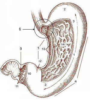
The stomach is a muscular, hollow organ in the gastrointestinal tract of humans and many other animals, including several invertebrates. The stomach has a dilated structure and functions as a vital organ in the digestive system. The stomach is involved in the gastric phase of digestion, following chewing. It performs a chemical breakdown by means of enzymes and hydrochloric acid. In humans and many other animals, the stomach is located between the oesophagus and the small intestine. The stomach secretes digestive enzymes and gastric acid to aid in food digestion. The pyloric sphincter controls the passage of partially digested food ( chyme) from the stomach into the duodenum, where peristalsis takes over to move this through the rest of intestines. Structure In the human digestive system, the stomach lies between the oesophagus and the duodenum (the first part of the small intestine). It is in the left upper quadrant of the abdominal cavity. The top of the stomach lies ag ... [...More Info...] [...Related Items...] OR: [Wikipedia] [Google] [Baidu] |
Vein
Veins are blood vessels in humans and most other animals that carry blood towards the heart. Most veins carry deoxygenated blood from the tissues back to the heart; exceptions are the pulmonary and umbilical veins, both of which carry oxygenated blood to the heart. In contrast to veins, arteries carry blood away from the heart. Veins are less muscular than arteries and are often closer to the skin. There are valves (called ''pocket valves'') in most veins to prevent backflow. Structure Veins are present throughout the body as tubes that carry blood back to the heart. Veins are classified in a number of ways, including superficial vs. deep, pulmonary vs. systemic, and large vs. small. * Superficial veins are those closer to the surface of the body, and have no corresponding arteries. *Deep veins are deeper in the body and have corresponding arteries. *Perforator veins drain from the superficial to the deep veins. These are usually referred to in the lower limbs and feet. *Communic ... [...More Info...] [...Related Items...] OR: [Wikipedia] [Google] [Baidu] |
Thoracic Diaphragm
The thoracic diaphragm, or simply the diaphragm ( grc, διάφραγμα, diáphragma, partition), is a sheet of internal Skeletal striated muscle, skeletal muscle in humans and other mammals that extends across the bottom of the thoracic cavity. The diaphragm is the most important Muscles of respiration, muscle of respiration, and separates the thoracic cavity, containing the heart and lungs, from the abdominal cavity: as the diaphragm contracts, the volume of the thoracic cavity increases, creating a negative pressure there, which draws air into the lungs. Its high oxygen consumption is noted by the many mitochondria and capillaries present; more than in any other skeletal muscle. The term ''diaphragm'' in anatomy, created by Gerard of Cremona, can refer to other flat structures such as the urogenital diaphragm or Pelvic floor, pelvic diaphragm, but "the diaphragm" generally refers to the thoracic diaphragm. In humans, the diaphragm is slightly asymmetric—its right half is h ... [...More Info...] [...Related Items...] OR: [Wikipedia] [Google] [Baidu] |
Lung
The lungs are the primary organs of the respiratory system in humans and most other animals, including some snails and a small number of fish. In mammals and most other vertebrates, two lungs are located near the backbone on either side of the heart. Their function in the respiratory system is to extract oxygen from the air and transfer it into the bloodstream, and to release carbon dioxide from the bloodstream into the atmosphere, in a process of gas exchange. Respiration is driven by different muscular systems in different species. Mammals, reptiles and birds use their different muscles to support and foster breathing. In earlier tetrapods, air was driven into the lungs by the pharyngeal muscles via buccal pumping, a mechanism still seen in amphibians. In humans, the main muscle of respiration that drives breathing is the diaphragm. The lungs also provide airflow that makes vocal sounds including human speech possible. Humans have two lungs, one on the left and on ... [...More Info...] [...Related Items...] OR: [Wikipedia] [Google] [Baidu] |


