|
List Of External Rotators Of The Human Body
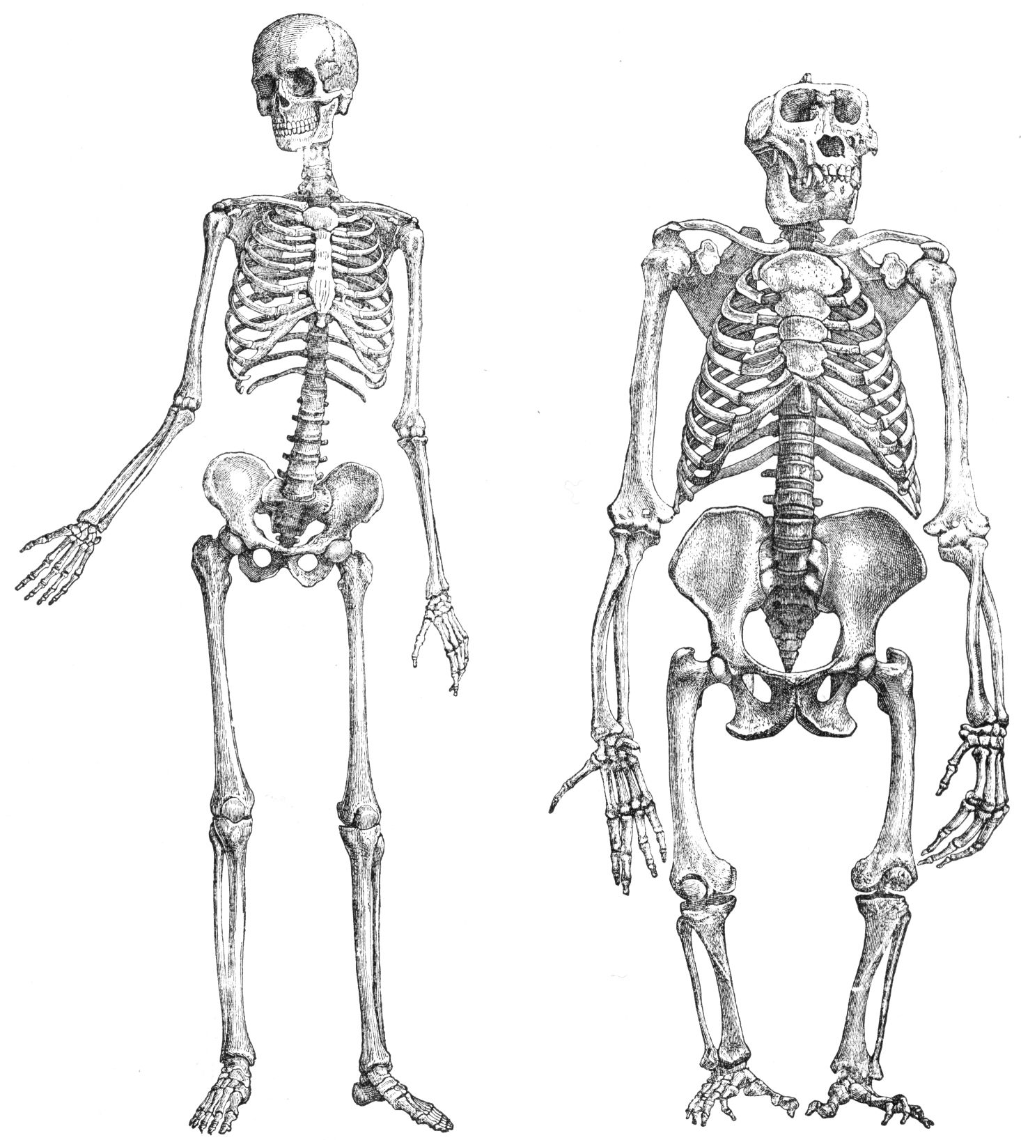
''External rotation'' (or ''extorsion'' or ''lateral rotation'') is an anatomical term of motion referring to rotation away from the center of the body. The external rotator muscles include: Muscles * of arm/ humerus at shoulder ** Deltoid muscle ** Supraspinatus ** Infraspinatus ** Teres minor * of thigh/femur at hip ** Gluteus maximus ** Lateral rotator group *** piriformis *** gemellus superior *** obturator internus *** gemellus inferior *** obturator externus *** quadratus femoris ** Sartorius * of leg at knee ** Biceps femoris * of eyeball (motion is also called "extorsion" or excyclotorsion) at Dictionary ** |
Anatomical Terms Of Motion
Motion, the process of movement, is described using specific anatomical terms. Motion includes movement of organs, joints, limbs, and specific sections of the body. The terminology used describes this motion according to its direction relative to the anatomical position of the body parts involved. Anatomists and others use a unified set of terms to describe most of the movements, although other, more specialized terms are necessary for describing unique movements such as those of the hands, feet, and eyes. In general, motion is classified according to the anatomical plane it occurs in. ''Flexion'' and ''extension'' are examples of ''angular'' motions, in which two axes of a joint are brought closer together or moved further apart. ''Rotational'' motion may occur at other joints, for example the shoulder, and are described as ''internal'' or ''external''. Other terms, such as ''elevation'' and ''depression'', describe movement above or below the horizontal plane. Many anatomica ... [...More Info...] [...Related Items...] OR: [Wikipedia] [Google] [Baidu] |
Gemellus Inferior
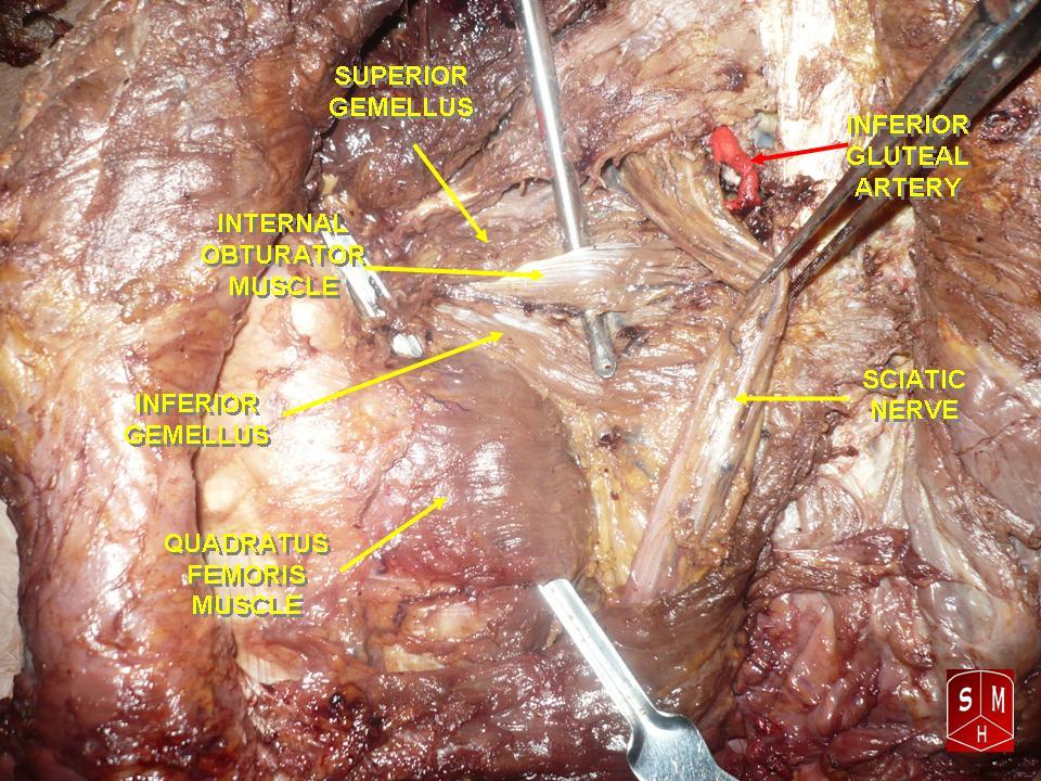
The gemelli muscles are the inferior gemellus muscle and the superior gemellus muscle, two small accessory fasciculi to the tendon of the internal obturator muscle. The gemelli muscles belong to the lateral rotator group of six muscles of the hip that rotate the femur in the hip joint. Superior gemellus muscle The gemelli muscles are two small muscular fasciculi, accessories to the tendon of the internal obturator muscle which is received into a groove between them. The superior gemellus muscle is the higher placed gemellus muscle that arises from the outer (gluteal) surface of the ischial spine, and blends with the upper part of the tendon of the internal obturator. It is smaller than the inferior gemellus. In some people, the fibres of the gemellus superior extend further than average, and are prolonged onto the medial surface of the greater trochanter of the femur. The superior and inferior gemelli are supplied by the inferior gluteal artery. Nerve supply to the superior geme ... [...More Info...] [...Related Items...] OR: [Wikipedia] [Google] [Baidu] |
Inferior Oblique Muscle
The inferior oblique muscle or obliquus oculi inferior is a thin, narrow muscle placed near the anterior margin of the floor of the orbit. The inferior oblique is one of the extraocular muscles, and is attached to the maxillary bone (origin) and the posterior, inferior, lateral surface of the eye (insertion). The inferior oblique is innervated by the inferior branch of the oculomotor nerve. Structure The inferior oblique arises from the orbital surface of the maxilla, lateral to the lacrimal groove. Unlike the other extraocular muscles (recti and superior oblique), the inferior oblique muscle does ''not'' originate from the common tendinous ring (annulus of Zinn). Passing lateralward, backward, and upward, between the inferior rectus and the floor of the orbit, and just underneath the lateral rectus muscle, the inferior oblique inserts onto the scleral surface between the inferior rectus and lateral rectus. In humans, the muscle is about 35 mm long. Innervation The i ... [...More Info...] [...Related Items...] OR: [Wikipedia] [Google] [Baidu] |
Inferior Rectus Muscle
The inferior rectus muscle is a muscle in the orbit near the eye. It is one of the four recti muscles in the group of extraocular muscles. It originates from the common tendinous ring, and inserts into the anteroinferior surface of the eye. It depresses the eye (downwards). Structure The inferior rectus muscle originates from the common tendinous ring (annulus of Zinn). It inserts into the anteroinferior surface of the eye. This insertion has a width of around 10.5 mm. It is around 7 mm from the corneal limbus. Blood supply The inferior rectus muscle is supplied by an inferior muscular branch of the ophthalmic artery. It may also be supplied by a branch of the infraorbital artery. It is drained by the corresponding veins: the inferior muscular branch of the ophthalmic vein, and sometimes a branch of the infraorbital vein. Nerve supply The inferior rectus muscle is supplied by the inferior division of the oculomotor nerve (III). Development The inferior rectus muscle devel ... [...More Info...] [...Related Items...] OR: [Wikipedia] [Google] [Baidu] |
EMedicine
eMedicine is an online clinical medical knowledge base founded in 1996 by doctors Scott Plantz and Jonathan Adler, and computer engineer Jeffrey Berezin. The eMedicine website consists of approximately 6,800 medical topic review articles, each of which is associated with a clinical subspecialty "textbook". The knowledge base includes over 25,000 clinically multimedia files. Each article is authored by board certified specialists in the subspecialty to which the article belongs and undergoes three levels of physician peer-review, plus review by a Doctor of Pharmacy. The article's authors are identified with their current faculty appointments. Each article is updated yearly, or more frequently as changes in practice occur, and the date is published on the article. eMedicine.com was sold to WebMD in January, 2006 and is available as the Medscape Reference. History Plantz, Adler and Berezin evolved the concept for eMedicine.com in 1996 and deployed the initial site via Boston Med ... [...More Info...] [...Related Items...] OR: [Wikipedia] [Google] [Baidu] |
Excyclotorsion
Eye movement includes the voluntary or involuntary movement of the eyes. Eye movements are used by a number of organisms (e.g. primates, rodents, flies, birds, fish, cats, crabs, octopus) to fixate, inspect and track visual objects of interests. A special type of eye movement, rapid eye movement, occurs during REM sleep. The eyes are the visual organs of the human body, and move using a system of six muscles. The retina, a specialised type of tissue containing photoreceptors, senses light. These specialised cells convert light into electrochemical signals. These signals travel along the optic nerve fibers to the brain, where they are interpreted as vision in the visual cortex. Primates and many other vertebrates use three types of voluntary eye movement to track objects of interest: smooth pursuit, vergence shifts and saccades. These types of movements appear to be initiated by a small cortical region in the brain's frontal lobe. This is corroborated by removal of the frontal ... [...More Info...] [...Related Items...] OR: [Wikipedia] [Google] [Baidu] |
Human Eyeball
The human eye is a sensory organ, part of the sensory nervous system, that reacts to visible light and allows humans to use visual information for various purposes including seeing things, keeping balance, and maintaining circadian rhythm. The eye can be considered as a living optical device. It is approximately spherical in shape, with its outer layers, such as the outermost, white part of the eye (the sclera) and one of its inner layers (the pigmented choroid) keeping the eye essentially light tight except on the eye's optic axis. In order, along the optic axis, the optical components consist of a first lens (the cornea—the clear part of the eye) that accomplishes most of the focussing of light from the outside world; then an aperture (the pupil) in a diaphragm (the iris—the coloured part of the eye) that controls the amount of light entering the interior of the eye; then another lens (the crystalline lens) that accomplishes the remaining focussing of light into ... [...More Info...] [...Related Items...] OR: [Wikipedia] [Google] [Baidu] |
Biceps Femoris Muscle
The biceps femoris () is a muscle of the thigh located to the posterior, or back. As its name implies, it has two parts, one of which (the long head) forms part of the hamstrings muscle group. Structure It has two heads of origin: *the ''long head'' arises from the lower and inner impression on the posterior part of the tuberosity of the ischium. This is a common tendon origin with the semitendinosus muscle, and from the lower part of the sacrotuberous ligament. *the ''short head'', arises from the lateral lip of the linea aspera, between the adductor magnus and vastus lateralis extending up almost as high as the insertion of the gluteus maximus, from the lateral prolongation of the linea aspera to within 5 cm. of the lateral condyle; and from the lateral intermuscular septum. The two muscle heads joint together distally and unite in an intricate fashion. The fibers of the long head form a fusiform belly, which passes obliquely downward and lateralward across the sciatic ... [...More Info...] [...Related Items...] OR: [Wikipedia] [Google] [Baidu] |
Knee
In humans and other primates, the knee joins the thigh with the leg and consists of two joints: one between the femur and tibia (tibiofemoral joint), and one between the femur and patella (patellofemoral joint). It is the largest joint in the human body. The knee is a modified hinge joint, which permits flexion and extension as well as slight internal and external rotation. The knee is vulnerable to injury and to the development of osteoarthritis. It is often termed a ''compound joint'' having tibiofemoral and patellofemoral components. (The fibular collateral ligament is often considered with tibiofemoral components.) Structure The knee is a modified hinge joint, a type of synovial joint, which is composed of three functional compartments: the patellofemoral articulation, consisting of the patella, or "kneecap", and the patellar groove on the front of the femur through which it slides; and the medial and lateral tibiofemoral articulations linking the femur, or thigh bone ... [...More Info...] [...Related Items...] OR: [Wikipedia] [Google] [Baidu] |
Human Leg
The human leg, in the general word sense, is the entire lower limb (anatomy), limb of the human body, including the foot, thigh or sometimes even the hip or Gluteal muscles, gluteal region. However, the definition in human anatomy refers only to the section of the lower limb extending from the knee to the ankle, also known as the crus or, especially in non-technical use, the shank. Legs are used for standing, and all forms of locomotion including recreational such as dancing, and constitute a significant portion of a person's mass. Female legs generally have greater hip anteversion and tibiofemoral angles, but shorter femur and tibial lengths than those in males. Structure In human anatomy, the lower leg is the part of the lower limb that lies between the knee and the ankle. Anatomists restrict the term ''leg'' to this use, rather than to the entire lower limb. The thigh is between the hip and knee and makes up the rest of the lower limb. The term ''lower limb'' or ''lower extre ... [...More Info...] [...Related Items...] OR: [Wikipedia] [Google] [Baidu] |
Sartorius Muscle
The sartorius muscle () is the longest muscle in the human body. It is a long, thin, superficial muscle that runs down the length of the thigh in the Anterior compartment of thigh, anterior compartment. Structure The sartorius muscle originates from the anterior superior iliac spine, and part of the notch between the anterior superior iliac spine and anterior inferior iliac spine. It runs obliquely across the upper and anterior part of the thigh in an inferomedial direction. It passes behind the medial condyle of the femur to end in a tendon. This tendon curves anteriorly to join the tendons of the Gracilis muscle, gracilis and semitendinosus muscles in the pes anserinus (leg), pes anserinus, where it inserts into the superomedial surface of the tibia. Its upper portion forms the lateral border of the femoral triangle, and the point where it crosses Adductor longus muscle, adductor longus marks the apex of the triangle. Deep to sartorius and its fascia is the adductor canal, throu ... [...More Info...] [...Related Items...] OR: [Wikipedia] [Google] [Baidu] |
Quadratus Femoris
The quadratus femoris is a flat, quadrilateral skeletal muscle. Located on the posterior side of the hip joint, it is a strong external rotator and adductor of the thigh, but also acts to stabilize the femoral head in the acetabulum. Quadratus femoris use in the Meyer's muscle pedicle grafting to prevent avascular necrosis of femur head. Course It originates on the lateral border of the ischial tuberosity of the ischium of the pelvis. From there, it passes laterally to its insertion on the posterior side of the head of the femur: the quadrate tubercle on the intertrochanteric crest and along the quadrate line, the vertical line which runs downward to bisect the lesser trochanter on the medial side of the femur. Along its course, quadratus is aligned edge to edge with the inferior gemellus above and the adductor magnus below, so that its upper and lower borders run horizontal and parallel. At its origin, the upper margin of the adductor magnus is separated from it by the te ... [...More Info...] [...Related Items...] OR: [Wikipedia] [Google] [Baidu] |


