|
Langerhan's Cell Histiocytosis
Langerhans cell histiocytosis (LCH) is an abnormal clonal proliferation of Langerhans cells, abnormal cells deriving from bone marrow and capable of migrating from skin to lymph nodes. Symptoms range from isolated bone lesions to multisystem disease. LCH is part of a group of syndromes called histiocytoses, which are characterized by an abnormal proliferation of histiocytes (an archaic term for activated dendritic cells and macrophages). These diseases are related to other forms of abnormal proliferation of white blood cells, such as leukemias and lymphomas. The disease has gone by several names, including Hand–Schüller–Christian disease, Abt-Letterer-Siwe disease, Hashimoto-Pritzker disease (a very rare self-limiting variant seen at birth) and histiocytosis X, until it was renamed in 1985 by the Histiocyte Society. Classification The disease spectrum results from clonal accumulation and proliferation of cells resembling the epidermal dendritic cells called Lange ... [...More Info...] [...Related Items...] OR: [Wikipedia] [Google] [Baidu] |
Micrograph
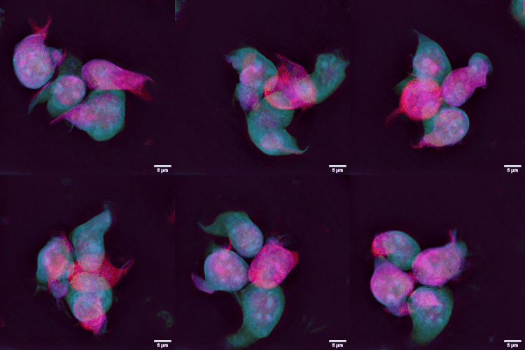
A micrograph or photomicrograph is a photograph or digital image taken through a microscope or similar device to show a magnified image of an object. This is opposed to a macrograph or photomacrograph, an image which is also taken on a microscope but is only slightly magnified, usually less than 10 times. Micrography is the practice or art of using microscopes to make photographs. A micrograph contains extensive details of microstructure. A wealth of information can be obtained from a simple micrograph like behavior of the material under different conditions, the phases found in the system, failure analysis, grain size estimation, elemental analysis and so on. Micrographs are widely used in all fields of microscopy. Types Photomicrograph A light micrograph or photomicrograph is a micrograph prepared using an optical microscope, a process referred to as ''photomicroscopy''. At a basic level, photomicroscopy may be performed simply by connecting a camera to a microscope, th ... [...More Info...] [...Related Items...] OR: [Wikipedia] [Google] [Baidu] |
Letterer–Siwe Disease
Letterer–Siwe disease, (LSD) or Abt-Letterer-Siwe disease, is one of the four recognized clinical syndromes of Langerhans cell histiocytosis (LCH) and is the most severe form, involving multiple organ systems such as the skin, bone marrow, spleen, liver, and lung. Oral cavity and gastrointestinal involvement may also be seen. Shahlaee, A. H. and Arceci, R. J. (2006). Histiocytic disorders, in LCH and all its subtypes are characterized by monoclonal migration and proliferation of specific dendritic cells. The subcategorization of Letterer-Siwe disease is a historical eponym. Designating the four subtypes of LCH as separate entities are mostly of historical significance, because they are varied manifestations of the same underlying disease process, and patients also often exhibit symptoms from more than one of the four syndromes. Letterer-Siwe causes approximately 10% of LCH disease. Prevalence is estimated at 1:500,000 and the disease almost exclusively occurs in children les ... [...More Info...] [...Related Items...] OR: [Wikipedia] [Google] [Baidu] |
Hand-Schüller-Christian Triad
Chronic multifocal Langerhans cell histiocytosis, previously known as Hand–Schüller–Christian disease, is a type of Langerhans cell histiocytosis (LCH), which can affect multiple organs. The condition is traditionally associated with a combination of three features; bulging eyes, breakdown of bone (lytic bone lesions often in the skull), and diabetes insipidus (excessive thirst and passing urine), although around 75% of cases do not have all three features. Other features may include a fever and weight loss, and depending on the organs involved there maybe rashes, asymmetry of the face, ear infections, signs in the mouth and the appearance of advanced gum disease. Features relating to lung and liver disease may occur. It is due to a genetic mutation in the MAPKinase pathway that occurs during early development. The diagnosis may be suspected based on symptoms and MRI and confirmed by tissue biopsy. Blood tests may show anaemia, and less commonly a low white blood cell count ... [...More Info...] [...Related Items...] OR: [Wikipedia] [Google] [Baidu] |
Exophthalmos
Exophthalmos (also called exophthalmus, exophthalmia, proptosis, or exorbitism) is a bulging of the eye anteriorly out of the orbit. Exophthalmos can be either bilateral (as is often seen in Graves' disease) or unilateral (as is often seen in an orbital tumor). Complete or partial dislocation from the orbit is also possible from trauma or swelling of surrounding tissue resulting from trauma. In the case of Graves' disease, the displacement of the eye results from abnormal connective tissue deposition in the orbit and extraocular muscles, which can be visualized by CT or MRI. If left untreated, exophthalmos can cause the eyelids to fail to close during sleep, leading to corneal dryness and damage. Another possible complication is a form of redness or irritation called superior limbic keratoconjunctivitis, in which the area above the cornea becomes inflamed as a result of increased friction when blinking. The process that is causing the displacement of the eye may also compre ... [...More Info...] [...Related Items...] OR: [Wikipedia] [Google] [Baidu] |
Diabetes Insipidus
Diabetes insipidus (DI), recently renamed to Arginine Vasopressin Deficiency (AVP-D) and Arginine Vasopressin Resistance (AVP-R), is a condition characterized by large amounts of dilute urine and increased thirst. The amount of urine produced can be nearly 20 liters per day. Reduction of fluid has little effect on the concentration of the urine. Complications may include dehydration or seizures. There are four types of DI, each with a different set of causes. Central DI (CDI) is due to a lack of the hormone vasopressin (antidiuretic hormone). This can be due to injury to the hypothalamus or pituitary gland or genetics. Nephrogenic DI (NDI) occurs when the kidneys do not respond properly to vasopressin. Dipsogenic DI is a result of excessive fluid intake due to damage to the hypothalamic thirst mechanism. It occurs more often in those with certain psychiatric disorders or on certain medications. Gestational DI occurs only during pregnancy. Diagnosis is often based on urine ... [...More Info...] [...Related Items...] OR: [Wikipedia] [Google] [Baidu] |
Pituitary Gland
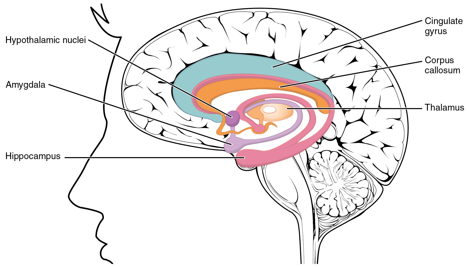
In vertebrate anatomy, the pituitary gland, or hypophysis, is an endocrine gland, about the size of a chickpea and weighing, on average, in humans. It is a protrusion off the bottom of the hypothalamus at the base of the brain. The hypophysis rests upon the hypophyseal fossa of the sphenoid bone in the center of the middle cranial fossa and is surrounded by a small bony cavity (sella turcica) covered by a dural fold (diaphragma sellae). The anterior pituitary (or adenohypophysis) is a lobe of the gland that regulates several physiological processes including stress, growth, reproduction, and lactation. The intermediate lobe synthesizes and secretes melanocyte-stimulating hormone. The posterior pituitary (or neurohypophysis) is a lobe of the gland that is functionally connected to the hypothalamus by the median eminence via a small tube called the pituitary stalk (also called the infundibular stalk or the infundibulum). Hormones secreted from the pituitary gland ... [...More Info...] [...Related Items...] OR: [Wikipedia] [Google] [Baidu] |
Misnomer
A misnomer is a name that is incorrectly or unsuitably applied. Misnomers often arise because something was named long before its correct nature was known, or because an earlier form of something has been replaced by a later form to which the name no longer suitably applies. A misnomer may also be simply a word that someone uses incorrectly or misleadingly. The word "misnomer" does not mean " misunderstanding" or " popular misconception", and a number of misnomers remain in common usage — which is to say that a word being a misnomer does not necessarily make usage of the word incorrect. Sources of misnomers Some of the sources of misnomers are: * An older name being retained after the thing named has changed (e.g., tin can, mince meat pie, steamroller, tin foil, clothes iron, digital darkroom). This is essentially a metaphorical extension with the older item standing for anything filling its role. * Transference of a well-known product brand name into a genericized tr ... [...More Info...] [...Related Items...] OR: [Wikipedia] [Google] [Baidu] |
Eosinophilic Granuloma
Humans Human eosinophilic granuloma is characterized by abnormal proliferation of Langerhans cells (LCs). LCs are antigen-presenting cells derived from dendritic cells. In humans, eosinophilic granulomas are considered as a benign tumors that occurs mainly in children and adolescents. EG is a quite rare condition, and its incidence is higher in white than in black population, also slightly more affecting males than females. EG develops in 4-5 children (aged under 15) per million/year and in 1 or 2 adults per million/year. The etiology of EG is not fully understood yet. However, the onset of abnormal LC proliferation may be triggered by viral stimuli ( EBV, Human Herpes virus 6), bacterial toxins or defective regulation of IL-1 and IL-10 production. Another possible explanation may be a defect in Ras/MAPK signaling pathway due to mutation of signaling proteins. Particularly, it was published that about 50% of the EG cases had mutated BRAF V600 E gene and about 21% displayed a ... [...More Info...] [...Related Items...] OR: [Wikipedia] [Google] [Baidu] |
Canine Histiocytic Diseases
Histiocytic diseases in dogs are a group of diseases in dogs which may involve the skin, and which can be difficult to differentiate from granulomatous, reactive inflammatory or lymphoproliferative diseases. The clinical presentation and behaviour as well as response to therapy vary greatly among the syndromes. There are at least four well-defined canine histiocytic diseases: :1. Canine cutaneous histiocytoma (derived from specialised epidermic dendritic cells, the Langerhans cells) :2. Reactive histiocytosis (immunohistochemical features show that interstitial/dermal DCs are involved) :2.a. Cutaneous histiocytosis (CH) :2.b. Systemic histiocytosis (SH) :3. Histiocytic sarcoma complex (immunohistochemical features of dendritic cells, possibly interdigitating or perivascular DCs) :3.a. Malignant histiocytosis :3.b. Histiocytic sarcoma *Localized histiocytic sarcoma *Diffuse histiocytic sarcoma Cutaneous histiocytoma Histiocytoma Histiocytoma is a common, benign, cutaneous neo ... [...More Info...] [...Related Items...] OR: [Wikipedia] [Google] [Baidu] |
Organ (anatomy)
In biology, an organ is a collection of tissues joined in a structural unit to serve a common function. In the hierarchy of life, an organ lies between tissue and an organ system. Tissues are formed from same type cells to act together in a function. Tissues of different types combine to form an organ which has a specific function. The intestinal wall for example is formed by epithelial tissue and smooth muscle tissue. Two or more organs working together in the execution of a specific body function form an organ system, also called a biological system or body system. An organ's tissues can be broadly categorized as parenchyma, the functional tissue, and stroma, the structural tissue with supportive, connective, or ancillary functions. For example, the gland's tissue that makes the hormones is the parenchyma, whereas the stroma includes the nerves that innervate the parenchyma, the blood vessels that oxygenate and nourish it and carry away its metabolic wastes, and the con ... [...More Info...] [...Related Items...] OR: [Wikipedia] [Google] [Baidu] |
Lymphocyte
A lymphocyte is a type of white blood cell (leukocyte) in the immune system of most vertebrates. Lymphocytes include natural killer cells (which function in cell-mediated, cytotoxic innate immunity), T cells (for cell-mediated, cytotoxic adaptive immunity), and B cells (for humoral, antibody-driven adaptive immunity). They are the main type of cell found in lymph, which prompted the name "lymphocyte". Lymphocytes make up between 18% and 42% of circulating white blood cells. Types The three major types of lymphocyte are T cells, B cells and natural killer (NK) cells. Lymphocytes can be identified by their large nucleus. T cells and B cells T cells (thymus cells) and B cells ( bone marrow- or bursa-derived cells) are the major cellular components of the adaptive immune response. T cells are involved in cell-mediated immunity, whereas B cells are primarily responsible for humoral immunity (relating to antibodies). The function of T cells and B cells is to recognize sp ... [...More Info...] [...Related Items...] OR: [Wikipedia] [Google] [Baidu] |


