|
Lysophosphatidic Acid
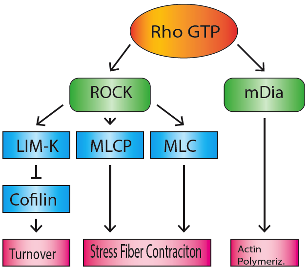
Lysophosphatidic acid (LPA) is a phospholipid derivative that can act as a signaling molecule. Function LPA acts as a potent mitogen due to its activation of three high-affinity G-protein-coupled receptors called LPAR1, LPAR2, and LPAR3 (also known as EDG2, EDG4, and EDG7). Additional, newly identified LPA receptors include LPAR4 (P2RY9, GPR23), LPAR5 (GPR92) and LPAR6 (P2RY5, GPR87). Clinical significance Because of its ability to stimulate cell proliferation, aberrant LPA-signaling has been linked to cancer in numerous ways. Dysregulation of autotaxin or the LPA receptors can lead to hyperproliferation, which may contribute to oncogenesis and metastasis. LPA may be the cause of pruritus (itching) in individuals with cholestatic (impaired bile flow) diseases. GTPase activation Downstream of LPA receptor activation, the small GTPase Rho can be activated, subsequently activating Rho kinase. This can lead to the formation of stress fibers and cell migration through the inhibiti ... [...More Info...] [...Related Items...] OR: [Wikipedia] [Google] [Baidu] |
Phospholipid
Phospholipids, are a class of lipids whose molecule has a hydrophilic "head" containing a phosphate group and two hydrophobic "tails" derived from fatty acids, joined by an alcohol residue (usually a glycerol molecule). Marine phospholipids typically have omega-3 fatty acids EPA and DHA integrated as part of the phospholipid molecule. The phosphate group can be modified with simple organic molecules such as choline, ethanolamine or serine. Phospholipids are a key component of all cell membranes. They can form lipid bilayers because of their amphiphilic characteristic. In eukaryotes, cell membranes also contain another class of lipid, sterol, interspersed among the phospholipids. The combination provides fluidity in two dimensions combined with mechanical strength against rupture. Purified phospholipids are produced commercially and have found applications in nanotechnology and materials science. The first phospholipid identified in 1847 as such in biological tissues was lecith ... [...More Info...] [...Related Items...] OR: [Wikipedia] [Google] [Baidu] |
Stress Fiber
Stress fibers are contractile actin bundles found in non-muscle cells. They are composed of actin (microfilaments) and non-muscle myosin II (NMMII), and also contain various crosslinking proteins, such as α-actinin, to form a highly regulated actomyosin structure within non-muscle cells. Stress fibers have been shown to play an important role in cellular contractility, providing force for a number of functions such as cell adhesion, migration and morphogenesis. Structure Stress fibers are primarily composed of actin and myosin. Actin is a ~43kDa globular protein, and can polymerize to form long filamentous structures. These filaments are made of two strands of actin monomers (or protofilaments) wrapping around each other, to create a single actin filament. Because actin monomers are not symmetrical molecules, their filaments have polarity based upon the structure of the actin monomer, which will allow one end of the actin filament to polymerize faster than the other. The end ... [...More Info...] [...Related Items...] OR: [Wikipedia] [Google] [Baidu] |
Sphingosine-1-phosphate
Sphingosine-1-phosphate (S1P) is a signaling sphingolipid, also known as lysosphingolipid. It is also referred to as a bioactive lipid mediator. Sphingolipids at large form a class of lipids characterized by a particular aliphatic aminoalcohol, which is sphingosine. Production S1P is formed from ceramide, which is composed of a sphingosine and a fatty acid. Ceramidase, an enzyme primarily present in plasma membrane, will convert ceramide to sphingosine. sphingosine is then phosphorylated by sphingosine kinase (SK) isoenzymes. There are two identified isoenzymes, SK1 and SK2. These two enzymes have different tissue distribution. SK1 is highly expressed in spleen, lung and leukocytes, while SK2 is highly expressed in liver and kidney. SK2 is located mainly in the mitochondria, nucleus and the endoplasmic reticulum whereas SK1 is mainly located in cytoplasm and the cell membrane. Metabolism and degradation S1P can be dephosphorylated to sphingosine by sphingosine phosphatases and ... [...More Info...] [...Related Items...] OR: [Wikipedia] [Google] [Baidu] |
Phosphatidic Acid
Phosphatidic acids are anionic phospholipids important to cell signaling and direct activation of lipid-gated ion channels. Hydrolysis of phosphatidic acid gives rise to one molecule each of glycerol and phosphoric acid and two molecules of fatty acids. They constitute about 0.25% of phospholipids in the bilayer. Structure Phosphatidic acid consists of a glycerol backbone, with, in general, a saturated fatty acid bonded to carbon-1, an unsaturated fatty acid bonded to carbon-2, and a phosphate group bonded to carbon-3. Formation and degradation Besides de novo synthesis, PA can be formed in three ways: * By phospholipase D (PLD), via the hydrolysis of the P-O bond of phosphatidylcholine (PC) to produce PA and choline. * By the phosphorylation of diglyceride, diacylglycerol (DAG) by diacylglycerol kinase, DAG kinase (DAGK). * By the acylation of lysophosphatidic acid by lysoPA-acyltransferase (LPAAT); this is the most common metabolic pathway, pathway.Devlin, T. M. 2004. ''Bioquími ... [...More Info...] [...Related Items...] OR: [Wikipedia] [Google] [Baidu] |
GPR35
G protein-coupled receptor 35 also known as GPR35 is a G protein-coupled receptor which in humans is encoded by the ''GPR35'' gene. Heightened expression of GPR35 is found in immune and gastrointestinal tissues, including the crypts of Lieberkühn. Ligands Endogenous ligands Although GPR35 is still considered an orphan receptor, there have been attempts to deorphanize it by identifying endogenous molecules that can activate the receptor. All of the currently proposed ligands are either unselective towards GPR35, or they lack high potency, a characteristic feature of natural ligands. The following list includes the most prominent examples: * kynurenic acidfree fulltext * LPA species [...More Info...] [...Related Items...] OR: [Wikipedia] [Google] [Baidu] |
Autotaxin
Autotaxin, also known as ectonucleotide pyrophosphatase/phosphodiesterase family member 2 (E-NPP 2), is an enzyme that in humans is encoded by the ''ENPP2'' gene. Function Autotaxin (ectonucleotide pyrophosphatase/phosphodiesterase 2 (NPP2 or ENPP2) is a secreted enzyme important for generating the lipid signaling molecule lysophosphatidic acid (LPA). Autotaxin has lyso phospholipase D activity that converts lysophosphatidylcholine into LPA. Autotaxin was originally identified as a tumor cell-motility-stimulating factor; later it was shown to be LPA (which signals through lysophospholipid receptors), the lipid product of the reaction catalyzed by autotaxin, which is responsible for its effects on cell-proliferation. The protein encoded by this gene functions as a phosphodiesterase. Autotaxin is secreted and further processed to make the biologically active form. Several alternatively spliced transcript variants have been identified. Autotaxin is able to cleave the phosphodie ... [...More Info...] [...Related Items...] OR: [Wikipedia] [Google] [Baidu] |
Autotaxin Rxn
Autotaxin, also known as ectonucleotide pyrophosphatase/phosphodiesterase family member 2 (E-NPP 2), is an enzyme that in humans is encoded by the ''ENPP2'' gene. Function Autotaxin (ectonucleotide pyrophosphatase/phosphodiesterase 2 (NPP2 or ENPP2) is a secreted enzyme important for generating the lipid signaling molecule lysophosphatidic acid (LPA). Autotaxin has lyso phospholipase D activity that converts lysophosphatidylcholine into LPA. Autotaxin was originally identified as a tumor cell-motility-stimulating factor; later it was shown to be LPA (which signals through lysophospholipid receptors), the lipid product of the reaction catalyzed by autotaxin, which is responsible for its effects on cell-proliferation. The protein encoded by this gene functions as a phosphodiesterase. Autotaxin is secreted and further processed to make the biologically active form. Several alternatively spliced transcript variants have been identified. Autotaxin is able to cleave the phosphodi ... [...More Info...] [...Related Items...] OR: [Wikipedia] [Google] [Baidu] |
Phosphatidic Acid
Phosphatidic acids are anionic phospholipids important to cell signaling and direct activation of lipid-gated ion channels. Hydrolysis of phosphatidic acid gives rise to one molecule each of glycerol and phosphoric acid and two molecules of fatty acids. They constitute about 0.25% of phospholipids in the bilayer. Structure Phosphatidic acid consists of a glycerol backbone, with, in general, a saturated fatty acid bonded to carbon-1, an unsaturated fatty acid bonded to carbon-2, and a phosphate group bonded to carbon-3. Formation and degradation Besides de novo synthesis, PA can be formed in three ways: * By phospholipase D (PLD), via the hydrolysis of the P-O bond of phosphatidylcholine (PC) to produce PA and choline. * By the phosphorylation of diglyceride, diacylglycerol (DAG) by diacylglycerol kinase, DAG kinase (DAGK). * By the acylation of lysophosphatidic acid by lysoPA-acyltransferase (LPAAT); this is the most common metabolic pathway, pathway.Devlin, T. M. 2004. ''Bioquími ... [...More Info...] [...Related Items...] OR: [Wikipedia] [Google] [Baidu] |
Lysophosphatidylcholine
Lysophosphatidylcholines (LPC, lysoPC), also called lysolecithins, are a class of chemical compounds which are derived from phosphatidylcholines. Overview Lysophosphatidylcholines are produced within cells mainly by the enzyme phospholipase A2, which removes one of the fatty acid groups from phosphatidylcholine to produce LPC. Among other properties, they activate endothelial cells during early atherosclerosis. LPC also acts as a find-me signal, released by apoptotic cells to recruit phagocytes, which then phagocytose the apoptotic cells Moreover, LPCs can be used in the lab to cause demyelination of brain slices, to mimic the effects of demyelinating diseases such as multiple sclerosis. Further, they are known to stimulate phagocytosis of the myelin sheath and can change the surface properties of erythrocytes. LPC-induced demyelination is thought to occur through the actions of recruited macrophages and microglia which phagocytose nearby myelin. Invading T cells are also t ... [...More Info...] [...Related Items...] OR: [Wikipedia] [Google] [Baidu] |
Choline
Choline is an essential nutrient for humans and many other animals. Choline occurs as a cation that forms various salts (X− in the depicted formula is an undefined counteranion). Humans are capable of some ''de novo synthesis'' of choline but require additional choline in the diet to maintain health. Dietary requirements can be met by choline per se or in the form of choline phospholipids, such as phosphatidylcholine. Choline is not formally classified as a vitamin despite being an essential nutrient with an amino acid–like structure and metabolism. In most animals, choline phospholipids are necessary components in cell membranes, in the membranes of cell organelles, and in very low-density lipoproteins. Choline is required to produce acetylcholine – a neurotransmitter – and ''S''-adenosylmethionine (SAM), a universal methyl donor. Upon methylation SAM is transformed into homocysteine. Symptomatic choline deficiency causes non-alcoholic fatty liver disease and muscle dama ... [...More Info...] [...Related Items...] OR: [Wikipedia] [Google] [Baidu] |
Phospholipase D
Phospholipase D (EC 3.1.4.4, lipophosphodiesterase II, lecithinase D, choline phosphatase, PLD; systematic name phosphatidylcholine phosphatidohydrolase) is an enzyme of the phospholipase superfamily that catalyses the following reaction : a phosphatidylcholine + H2O = choline + a phosphatidate Phospholipases occur widely, and can be found in a wide range of organisms, including bacteria, yeast, plants, animals, and viruses. Phospholipase D's principal substrate is phosphatidylcholine, which it hydrolyzes to produce the signal molecule phosphatidic acid (PA), and soluble choline in a cholesterol dependent process called substrate presentation. Plants contain numerous genes that encode various PLD isoenzymes, with molecular weights ranging from 90 to 125 kDa. Mammalian cells encode two isoforms of phospholipase D: PLD1 and PLD2. Phospholipase D is an important player in many physiological processes, including membrane trafficking, cytoskeletal reorganization, receptor-mediated e ... [...More Info...] [...Related Items...] OR: [Wikipedia] [Google] [Baidu] |
Myosin Light-chain Phosphatase
Myosin light-chain phosphatase, also called myosin phosphatase (EC 3.1.3.53; systematic name yosin-light-chainphosphate phosphohydrolase), is an enzyme (specifically a serine/threonine-specific protein phosphatase) that dephosphorylates the regulatory light chain of myosin II: : yosin light-chainphosphate + H2O = yosin light-chain+ phosphate This dephosphorylation reaction occurs in smooth muscle tissue and initiates the relaxation process of the muscle cells. Thus, myosin phosphatase undoes the muscle contraction process initiated by myosin light-chain kinase. The enzyme is composed of three subunits: the catalytic region (protein phosphatase 1, or PP1), the myosin binding subunit (MYPT1), and a third subunit (M20) of unknown function. The catalytic region uses two manganese ions as catalysts to dephosphorylate the light-chains on myosin, which causes a conformational change in the myosin and relaxes the muscle. The enzyme is highly conserved and is found in all organis ... [...More Info...] [...Related Items...] OR: [Wikipedia] [Google] [Baidu] |


