|
List Of MeSH Codes (C14)
The following is a partial list of the "C" codes for Medical Subject Headings (MeSH), as defined by the United States National Library of Medicine (NLM). This list continues the information at List of MeSH codes (C13). Codes following these are found at List of MeSH codes (C15). For other MeSH codes, see List of MeSH codes. The source for this content is the set o2006 MeSH Treesfrom the NLM. – cardiovascular diseases – cardiovascular abnormalities – arterio-arterial fistula – arteriovenous malformations * – arteriovenous fistula * – intracranial arteriovenous malformations – central nervous system vascular malformations – heart defects, congenital * – alagille syndrome * – aortic coarctation * – arrhythmogenic right ventricular dysplasia * – cor triatriatum * – coronary vessel anomalies * – crisscross heart * – dextrocardia * – kartagener syndrome * – ductus arteriosus, patent * – Ebstein's anomaly * – Eisenmenger com ... [...More Info...] [...Related Items...] OR: [Wikipedia] [Google] [Baidu] |
Medical Subject Headings
Medical Subject Headings (MeSH) is a comprehensive controlled vocabulary for the purpose of indexing journal articles and books in the life sciences. It serves as a thesaurus that facilitates searching. Created and updated by the United States National Library of Medicine (NLM), it is used by the MEDLINE/PubMed article database and by NLM's catalog of book holdings. MeSH is also used by ClinicalTrials.gov registry to classify which diseases are studied by trials registered in ClinicalTrials. MeSH was introduced in the 1960s, with the NLM's own index catalogue and the subject headings of the Quarterly Cumulative Index Medicus (1940 edition) as precursors. The yearly printed version of MeSH was discontinued in 2007; MeSH is now available only online. It can be browsed and downloaded free of charge through PubMed. Originally in English, MeSH has been translated into numerous other languages and allows retrieval of documents from different origins. Structure MeSH vocabulary is divi ... [...More Info...] [...Related Items...] OR: [Wikipedia] [Google] [Baidu] |
Coronary Vessel Anomalies
Coronary artery anomalies are variations of the coronary circulation, affecting 1% of an unselected population - ''normal variant'': an alternative, unusual but benign morphological feature identified in >1% of the same population (e.g. left main is absent in 1-2% of the general population with LAD and LCx originating from separate ostia - “absent left trunk” variant) - ''coronary artery anomaly (CAA)'': a morphological feature seen in 50% increase of the vessel diameter. Some cases are congenital/idiopathic, but most are secondary to atherosclerosis or Kawasaki disease (an immuno-inflammatory disease especially targeting coronary vessels wall). Potential complications include localized thrombosis, distal embolization, rupture, or late lipid deposits. ''Coronary arteriovenous fistulas'' are anomalies at the termination consisting of an anomalous connection of coronary arteries to coronary veins, veins of the pulmonary or systemic circulations, or to any cardi ... [...More Info...] [...Related Items...] OR: [Wikipedia] [Google] [Baidu] |
Heart Septal Defects, Ventricular
A ventricular septal defect (VSD) is a defect in the ventricular septum, the wall dividing the left and right ventricles of the heart. The extent of the opening may vary from pin size to complete absence of the ventricular septum, creating one common ventricle. The ventricular septum consists of an inferior muscular and superior membranous portion and is extensively innervated with conducting cardiomyocytes. The membranous portion, which is close to the atrioventricular node, is most commonly affected in adults and older children in the United States. It is also the type that will most commonly require surgical intervention, comprising over 80% of cases. Membranous ventricular septal defects are more common than muscular ventricular septal defects, and are the most common congenital cardiac anomaly. Signs and symptoms Ventricular septal defect is usually symptomless at birth. It usually manifests a few weeks after birth. VSD is an acyanotic congenital heart defect, aka a ... [...More Info...] [...Related Items...] OR: [Wikipedia] [Google] [Baidu] |
Trilogy Of Fallot
The Trilogy of Fallot also called Fallot's trilogy is a rare congenital heart disease consisting of the following defects: pulmonary valve stenosis, right ventricular hypertrophy and atrial septal defect. It occurs in 1.2% of all congenital heart defects. A 1960 case report of 22 patients who underwent surgery showed an excess of females with a ratio of 3:2, with the youngest person being 7 months old and the oldest being 50 years old. Symptoms and signs Mechanism Trilogy of Fallot is a combination of three congenital heart defects: pulmonary stenosis, right ventricular hypertrophy, and an atrial septal defect. The first two of these are also found in the more common tetralogy of Fallot. However, the tetralogy has a ventricular septal defect instead of an atrial one, and it also involves an overriding aorta The Three Malformations Diagnosis Diagnosis is done via echocardiography or angiography. Treatment It is treated using surgery to repair the atrial septal defe ... [...More Info...] [...Related Items...] OR: [Wikipedia] [Google] [Baidu] |
Lutembacher's Syndrome
Lutembacher's syndrome is a very rare form of congenital heart disease that affects one of the chambers of the heart (commonly the atria) as well as a valve (commonly the mitral valve). It is commonly known as both congenital atrial septal defect (ASD) and acquired mitral stenosis (MS). Congenital (at birth) atrial septal defect refers to a hole being in the septum or wall that separates the two atria; this condition is usually seen in fetuses and infants. Mitral stenosis refers to mitral valve leaflets (or valve flaps) sticking to each other making the opening for blood to pass from the atrium to the ventricles very small. With the valve being so small, blood has difficulty passing from the left atrium into the left ventricle. Septal defects that may occur with Lutembacher's syndrome include: Ostium primum atrial septal defect or ostium secundum which is more prevalent. Lutembacher's syndrome affects females more often than males. It can affect children or adults; the person ca ... [...More Info...] [...Related Items...] OR: [Wikipedia] [Google] [Baidu] |
Heart Septal Defects, Atrial
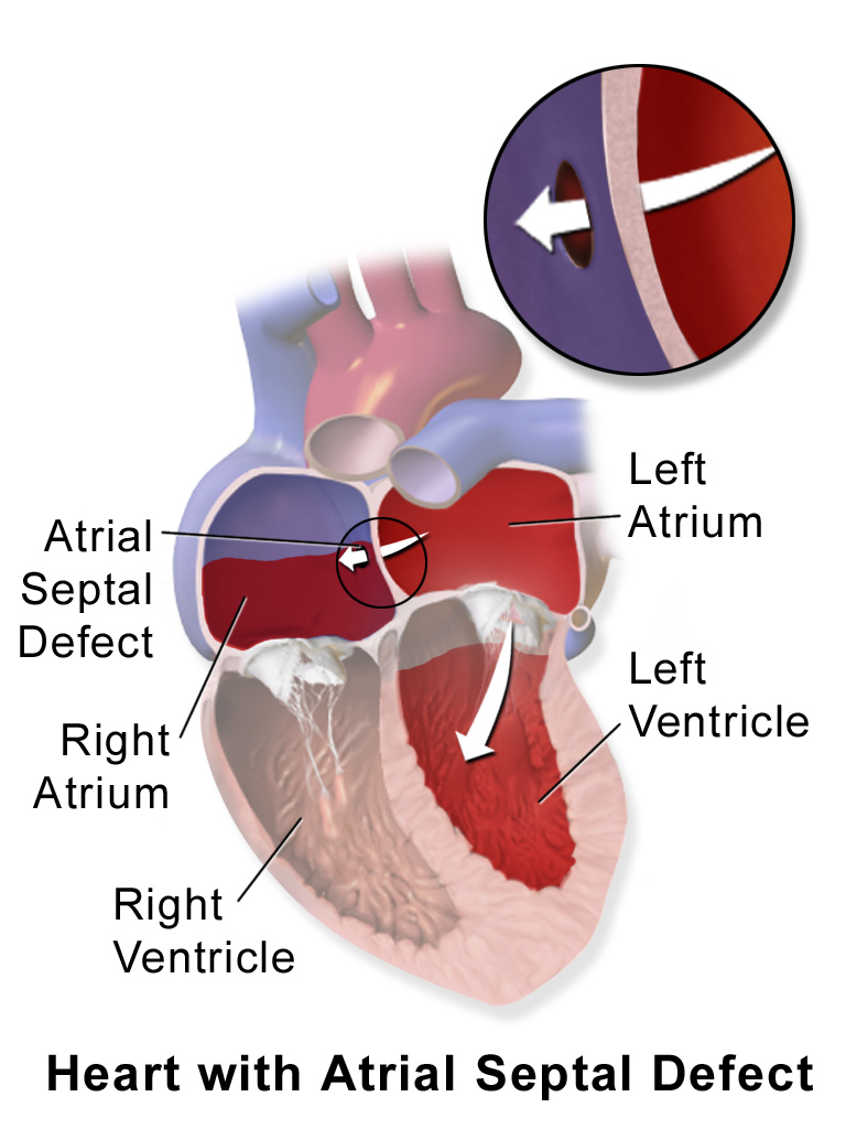
Atrial septal defect (ASD) is a congenital heart defect in which blood flows between the atria (upper chambers) of the heart. Some flow is a normal condition both pre-birth and immediately post-birth via the foramen ovale; however, when this does not naturally close after birth it is referred to as a patent (open) foramen ovale (PFO). It is common in patients with a congenital atrial septal aneurysm (ASA). After PFO closure the atria normally are separated by a dividing wall, the interatrial septum. If this septum is defective or absent, then oxygen-rich blood can flow directly from the left side of the heart to mix with the oxygen-poor blood in the right side of the heart; or the opposite, depending on whether the left or right atrium has the higher blood pressure. In the absence of other heart defects, the left atrium has the higher pressure. This can lead to lower-than-normal oxygen levels in the arterial blood that supplies the brain, organs, and tissues. However, an A ... [...More Info...] [...Related Items...] OR: [Wikipedia] [Google] [Baidu] |
Endocardial Cushion Defects
Atrioventricular septal defect (AVSD) or atrioventricular canal defect (AVCD), also known as "common atrioventricular canal" (CAVC) or "endocardial cushion defect" (ECD), is characterized by a deficiency of the atrioventricular septum of the heart that creates connections between all four of its chambers. It is caused by an abnormal or inadequate fusion of the superior and inferior endocardial cushions with the mid portion of the atrial septum and the muscular portion of the ventricular septum. Symptoms and signs Symptoms include difficulty breathing (dyspnea) and bluish discoloration on skin, fingernails, and lips (cyanosis). A newborn baby will show signs of heart failure such as edema, fatigue, wheezing, sweating and irregular heartbeat. Complications Normally, the four chambers of the heart divide oxygenated and de-oxygenated blood into separate pools. When holes form between the chambers, as in AVSD, the pools can mix. Consequently, arterial blood supplies become less oxygena ... [...More Info...] [...Related Items...] OR: [Wikipedia] [Google] [Baidu] |
Aortopulmonary Septal Defect
Aortopulmonary septal defect is a rare congenital heart disorder accounting for only 0.1-0.3% of congenital heart defects worldwide. It is characterized by a communication between the aortic and pulmonary arteries, with preservation of two normal semilunar valves. It is the result of an incomplete separation of the aorticopulmonary trunk that normally occurs in early fetal development with formation of the spiral septum.Burakovsky, V. I., Falkovsky, G. E., & Ivanitsky, A. V. (1984). Surgical repair of truncus arteriosus. ''Pediatric cardiology'', ''5''(2), 111-114. Aortopulmonary septal defects occur in isolation in about half of cases, the remainder are associated with more complex heart abnormalities. Causes Diagnosis Subtypes There are numerous types, differentiated by the extent of the defect. These types are: * Type I: simple defects leading to communication between the ascending aorta and pulmonic trunk * Type II: defects that extend to the origin of the right pulmona ... [...More Info...] [...Related Items...] OR: [Wikipedia] [Google] [Baidu] |
Heart Septal Defects
Heart septal defect refers to a congenital heart defect of one of the septa of the heart. * Atrial septal defect * Atrioventricular septal defect * Ventricular septal defect Although aortopulmonary septal defects are defects of the aorticopulmonary septum The aorticopulmonary septum is developmentally formed from neural crest, specifically the cardiac neural crest, and actively separates the aorta and pulmonary arteries and fuses with the interventricular septum within the heart during heart develop ..., which is not technically part of the heart, they are sometimes grouped with the heart septal defects. References External links Congenital heart defects {{circulatory-stub ... [...More Info...] [...Related Items...] OR: [Wikipedia] [Google] [Baidu] |
Eisenmenger Complex
Eisenmenger syndrome or Eisenmenger's syndrome is defined as the process in which a long-standing left-to-right cardiac shunt caused by a congenital heart defect (typically by a ventricular septal defect, atrial septal defect, or less commonly, patent ductus arteriosus) causes pulmonary hypertension and eventual reversal of the shunt into a cyanotic right-to-left shunt. Because of the advent of fetal screening with echocardiography early in life, the incidence of heart defects progressing to Eisenmenger syndrome has decreased. Eisenmenger syndrome in a pregnant mother can cause serious complications, though successful delivery has been reported. Maternal mortality ranges from 30% to 60%, and may be attributed to fainting spells, blood clots forming in the veins and traveling to distant sites, hypovolemia, coughing up blood or preeclampsia. Most deaths occur either during or within the first weeks after delivery.Curr Cardiol Rev. 2010 November; 6(4): 363–372.The Adult P ... [...More Info...] [...Related Items...] OR: [Wikipedia] [Google] [Baidu] |
Ebstein's Anomaly
Ebstein's anomaly is a congenital heart defect in which the septal and posterior leaflets of the tricuspid valve are displaced towards the apex of the right ventricle of the heart. It is classified as a critical congenital heart defect accounting for less than 1% of all congenital heart defects presenting in around per 200,000 live births. Ebstein anomaly is the congenital heart lesion most commonly associated with supraventricular tachycardia. Signs and symptoms The annulus of the valve is still in the normal position. The valve leaflets, however, are to a varying degree, attached to the walls and septum of the right ventricle. A subsequent "atrialization" of a portion of the morphologic right ventricle (which is then contiguous with the right atrium) is seen. This causes the right atrium to be large and the anatomic right ventricle to be small in size. * S3 heart sound * S4 heart sound * Triple or quadruple gallop due to widely split S1 and S2 sounds plus a loud S3 and/or S4 ... [...More Info...] [...Related Items...] OR: [Wikipedia] [Google] [Baidu] |
Ductus Arteriosus, Patent
''Patent ductus arteriosus'' (PDA) is a medical condition in which the '' ductus arteriosus'' fails to close after birth: this allows a portion of oxygenated blood from the left heart to flow back to the lungs by flowing from the aorta, which has a higher pressure, to the pulmonary artery. Symptoms are uncommon at birth and shortly thereafter, but later in the first year of life there is often the onset of an increased work of breathing and failure to gain weight at a normal rate. With time, an uncorrected PDA usually leads to pulmonary hypertension followed by right-sided heart failure. The ''ductus arteriosus'' is a fetal blood vessel that normally closes soon after birth. In a PDA, the vessel does not close, but remains ''patent'' (open), resulting in an abnormal transmission of blood from the aorta to the pulmonary artery. PDA is common in newborns with persistent respiratory problems such as hypoxia, and has a high occurrence in premature newborns. Premature newborns ... [...More Info...] [...Related Items...] OR: [Wikipedia] [Google] [Baidu] |


