|
List Of Human Clusters Of Differentiation
The following is a list of human clusters of differentiation The cluster of differentiation (also known as cluster of designation or classification determinant and often abbreviated as CD) is a protocol used for the identification and investigation of cell surface molecules providing targets for immunophen ... (or ''CD'') molecules. * = group; ** = not listed on hcdm References External links AbCam antigen poster {{Clusters of differentiation Human clusters of differentiation ... [...More Info...] [...Related Items...] OR: [Wikipedia] [Google] [Baidu] |
Cluster Of Differentiation
The cluster of differentiation (also known as cluster of designation or classification determinant and often abbreviated as CD) is a protocol used for the identification and investigation of cell surface molecules providing targets for immunophenotyping of cells. In terms of physiology, CD molecules can act in numerous ways, often acting as receptors or ligands important to the cell. A signal cascade is usually initiated, altering the behavior of the cell (see cell signaling). Some CD proteins do not play a role in cell signaling, but have other functions, such as cell adhesion. CD for humans is numbered up to 371 (). Nomenclature The CD nomenclature was proposed and established in the 1st International Workshop and Conference on Human Leukocyte Differentiation Antigens (HLDA), which was held in Paris in 1982. This system was intended for the classification of the many monoclonal antibodies (mAbs) generated by different laboratories around the world against epitopes on the ... [...More Info...] [...Related Items...] OR: [Wikipedia] [Google] [Baidu] |
Adult T-cell Leukemia/lymphoma
Adult T-cell leukemia/lymphoma (ATL or ATLL) is a rare cancer of the immune system's T-cells caused by human T cell leukemia/lymphotropic virus type 1 (HTLV-1). All ATL cells contain integrated HTLV-1 provirus further supporting that causal role of the virus in the cause of the neoplasm. A small amount of HTLV-1 individuals progress to develop ATL with a long latency period between infection and ATL development. ATL is categorized into 4 subtypes: acute, smoldering, lymphoma-type, chronic. Acute and Lymphoma-type are known to particularity be aggressive with poorer prognosis. Globally, the retrovirus HTLV-1 is estimated to infect 20 million people with the incidence of ATL approximately 0.05 per 100,000 with endemic regions such as regions of Japan, as high as 27 per 100,000. However, cases have increased in non-endemic regions with highest incidence of HTLV-1 in southern/northern islands of Japan, Caribbean, Central and South America, intertropical Africa, Romania, northern Iran ... [...More Info...] [...Related Items...] OR: [Wikipedia] [Google] [Baidu] |
B Cell
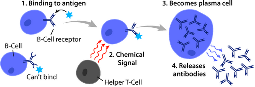
B cells, also known as B lymphocytes, are a type of white blood cell of the lymphocyte subtype. They function in the humoral immunity component of the adaptive immune system. B cells produce antibody molecules which may be either secreted or inserted into the plasma membrane where they serve as a part of B-cell receptors. When a naïve or memory B cell is activated by an antigen, it proliferates and differentiates into an antibody-secreting effector cell, known as a plasmablast or plasma cell. Additionally, B cells present antigens (they are also classified as professional antigen-presenting cells (APCs)) and secrete cytokines. In mammals, B cells mature in the bone marrow, which is at the core of most bones. In birds, B cells mature in the bursa of Fabricius, a lymphoid organ where they were first discovered by Chang and Glick, which is why the 'B' stands for bursa and not bone marrow as commonly believed. B cells, unlike the other two classes of lymphocytes, T cells and ... [...More Info...] [...Related Items...] OR: [Wikipedia] [Google] [Baidu] |
CD5 (protein)
CD5 is a cluster of differentiation expressed on the surface of T cells (various species) and in a subset of murine B cells known as B-1a. The expression of this receptor in human B cells has been a controversial topic and to date there is no consensus regarding the role of this receptor as a marker of human B cells. B-1 cells have limited diversity of their B-cell receptor due to their lack of the enzyme terminal deoxynucleotidyl transferase (TdT) and are potentially self-reactive. CD5 serves to mitigate activating signals from the BCR so that the B-1 cells can only be activated by very strong stimuli (such as bacterial proteins) and not by normal tissue proteins. CD5 was used as a T-cell marker until monoclonal antibodies against CD3 were developed. In humans, the gene is located on the long arm of chromosome 11. There is no confirmed ligand for CD5 but there is evidence that CD72, a C-type lectin, may be a ligand or that CD5 may be homophilic, binding CD5 on the surface of ot ... [...More Info...] [...Related Items...] OR: [Wikipedia] [Google] [Baidu] |
Cytotoxic T Cell
A cytotoxic T cell (also known as TC, cytotoxic T lymphocyte, CTL, T-killer cell, cytolytic T cell, CD8+ T-cell or killer T cell) is a T lymphocyte (a type of white blood cell) that kills cancer cells, cells that are infected by intracellular pathogens (such as viruses or bacteria), or cells that are damaged in other ways. Most cytotoxic T cells express T-cell receptors (TCRs) that can recognize a specific antigen. An antigen is a molecule capable of stimulating an immune response and is often produced by cancer cells, viruses, bacteria or intracellular signals. Antigens inside a cell are bound to class I MHC molecules, and brought to the surface of the cell by the class I MHC molecule, where they can be recognized by the T cell. If the TCR is specific for that antigen, it binds to the complex of the class I MHC molecule and the antigen, and the T cell destroys the cell. In order for the TCR to bind to the class I MHC molecule, the former must be accompanied by a glycoprotein ... [...More Info...] [...Related Items...] OR: [Wikipedia] [Google] [Baidu] |
Dendritic Cell
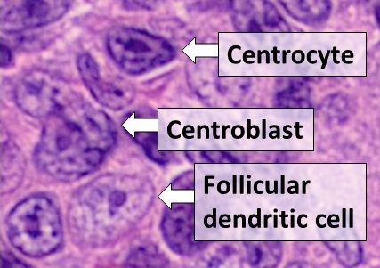
Dendritic cells (DCs) are antigen-presenting cells (also known as ''accessory cells'') of the mammalian immune system. Their main function is to process antigen material and present it on the cell surface to the T cells of the immune system. They act as messengers between the innate and the adaptive immune systems. Dendritic cells are present in those tissues that are in contact with the external environment, such as the skin (where there is a specialized dendritic cell type called the Langerhans cell) and the inner lining of the nose, lungs, stomach and intestines. They can also be found in an immature state in the blood. Once activated, they migrate to the lymph nodes where they interact with T cells and B cells to initiate and shape the adaptive immune response. At certain development stages they grow branched projections, the ''dendrites'' that give the cell its name (δένδρον or déndron being Greek for 'tree'). While similar in appearance, these are structures ... [...More Info...] [...Related Items...] OR: [Wikipedia] [Google] [Baidu] |
Macrophage
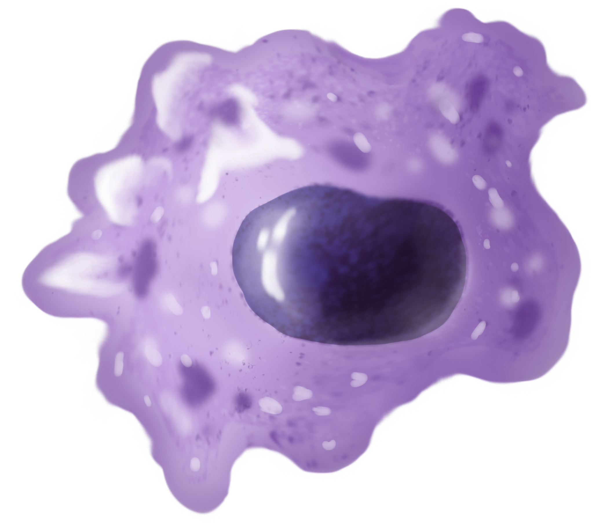
Macrophages (abbreviated as M φ, MΦ or MP) ( el, large eaters, from Greek ''μακρός'' (') = large, ''φαγεῖν'' (') = to eat) are a type of white blood cell of the immune system that engulfs and digests pathogens, such as cancer cells, microbes, cellular debris, and foreign substances, which do not have proteins that are specific to healthy body cells on their surface. The process is called phagocytosis, which acts to defend the host against infection and injury. These large phagocytes are found in essentially all tissues, where they patrol for potential pathogens by amoeboid movement. They take various forms (with various names) throughout the body (e.g., histiocytes, Kupffer cells, alveolar macrophages, microglia, and others), but all are part of the mononuclear phagocyte system. Besides phagocytosis, they play a critical role in nonspecific defense (innate immunity) and also help initiate specific defense mechanisms (adaptive immunity) by recruiting other immune ... [...More Info...] [...Related Items...] OR: [Wikipedia] [Google] [Baidu] |
Monocyte
Monocytes are a type of leukocyte or white blood cell. They are the largest type of leukocyte in blood and can differentiate into macrophages and conventional dendritic cells. As a part of the vertebrate innate immune system monocytes also influence adaptive immune responses and exert tissue repair functions. There are at least three subclasses of monocytes in human blood based on their phenotypic receptors. Structure Monocytes are amoeboid in appearance, and have nongranulated cytoplasm. Thus they are classified as agranulocytes, although they might occasionally display some azurophil granules and/or vacuoles. With a diameter of 15–22 μm, monocytes are the largest cell type in peripheral blood. Monocytes are mononuclear cells and the ellipsoidal nucleus is often lobulated/indented, causing a bean-shaped or kidney-shaped appearance. Monocytes compose 2% to 10% of all leukocytes in the human body. Development Monocytes are produced by the bone marrow from precursors ca ... [...More Info...] [...Related Items...] OR: [Wikipedia] [Google] [Baidu] |
T Helper Cell
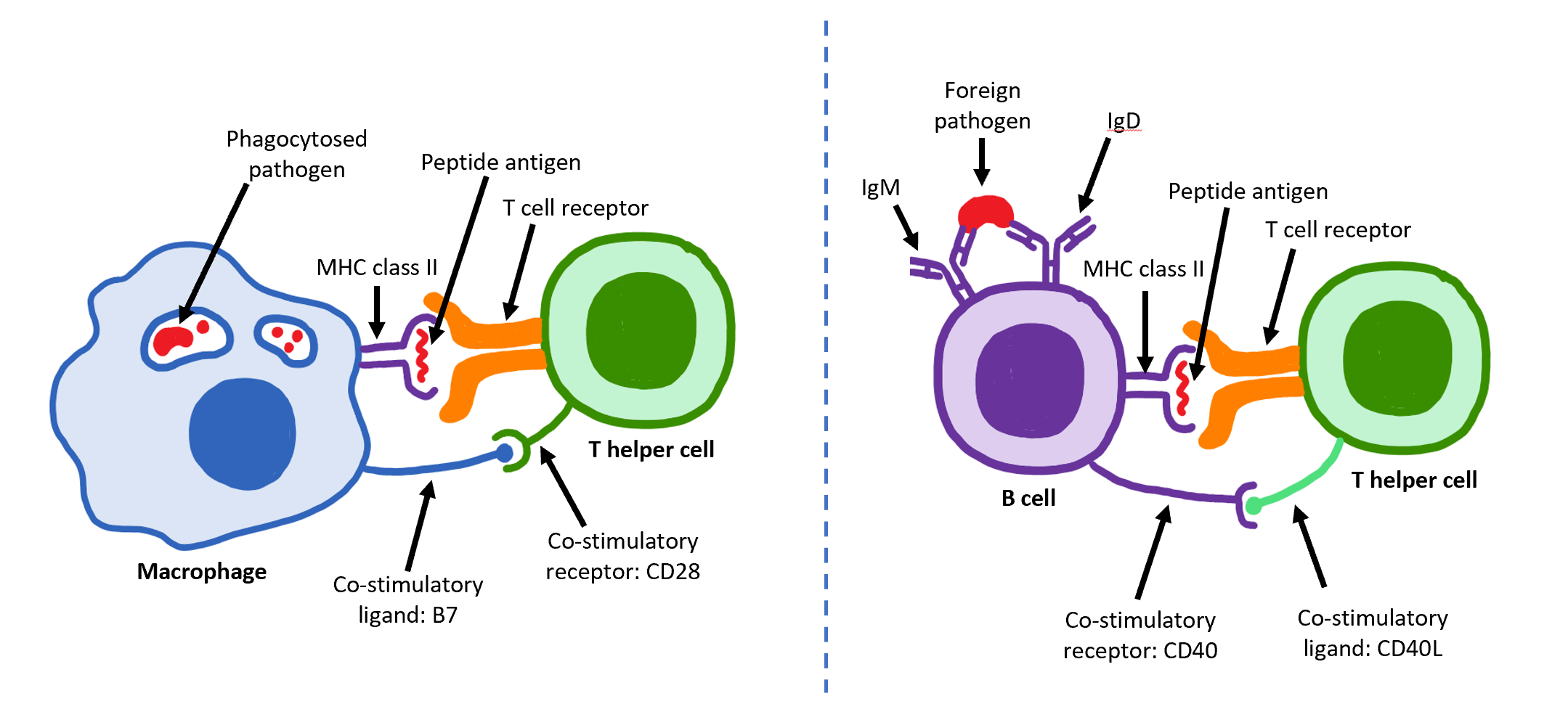
The T helper cells (Th cells), also known as CD4+ cells or CD4-positive cells, are a type of T cell that play an important role in the adaptive immune system. They aid the activity of other immune cells by releasing cytokines. They are considered essential in B cell antibody class switching, breaking cross-tolerance in dendritic cells, in the activation and growth of cytotoxic T cells, and in maximizing bactericidal activity of phagocytes such as macrophages and neutrophils. CD4+ cells are mature Th cells that express the surface protein CD4. Genetic variation in regulatory elements expressed by CD4+ cells determines susceptibility to a broad class of autoimmune diseases. Structure and function Th cells contain and release cytokines to aid other immune cells. Cytokines are small protein mediators that alter the behavior of target cells that express receptors for those cytokines. These cells help polarize the immune response depending on the nature of the immunological insult ... [...More Info...] [...Related Items...] OR: [Wikipedia] [Google] [Baidu] |
MHC Class II
MHC Class II molecules are a class of major histocompatibility complex (MHC) molecules normally found only on professional antigen-presenting cells such as dendritic cells, mononuclear phagocytes, some endothelial cells, thymic epithelial cells, and B cells. These cells are important in initiating immune responses. The antigens presented by class II peptides are derived from extracellular proteins (not cytosolic as in MHC class I). Loading of a MHC class II molecule occurs by phagocytosis; extracellular proteins are endocytosed, digested in lysosomes, and the resulting epitopic peptide fragments are loaded onto MHC class II molecules prior to their migration to the cell surface. In humans, the MHC class II protein complex is encoded by the human leukocyte antigen gene complex (HLA). HLAs corresponding to MHC class II are HLA-DP, HLA-DM, HLA-DOA, HLA-DOB, HLA-DQ, and HLA-DR. Mutations in the HLA gene complex can lead to bare lymphocyte syndrome (BLS), which is a type of MHC ... [...More Info...] [...Related Items...] OR: [Wikipedia] [Google] [Baidu] |
CD3G
T-cell surface glycoprotein CD3 gamma chain is a protein that in humans is encoded by the ''CD3G'' gene. T cell antigen receptor (TCR) is associated on the T cell surface with a complex of protein called CD3. CD3G (gamma chain) is one of the four peptides (gamma, delta, epsilon and zeta) that form CD3. Defects in CD3G are associated with T cell immunodeficiency. See also * CD3 (immunology) * Cluster of differentiation The cluster of differentiation (also known as cluster of designation or classification determinant and often abbreviated as CD) is a protocol used for the identification and investigation of cell surface molecules providing targets for immunophen ... References Further reading * * * * * * * * * * * * * * * * * * * * External links * * Clusters of differentiation {{membrane-protein-stub ... [...More Info...] [...Related Items...] OR: [Wikipedia] [Google] [Baidu] |
CD3e
CD3e molecule, epsilon also known as CD3E is a polypeptide which in humans is encoded by the ''CD3E'' gene which resides on chromosome 11. Function The protein encoded by this gene is the CD3-epsilon polypeptide, which together with CD3- gamma, -delta and -zeta, and the T-cell receptor alpha/beta and gamma/delta heterodimers, forms the T cell receptor- CD3 complex. This complex plays an important role in coupling antigen recognition to several intracellular signal-transduction pathways. The genes encoding the epsilon, gamma and delta polypeptides are located in the same cluster on chromosome 11. The epsilon polypeptide plays an essential role in T-cell development. Clinical significance Defects in this gene cause severe immunodeficiency. This gene has also been linked to a susceptibility to type I diabetes in women. Interactions T-cell surface glycoprotein CD3 epsilon chain has been shown to interact with TOP2B, CD3EAP and NCK2. See also * CD3 (immunology) * Cluster ... [...More Info...] [...Related Items...] OR: [Wikipedia] [Google] [Baidu] |


.png)

