|
Late Positive Component
The late positive component or late positive complex (LPC) is a positive-going event-related brain potential (ERP) component that has been important in studies of explicit recognition memory.Munte, T. F., Urbach, T. P., Duzel, E., & Kutas, M., (2000). Event-related brain potentials in the study of human cognition and neuropsychology, In: F. Boller, J. Grafman, and G. Rizzolatti (Eds.) Handbook of Neuropsychology, Vol. 1, 2nd edition, Elsevier Science Publishers B.V., 97. It is generally found to be largest over parietal scalp sites (relative to reference electrodes placed on the mastoid processes), beginning around 400–500 ms after the onset of a stimulus and lasting for a few hundred milliseconds. It is an important part of the ERP "old/new" effect, which may also include modulations of an earlier component similar to an N400. Similar positivities have sometimes been referred to as the P3b, P300, and P600.Finnigan, S., Humphreys, M.S., Dennis, S., Geffen, G. (2002). ERP 'o ... [...More Info...] [...Related Items...] OR: [Wikipedia] [Google] [Baidu] |
Brain
The brain is an organ that serves as the center of the nervous system in all vertebrate and most invertebrate animals. It consists of nervous tissue and is typically located in the head ( cephalization), usually near organs for special senses such as vision, hearing and olfaction. Being the most specialized organ, it is responsible for receiving information from the sensory nervous system, processing those information (thought, cognition, and intelligence) and the coordination of motor control (muscle activity and endocrine system). While invertebrate brains arise from paired segmental ganglia (each of which is only responsible for the respective body segment) of the ventral nerve cord, vertebrate brains develop axially from the midline dorsal nerve cord as a vesicular enlargement at the rostral end of the neural tube, with centralized control over all body segments. All vertebrate brains can be embryonically divided into three parts: the forebrain (prosencep ... [...More Info...] [...Related Items...] OR: [Wikipedia] [Google] [Baidu] |
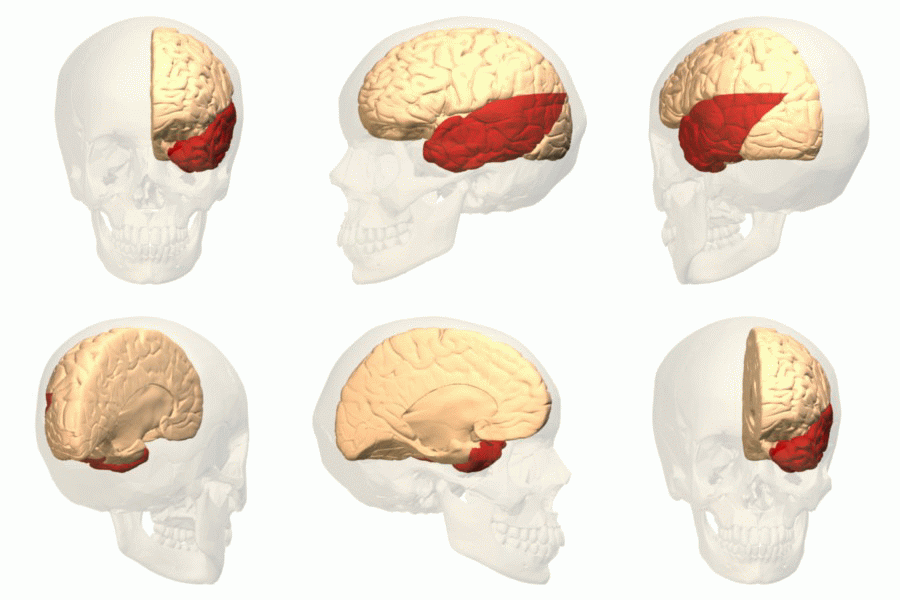
Medial Temporal Lobe
The temporal lobe is one of the four major lobes of the cerebral cortex in the brain of mammals. The temporal lobe is located beneath the lateral fissure on both cerebral hemispheres of the mammalian brain. The temporal lobe is involved in processing sensory input into derived meanings for the appropriate retention of visual memory, language comprehension, and emotion association. ''Temporal'' refers to the head's temples. Structure The temporal lobe consists of structures that are vital for declarative or long-term memory. Declarative (denotative) or explicit memory is conscious memory divided into semantic memory (facts) and episodic memory (events). Medial temporal lobe structures that are critical for long-term memory include the hippocampus, along with the surrounding hippocampal region consisting of the perirhinal, parahippocampal, and entorhinal neocortical regions. The hippocampus is critical for memory formation, and the surrounding medial temporal cortex is ... [...More Info...] [...Related Items...] OR: [Wikipedia] [Google] [Baidu] |
Somatosensory Evoked Potential
Somatosensory evoked potential (SEP or SSEP) is the electrical activity of the brain that results from the stimulation of touch. SEP tests measure that activity and are a useful, noninvasive means of assessing somatosensory system functioning. By combining SEP recordings at different levels of the somatosensory pathways, it is possible to assess the transmission of the afferent volley from the periphery up to the cortex. SEP components include a series of positive and negative deflections that can be elicited by virtually any sensory stimuli. For example, SEPs can be obtained in response to a brief mechanical impact on the fingertip or to air puffs. However, SEPs are most commonly elicited by bipolar transcutaneous electrical stimulation applied on the skin over the trajectory of peripheral nerves of the upper limb (e.g., the median nerve) or lower limb (e.g., the posterior tibial nerve), and then recorded from the scalp. In general, somatosensory stimuli evoke early cortical compone ... [...More Info...] [...Related Items...] OR: [Wikipedia] [Google] [Baidu] |
P200
In neuroscience, the visual P200 or P2 is a waveform component or feature of the event-related potential (ERP) measured at the human scalp. Like other potential changes measurable from the scalp, this effect is believed to reflect the post-synaptic activity of a specific neural process. The P2 component, also known as the P200, is so named because it is a positive going electrical potential that peaks at about 200 milliseconds (varying between about 150 and 275 ms) after the onset of some external stimulus. This component is often distributed around the centro-frontal and the parieto-occipital areas of the scalp. It is generally found to be maximal around the vertex (frontal region) of the scalp, however there have been some topographical differences noted in ERP studies of the P2 in different experimental conditions. Research on the visual P2 is at an early stage compared to other more established ERP components and there is much that we still do not know about it. Part of the di ... [...More Info...] [...Related Items...] OR: [Wikipedia] [Google] [Baidu] |
Lateralized Readiness Potential
In neuroscience, the lateralized readiness potential (LRP) is an event-related brain potential, or increase in electrical activity at the surface of the brain, that is thought to reflect the preparation of motor activity on a certain side of the body; in other words, it is a spike in the electrical activity of the brain that happens when a person gets ready to move one arm, leg, or foot. It is a special form of bereitschaftspotential (a general pre-motor potential). LRPs are recorded using electroencephalography (EEG) and have numerous applications in cognitive neuroscience. History Kornhuber and Deecke's discovery of the Bereitschaftspotential (German for readiness potential) led to research on the now extensively used LRP, which has often been investigated in the context of the mental chronometry paradigm.Coles, M. G. H., 1988. Modern Mind-brain Reading: Psychophysiology, Physiology, and Cognition. 26, 251–269. In the basic chronometric paradigm, the subject experiences a w ... [...More Info...] [...Related Items...] OR: [Wikipedia] [Google] [Baidu] |
Difference Due To Memory
Difference due to memory (Dm) indexes differences in neural activity during the study phase of an experiment for items that subsequently are remembered compared to items that are later forgotten. It is mainly discussed as an event-related potential (ERP) effect that appears in studies employing a subsequent memory paradigm, in which ERPs are recorded when a participant is studying a list of materials and trials are sorted as a function of whether they go on to be remembered or not in the test phase. For meaningful study material, such as words or line drawings, items that are subsequently remembered typically elicit a more positive waveform during the study phase (see Main Paradigms for further information on subsequent memory). This difference typically occurs in the range of 400–800 milliseconds (ms) and is generally greatest over centro-parietal recording sites, although these characteristics are modulated by many factors.Wagner, AD., Koutstaal, W., & Schacter, D.L. (1999)When e ... [...More Info...] [...Related Items...] OR: [Wikipedia] [Google] [Baidu] |
C1 And P1 (neuroscience)
The C1 and P1 (also called the P100) are two human scalp-recorded event-related brain potential ( event-related potential (ERP)) components, collected by means of a technique called electroencephalography (EEG). The C1 is named so because it was the first component in a series of components found to respond to visual stimuli when it was first discovered. It can be a negative-going component (when using a mastoid reference point) or a positive going component with its peak normally observed in the 65–90 ms range post-stimulus onset. The P1 is called the P1 because it is the first positive-going component (when also using a mastoid reference point) and its peak is normally observed in around 100 ms. Both components are related to processing of visual stimuli and are under the category of potentials called visually evoked potentials (VEPs). Both components are theorized to be evoked within the visual cortices of the brain with C1 being linked to the primary visual cort ... [...More Info...] [...Related Items...] OR: [Wikipedia] [Google] [Baidu] |
Bereitschaftspotential
In neurology, the Bereitschaftspotential or BP (German for "readiness potential"), also called the pre-motor potential or readiness potential (RP), is a measure of activity in the motor cortex and supplementary motor area of the brain leading up to voluntary muscle movement. The BP is a manifestation of cortical contribution to the pre-motor planning of volitional movement. It was first recorded and reported in 1964 by Hans Helmut Kornhuber and Lüder Deecke at the University of Freiburg in Germany. In 1965 the full publication appeared after many control experiments.; Englisch translation: PDF(accessed October 21, 2016). Discovery In the spring of 1964 Hans Helmut Kornhuber (then docent and chief physician at the department of neurology, head Professor Richard Jung, university hospital Freiburg im Breisgau) and Lüder Deecke (his doctoral student) went for lunch to the 'Gasthaus zum Schwanen' at the foot of the Schlossberg hill in Freiburg. Sitting alone in the beautiful garden t ... [...More Info...] [...Related Items...] OR: [Wikipedia] [Google] [Baidu] |
FMRI
Functional magnetic resonance imaging or functional MRI (fMRI) measures brain activity by detecting changes associated with blood flow. This technique relies on the fact that cerebral blood flow and neuronal activation are coupled. When an area of the brain is in use, blood flow to that region also increases. The primary form of fMRI uses the blood-oxygen-level dependent (BOLD) contrast, discovered by Seiji Ogawa in 1990. This is a type of specialized brain and body scan used to map neural activity in the brain or spinal cord of humans or other animals by imaging the change in blood flow ( hemodynamic response) related to energy use by brain cells. Since the early 1990s, fMRI has come to dominate brain mapping research because it does not involve the use of injections, surgery, the ingestion of substances, or exposure to ionizing radiation. This measure is frequently corrupted by noise from various sources; hence, statistical procedures are used to extract the underlying signal ... [...More Info...] [...Related Items...] OR: [Wikipedia] [Google] [Baidu] |
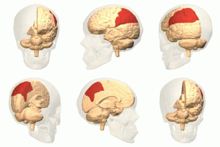
Parietal Cortex
The parietal lobe is one of the four major lobes of the cerebral cortex in the brain of mammals. The parietal lobe is positioned above the temporal lobe and behind the frontal lobe and central sulcus. The parietal lobe integrates sensory information among various modalities, including spatial sense and navigation (proprioception), the main sensory receptive area for the sense of touch in the somatosensory cortex which is just posterior to the central sulcus in the postcentral gyrus, and the dorsal stream of the visual system. The major sensory inputs from the skin (touch, temperature, and pain receptors), relay through the thalamus to the parietal lobe. Several areas of the parietal lobe are important in language processing. The somatosensory cortex can be illustrated as a distorted figure – the cortical homunculus (Latin: "little man") in which the body parts are rendered according to how much of the somatosensory cortex is devoted to them. The superior parietal lobule and i ... [...More Info...] [...Related Items...] OR: [Wikipedia] [Google] [Baidu] |
Amplitude
The amplitude of a periodic variable is a measure of its change in a single period (such as time or spatial period). The amplitude of a non-periodic signal is its magnitude compared with a reference value. There are various definitions of amplitude (see below), which are all functions of the magnitude of the differences between the variable's extreme values. In older texts, the phase of a periodic function is sometimes called the amplitude. Definitions Peak amplitude & semi-amplitude For symmetric periodic waves, like sine wave A sine wave, sinusoidal wave, or just sinusoid is a curve, mathematical curve defined in terms of the ''sine'' trigonometric function, of which it is the graph of a function, graph. It is a type of continuous wave and also a Smoothness, smooth p ...s, square waves or triangle waves ''peak amplitude'' and ''semi amplitude'' are the same. Peak amplitude In audio system measurements, telecommunications and others where the wikt:measurand, measu ... [...More Info...] [...Related Items...] OR: [Wikipedia] [Google] [Baidu] |
Hippocampus
The hippocampus (via Latin from Greek , ' seahorse') is a major component of the brain of humans and other vertebrates. Humans and other mammals have two hippocampi, one in each side of the brain. The hippocampus is part of the limbic system, and plays important roles in the consolidation of information from short-term memory to long-term memory, and in spatial memory that enables navigation. The hippocampus is located in the allocortex, with neural projections into the neocortex in humans, as well as primates. The hippocampus, as the medial pallium, is a structure found in all vertebrates. In humans, it contains two main interlocking parts: the hippocampus proper (also called ''Ammon's horn''), and the dentate gyrus. In Alzheimer's disease (and other forms of dementia), the hippocampus is one of the first regions of the brain to suffer damage; short-term memory loss and disorientation are included among the early symptoms. Damage to the hippocampus can also result ... [...More Info...] [...Related Items...] OR: [Wikipedia] [Google] [Baidu] |


