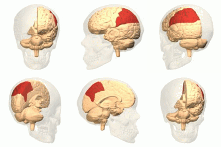
Parietal cortex on:
[Wikipedia]
[Google]
[Amazon]
The parietal lobe is one of the four major lobes of the
 The parietal lobe is defined by three anatomical boundaries: The central sulcus separates the parietal lobe from the frontal lobe; the parieto-occipital sulcus separates the parietal and
cerebral cortex
The cerebral cortex, also known as the cerebral mantle, is the outer layer of neural tissue of the cerebrum of the brain in humans and other mammals. It is the largest site of Neuron, neural integration in the central nervous system, and plays ...
in the brain of mammals. The parietal lobe is positioned above the temporal lobe
The temporal lobe is one of the four major lobes of the cerebral cortex in the brain of mammals. The temporal lobe is located beneath the lateral fissure on both cerebral hemispheres of the mammalian brain.
The temporal lobe is involved in pr ...
and behind the frontal lobe
The frontal lobe is the largest of the four major lobes of the brain in mammals, and is located at the front of each cerebral hemisphere (in front of the parietal lobe and the temporal lobe). It is parted from the parietal lobe by a Sulcus (neur ...
and central sulcus
In neuroanatomy, the central sulcus (also central fissure, fissure of Rolando, or Rolandic fissure, after Luigi Rolando) is a sulcus, or groove, in the cerebral cortex in the brains of vertebrates. It is sometimes confused with the longitudinal ...
.
The parietal lobe integrates sensory information among various modalities, including spatial sense and navigation (proprioception
Proprioception ( ) is the sense of self-movement, force, and body position.
Proprioception is mediated by proprioceptors, a type of sensory receptor, located within muscles, tendons, and joints. Most animals possess multiple subtypes of propri ...
), the main sensory receptive area for the sense of touch in the somatosensory cortex
The somatosensory system, or somatic sensory system is a subset of the sensory nervous system. The main functions of the somatosensory system are the perception of external stimuli, the perception of internal stimuli, and the regulation of bod ...
which is just posterior to the central sulcus in the postcentral gyrus
In neuroanatomy, the postcentral gyrus is a prominent gyrus in the lateral parietal lobe of the human brain. It is the location of the primary somatosensory cortex, the main sensory receptive area for the sense of touch. Like other sensory area ...
, and the dorsal stream of the visual system. The major sensory inputs from the skin (touch
The somatosensory system, or somatic sensory system is a subset of the sensory nervous system. The main functions of the somatosensory system are the perception of external stimuli, the perception of internal stimuli, and the regulation of bo ...
, temperature
Temperature is a physical quantity that quantitatively expresses the attribute of hotness or coldness. Temperature is measurement, measured with a thermometer. It reflects the average kinetic energy of the vibrating and colliding atoms making ...
, and pain
Pain is a distressing feeling often caused by intense or damaging Stimulus (physiology), stimuli. The International Association for the Study of Pain defines pain as "an unpleasant sense, sensory and emotional experience associated with, or res ...
receptors), relay through the thalamus
The thalamus (: thalami; from Greek language, Greek Wikt:θάλαμος, θάλαμος, "chamber") is a large mass of gray matter on the lateral wall of the third ventricle forming the wikt:dorsal, dorsal part of the diencephalon (a division of ...
to the parietal lobe.
Several areas of the parietal lobe are important in language processing. The somatosensory cortex can be illustrated as a distorted figure – the cortical homunculus (Latin: "little man") in which the body parts are rendered according to how much of the somatosensory cortex is devoted to them. The superior parietal lobule and inferior parietal lobule
The inferior parietal lobule (subparietal district) lies below the horizontal portion of the intraparietal sulcus, and behind the lower part of the postcentral sulcus. Also known as Geschwind's territory after Norman Geschwind, an American neu ...
are the primary areas of body or spatial awareness. A lesion commonly in the right superior or inferior parietal lobule leads to hemispatial neglect
Hemispatial neglect is a neuropsychological condition in which, after damage to one hemisphere of the brain (e.g. after a stroke), a deficit in attention and awareness towards the side of space opposite brain damage (contralesional space) is obs ...
.
The name comes from the parietal bone
The parietal bones ( ) are two bones in the skull which, when joined at a fibrous joint known as a cranial suture, form the sides and roof of the neurocranium. In humans, each bone is roughly quadrilateral in form, and has two surfaces, four bord ...
, which is named from the Latin ''paries-,'' meaning "wall".
Structure
 The parietal lobe is defined by three anatomical boundaries: The central sulcus separates the parietal lobe from the frontal lobe; the parieto-occipital sulcus separates the parietal and
 occipital lobe
The occipital lobe is one of the four Lobes of the brain, major lobes of the cerebral cortex in the brain of mammals. The name derives from its position at the back of the head, from the Latin , 'behind', and , 'head'.
The occipital lobe is the ...
s; the lateral sulcus
The lateral sulcus (or lateral fissure, also called Sylvian fissure, after Franciscus Sylvius) is the most prominent sulcus (neuroanatomy), sulcus of each cerebral hemisphere in the human brain. The lateral sulcus (neuroanatomy), sulcus is a deep ...
(sylvian fissure) is the most lateral boundary, separating it from the temporal lobe; and the longitudinal fissure divides the two hemispheres. Within each hemisphere, the somatosensory cortex represents the skin area on the contralateral surface of the body.
Immediately posterior to the central sulcus, and the most anterior part of the parietal lobe, is the postcentral gyrus (Brodmann area
A Brodmann area is a region of the cerebral cortex, in the human or other primate brain, defined by its cytoarchitecture, or histological structure and organization of cells. The concept was first introduced by the German anatomist Korbinian B ...
3), the primary somatosensory cortical area. Separating this from the posterior parietal cortex is the postcentral sulcus.
The posterior parietal cortex can be subdivided into the superior parietal lobule (Brodmann areas 5 + 7) and the inferior parietal lobule
The inferior parietal lobule (subparietal district) lies below the horizontal portion of the intraparietal sulcus, and behind the lower part of the postcentral sulcus. Also known as Geschwind's territory after Norman Geschwind, an American neu ...
( 39 + 40), separated by the intraparietal sulcus (IPS). The intraparietal sulcus and adjacent gyri
In neuroanatomy, a gyrus (: gyri) is a ridge on the cerebral cortex. It is generally surrounded by one or more sulcus (neuroanatomy), sulci (depressions or furrows; : sulcus). Gyri and sulci create the folded appearance of the brain in huma ...
are essential in guidance of limb and eye movement
Eye movement includes the voluntary or involuntary movement of the eyes. Eye movements are used by a number of organisms (e.g. primates, rodents, flies, birds, fish, cats, crabs, octopus) to fixate, inspect and track visual objects of inte ...
, and—based on cytoarchitectural and functional differences—is further divided into medial (MIP), lateral (LIP), ventral (VIP), and anterior (AIP) areas.
Function
Functions of the parietal lobe include: * Two point discrimination – through touch alone without other sensory input (e.g. visual) * Graphesthesia – recognizing writing on skin by touch alone * Touch localization (bilateral simultaneous stimulation) The parietal lobe plays important roles in integrating sensory information from various parts of the body, knowledge of numbers and their relations, and in the manipulation of objects. Its function also includes processing information relating to the sense of touch. Portions of the parietal lobe are involved with visuospatial processing. Although multisensory in nature, the posterior parietal cortex is often referred to by vision scientists as the dorsal stream of vision (as opposed to the ventral stream in the temporal lobe). This dorsal stream has been called both the "where" stream (as in spatial vision) and the "how" stream (as in vision for action). The posterior parietal cortex (PPC) receives somatosensory and visual input, which then, through motor signals, controls movement of the arm, hand, and eyes. Various studies in the 1990s found that different regions of the posterior parietal cortex inmacaque
The macaques () constitute a genus (''Macaca'') of gregarious Old World monkeys of the subfamily Cercopithecinae. The 23 species of macaques inhabit ranges throughout Asia, North Africa, and Europe (in Gibraltar). Macaques are principally f ...
s represent different parts of space.
* The lateral intraparietal (LIP) area contains a map of neurons (retinotopically-coded when the eyes are fixed) representing the saliency of spatial locations, and attention to these spatial locations. It can be used by the oculomotor system for targeting eye movements, when appropriate.
* The ventral intraparietal (VIP) area receives input from a number of senses (visual, somatosensory
The somatosensory system, or somatic sensory system is a subset of the sensory nervous system. The main functions of the somatosensory system are the perception of external stimuli, the perception of internal stimuli, and the regulation of bod ...
, auditory, and vestibular). Neurons with tactile receptive fields represent space in a head-centered reference frame. The cells with visual receptive fields also fire with head-centered reference frames but possibly also with eye-centered coordinates
* The medial intraparietal (MIP) area neurons encode the location of a reach target in eye-centered coordinates.
* The anterior intraparietal (AIP) area contains neurons responsive to shape, size, and orientation of objects to be grasped as well as for manipulation of the hands themselves, both to viewed and remembered stimuli. The AIP has neurons that are responsible for grasping and manipulating objects through motor and visual inputs. The AIP and ventral premotor together are responsible for visuomotor transformations for actions of the hand.
More recent fMRI
Functional magnetic resonance imaging or functional MRI (fMRI) measures brain activity by detecting changes associated with blood flow. This technique relies on the fact that cerebral blood flow and neuronal activation are coupled. When an area o ...
studies have shown that humans have similar functional regions in and around the intraparietal sulcus and parietal-occipital junction. The human "parietal eye fields" and " parietal reach region", equivalent to LIP and MIP in the monkey, also appear to be organized in gaze-centered coordinates so that their goal-related activity is "remapped" when the eyes move.
Emerging evidence has linked processing in the inferior parietal lobe to declarative memory. Bilateral damage to this brain region does not cause amnesia however the strength of memory is diminished, details of complex events become harder to retrieve, and subjective confidence in memory is very low. This has been interpreted as reflecting either deficits in internal attention, deficits in subjective memory states, or problems with the computation that allows evidence to accumulate, thus allowing decisions to be made about internal representations.
Clinical significance
Features of parietal lobe lesions are as follows: * Unilateral parietal lobe ** Contralateral hemisensory loss ** Astereognosis – inability to determine 3-D shape by touch. ** Agraphaesthesia – inability to ''read'' numbers or letters drawn on hand, with eyes shut. ** Contralateral homonymous inferior quadrantanopia ** Asymmetry of optokinetic nystagmus (OKN) ** Sensory seizures * Dominant hemisphere **Conduction aphasia
Conduction aphasia, also called associative aphasia, is an uncommon form of aphasia caused by damage to the parietal lobe of the brain. An acquired language disorder, it is characterized by intact auditory comprehension, coherent (yet paraphasi ...
** Dyslexia
Dyslexia (), previously known as word blindness, is a learning disability that affects either reading or writing. Different people are affected to different degrees. Problems may include difficulties in spelling words, reading quickly, wri ...
– a general term for disorders that can involve difficulty in learning to read or interpret words, letters, and other symbols
** Apraxia
Apraxia is a motor disorder caused by damage to the brain (specifically the posterior parietal cortex or corpus callosum), which causes difficulty with motor planning to perform tasks or movements. The nature of the damage determines the di ...
– inability to perform complex movements in the presence of normal motor, sensory and cerebellar function
** Gerstmann syndrome – characterized by acalculia, agraphia, finger agnosia, and left-right disorientation
* Non-dominant hemisphere
** Contralateral hemispatial neglect
Hemispatial neglect is a neuropsychological condition in which, after damage to one hemisphere of the brain (e.g. after a stroke), a deficit in attention and awareness towards the side of space opposite brain damage (contralesional space) is obs ...
** Constructional apraxia
** Dress apraxia
** Anosognosia – lack of awareness of the existence of one's disability
* Bilateral hemispheres
** Bálint's syndrome
Damage to this lobe in the right hemisphere results in the loss of imagery, visualization of spatial relationships and neglect of left-side space and left side of the body. Even drawings may be neglected on the left side. Damage to this lobe in the left hemisphere will result in problems in mathematics, long reading, writing, and understanding symbols. The parietal association cortex enables individuals to read, write, and solve mathematical problems. The sensory inputs from the right side of the body go to the left side of the brain and vice versa.
The syndrome of hemispatial neglect
Hemispatial neglect is a neuropsychological condition in which, after damage to one hemisphere of the brain (e.g. after a stroke), a deficit in attention and awareness towards the side of space opposite brain damage (contralesional space) is obs ...
is usually associated with large deficits of attention of the non-dominant hemisphere. Optic ataxia is associated with difficulties reaching toward objects in the visual field opposite to the side of the parietal damage. Some aspects of optic ataxia have been explained in terms of the functional organization described above.
Apraxia
Apraxia is a motor disorder caused by damage to the brain (specifically the posterior parietal cortex or corpus callosum), which causes difficulty with motor planning to perform tasks or movements. The nature of the damage determines the di ...
is a disorder of motor control which can be referred neither to "elemental" motor deficits nor to general cognitive impairment. The concept of apraxia was shaped by Hugo Liepmann. Apraxia is predominantly a symptom of left brain damage, but some symptoms of apraxia can also occur after right brain damage.
Amorphosynthesis is a loss of perception on one side of the body caused by a lesion in the parietal lobe. Usually, left-sided lesions cause agnosia, a full-body loss of perception, while right-sided lesions cause lack of recognition of the person's left side and extrapersonal space. The term amorphosynthesis was coined by D. Denny-Brown to describe patients he studied in the 1950s.
Can also result in sensory impairment where one of the affected person's senses (sight, hearing, smell, touch, taste and spatial awareness) is no longer normal.
See also
*Lobes of the brain
The lobes of the brain are the four major identifiable regions of the human cerebral cortex, and they comprise the surface of each hemisphere of the cerebrum. The two hemispheres are roughly symmetrical in structure, and are connected by the c ...
* Temporoparietal junction
References
{{DEFAULTSORT:Parietal Lobe Cerebrum