|
Posterior Parietal Cortex
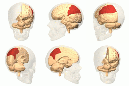
The posterior parietal cortex (the portion of parietal neocortex posterior to the primary somatosensory cortex) plays an important role in planned movements, spatial reasoning, and attention. Damage to the posterior parietal cortex can produce a variety of sensorimotor deficits, including deficits in the perception and memory of spatial relationships, inaccurate reaching and grasping, in the control of eye movement, and inattention. The two most striking consequences of PPC damage are apraxia and hemispatial neglect. Anatomy The posterior parietal cortex receives input from the three sensory systems that play roles in the localization of the body and external objects in space: the visual system, the auditory system, and the somatosensory system. In turn, much of the output of the posterior parietal cortex goes to areas of frontal motor cortex: the dorsolateral prefrontal cortex, various areas of the secondary motor cortex, and the frontal eye field. The posterior parietal corte ... [...More Info...] [...Related Items...] OR: [Wikipedia] [Google] [Baidu] |
Parietal Lobe
The parietal lobe is one of the four major lobes of the cerebral cortex in the brain of mammals. The parietal lobe is positioned above the temporal lobe and behind the frontal lobe and central sulcus. The parietal lobe integrates sensory information among various modalities, including spatial sense and navigation (proprioception), the main sensory receptive area for the sense of touch in the somatosensory cortex which is just posterior to the central sulcus in the postcentral gyrus, and the dorsal stream of the visual system. The major sensory inputs from the skin (touch, temperature, and pain receptors), relay through the thalamus to the parietal lobe. Several areas of the parietal lobe are important in language processing. The somatosensory cortex can be illustrated as a distorted figure – the cortical homunculus (Latin: "little man") in which the body parts are rendered according to how much of the somatosensory cortex is devoted to them. The superior parietal lobule an ... [...More Info...] [...Related Items...] OR: [Wikipedia] [Google] [Baidu] |
Postcentral Gyrus
In neuroanatomy, the postcentral gyrus is a prominent gyrus in the lateral parietal lobe of the human brain. It is the location of the primary somatosensory cortex, the main sensory receptive area for the sense of touch. Like other sensory areas, there is a map of sensory space in this location, called the '' sensory homunculus''. The primary somatosensory cortex was initially defined from surface stimulation studies of Wilder Penfield, and parallel surface potential studies of Bard, Woolsey, and Marshall. Although initially defined to be roughly the same as Brodmann areas 3, 1 and 2, more recent work by Kaas has suggested that for homogeny with other sensory fields only area 3 should be referred to as "primary somatosensory cortex", as it receives the bulk of the thalamocortical projections from the sensory input fields. Structure The lateral postcentral gyrus is bounded by: * medial longitudinal fissure medially (to the middle) *central sulcus rostrally (in front) * post ... [...More Info...] [...Related Items...] OR: [Wikipedia] [Google] [Baidu] |
Attention
Attention is the behavioral and cognitive process of selectively concentrating on a discrete aspect of information, whether considered subjective or objective, while ignoring other perceivable information. William James (1890) wrote that "Attention is the taking possession by the mind, in clear and vivid form, of one out of what seem several simultaneously possible objects or trains of thought. Focalization, concentration, of consciousness are of its essence." Attention has also been described as the allocation of limited cognitive processing resources. Attention is manifested by an attentional bottleneck, in terms of the amount of data the brain can process each second; for example, in human vision, only less than 1% of the visual input data (at around one megabyte per second) can enter the bottleneck, leading to inattentional blindness. Attention remains a crucial area of investigation within education, psychology, neuroscience, cognitive neuroscience, and neuropsycho ... [...More Info...] [...Related Items...] OR: [Wikipedia] [Google] [Baidu] |
Apraxia
Apraxia is a motor disorder caused by damage to the brain (specifically the posterior parietal cortex or corpus callosum), which causes difficulty with motor planning to perform tasks or movements. The nature of the damage determines the disorder's severity, and the absence of sensory loss or paralysis helps to explain the level of difficulty. Children may be born with apraxia; its cause is unknown, and symptoms are usually noticed in the early stages of development. Apraxia occurring later in life, known as ''acquired apraxia'', is typically caused by traumatic brain injury, stroke, dementia, Alzheimer's disease, brain tumor, or other neurodegenerative disorders. The multiple types of apraxia are categorized by the specific ability and/or body part affected. The term "apraxia" comes from the Greek ἀ- ''a-'' ("without") and πρᾶξις ''praxis'' ("action"). Types The several types of apraxia include: * Apraxia of speech (AOS) is having difficulty planning and coordinatin ... [...More Info...] [...Related Items...] OR: [Wikipedia] [Google] [Baidu] |
Hemispatial Neglect
Hemispatial neglect is a neuropsychological condition in which, after damage to one hemisphere of the brain (e.g. after a stroke), a deficit in attention and awareness towards the side of space opposite brain damage (contralesional space) is observed. It is defined by the inability of a person to process and perceive stimuli towards the contralesional side of the body or environment. Hemispatial neglect is very commonly contralateral to the damaged hemisphere, but instances of ipsilesional neglect (on the same side as the lesion) have been reported. Presentation Hemispatial neglect results most commonly from strokes and brain unilateral injury to the right cerebral hemisphere, with rates in the critical stage of up to 80% causing visual neglect of the left-hand side of space. Neglect is often produced by massive strokes in the middle cerebral artery region and is variegated, so that most sufferers do not exhibit all of the syndrome's traits. Right-sided spatial neglect is rare ... [...More Info...] [...Related Items...] OR: [Wikipedia] [Google] [Baidu] |
Dorsolateral Prefrontal Cortex
The dorsolateral prefrontal cortex (DLPFC or DL-PFC) is an area in the prefrontal cortex of the primate brain. It is one of the most recently derived parts of the human brain. It undergoes a prolonged period of maturation which lasts until adulthood. The DLPFC is not an anatomical structure, but rather a functional one. It lies in the middle frontal gyrus of humans (i.e., lateral part of Brodmann's area (BA) 9 and 46). In macaque monkeys, it is around the principal sulcus (i.e., in Brodmann's area 46). Other sources consider that DLPFC is attributed anatomically to BA 9 and 46 and BA 8, 9 and 10. The DLPFC has connections with the orbitofrontal cortex, as well as the thalamus, parts of the basal ganglia (specifically, the dorsal caudate nucleus), the hippocampus, and primary and secondary association areas of neocortex (including posterior temporal, parietal, and occipital areas). The DLPFC is also the end point for the dorsal pathway (stream), which is concerned with how t ... [...More Info...] [...Related Items...] OR: [Wikipedia] [Google] [Baidu] |
Intraparietal Sulcus
The intraparietal sulcus (IPS) is located on the lateral surface of the parietal lobe, and consists of an oblique and a horizontal portion. The IPS contains a series of functionally distinct subregions that have been intensively investigated using both single cell neurophysiology in primates and human functional neuroimaging. Its principal functions are related to perceptual-motor coordination (e.g., directing eye movements and reaching) and visual attention, which allows for visually-guided pointing, grasping, and object manipulation that can produce a desired effect. The IPS is also thought to play a role in other functions, including processing symbolic numerical information, visuospatial working memory and interpreting the intent of others. Function Five regions of the intraparietal sulcus (IPS): anterior, lateral, ventral, caudal, and medial * LIP & VIP: involved in visual attention and saccadic eye movements * VIP & MIP: visual control of reaching and pointing * AIP: visu ... [...More Info...] [...Related Items...] OR: [Wikipedia] [Google] [Baidu] |
Superior Parietal Lobule
The superior parietal lobule is bounded in front by the upper part of the postcentral sulcus, but is usually connected with the postcentral gyrus above the end of the sulcus. The superior parietal lobule contains Brodmann's areas 5 and 7. Behind it is the lateral part of the parietooccipital fissure, around the end of which it is joined to the occipital lobe by a curved gyrus, the arcus parietooccipitalis. Below, it is separated from the inferior parietal lobule by the horizontal portion of the intraparietal sulcus. The superior parietal lobule is involved with spatial orientation, and receives a great deal of visual input as well as sensory input from one's hand. It is also involved with other functions of the parietal lobe in general. There are major white matter pathway connections with the superior parietal lobule such as the Cingulum, SLF I, superior parietal lobule connections of the Medial longitudinal fasciculus and other newly described superior parietal white matte ... [...More Info...] [...Related Items...] OR: [Wikipedia] [Google] [Baidu] |
Inferior Parietal Lobule
The inferior parietal lobule (subparietal district) lies below the horizontal portion of the intraparietal sulcus, and behind the lower part of the postcentral sulcus. Also known as Geschwind's territory after Norman Geschwind, an American neurologist, who in the early 1960s recognised its importance. It is a part of the parietal lobe. Structure It is divided from rostral to caudal into two gyri: * One, the supramarginal gyrus, arches over the upturned end of the lateral fissure; it is continuous in front with the postcentral gyrus, and behind with the superior temporal gyrus. * The second, the angular gyrus, arches over the posterior end of the superior temporal sulcus, behind which it is continuous with the middle temporal gyrus. In macaque neuroanatomy, this region is often divided into caudal and rostral portions, cIPL and rIPL, respectively. The cIPL is further divided into areas Opt and PG whereas rIPL is divided into PFG and PF areas. Function Inferior parietal lobule ... [...More Info...] [...Related Items...] OR: [Wikipedia] [Google] [Baidu] |
Supramarginal Gyrus
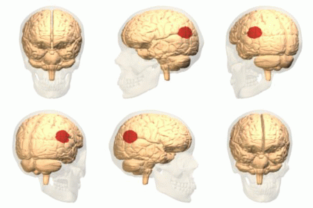
The supramarginal gyrus is a portion of the parietal lobe. This area of the brain is also known as Brodmann area 40 based on the brain map created by Korbinian Brodmann to define the structures in the cerebral cortex. It is probably involved with language perception and processing, and lesions in it may cause receptive aphasia. Important functions The supramarginal gyrus is part of the somatosensory association cortex, which interprets tactile sensory data and is involved in perception of space and limbs location. It is also involved in identifying postures and gestures of other people and is thus a part of the mirror neuron system. The right-hemisphere supramarginal gyrus appears to play a central role in controlling empathy towards other people. When this structure is not working properly or when individuals have to make very quick judgements, empathy becomes severely limited. Research has shown that disrupting the neurons in the right supramarginal gyrus causes humans to p ... [...More Info...] [...Related Items...] OR: [Wikipedia] [Google] [Baidu] |
Temporoparietal Junction
The temporoparietal junction (TPJ) is an area of the brain where the temporal and parietal lobes meet, at the posterior end of the lateral sulcus (Sylvian fissure). The TPJ incorporates information from the thalamus and the limbic system as well as from the visual, auditory, and somatosensory systems. The TPJ also integrates information from both the external environment as well as from within the body. The TPJ is responsible for collecting all of this information and then processing it. This area is also known to play a crucial role in self–other distinctions processes and theory of mind (ToM). Furthermore, damage to the TPJ has been implicated in having adverse effects on an individual's ability to make moral decisions and has been known to produce out-of-body experiences (OBEs). Electromagnetic stimulation of the TPJ can also cause these effects. Apart from these diverse roles that the TPJ plays, it is also known for its involvement in a variety of widespread disorder ... [...More Info...] [...Related Items...] OR: [Wikipedia] [Google] [Baidu] |
Angular Gyrus
The angular gyrus is a region of the brain lying mainly in the posteroinferior region of the parietal lobe, occupying the posterior part of the inferior parietal lobule. It represents the Brodmann area 39. Its significance is in transferring visual information to Wernicke's area, in order to make meaning out of visually perceived words. It is also involved in a number of processes related to language, number processing and spatial cognition, memory retrieval, attention, and theory of mind. Anatomy Connections Left and right angular gyri are connected by the dorsal splenium and isthmus of the corpus callosum. Boundaries * Anteriorly by the Supramarginal gyrus. * Superiorly by the Intraparietal sulcus. * Posteriorly by the Parieto-occipital sulcus. * Inferiorly the angular gyrus of the parietal lobe is continuous as the superior and middle temporal gyri. Also, the angular sulcus, which is capped by the angular gyrus, is continuous as the superior temporal sulcu ... [...More Info...] [...Related Items...] OR: [Wikipedia] [Google] [Baidu] |


