|
Keratin 6B
Keratin 6B is a type II cytokeratin, one of a number of isoforms of keratin 6. It is found with keratin 16 and/or keratin 17 in the hair follicles, the filiform papillae of the tongue and the epithelial lining of oral mucosa and esophagus. This keratin 6 isoform is thought be less abundant than the closely related keratin 6A protein. Mutations in the gene encoding this protein have been associated with pachyonychia congenita, an inherited disorder of the epithelial tissues in which this keratin is expressed, particularly leading to structural abnormalities of the nails, the epidermis of the palms and soles, and oral epithelia. Keratin 6B is associated with the PC-K6B subtype of pachyonychia congenita Pachyonychia congenita (often abbreviated as "PC") is a rare group of autosomal dominant skin disorders that are caused by a mutation in one of five different keratin genes. Pachyonychia congenita is often associated with thickened toenails, plant .... References External links G ... [...More Info...] [...Related Items...] OR: [Wikipedia] [Google] [Baidu] |
Keratin
Keratin () is one of a family of structural fibrous proteins also known as ''scleroproteins''. Alpha-keratin (α-keratin) is a type of keratin found in vertebrates. It is the key structural material making up scales, hair, nails, feathers, horns, claws, hooves, and the outer layer of skin among vertebrates. Keratin also protects epithelial cells from damage or stress. Keratin is extremely insoluble in water and organic solvents. Keratin monomers assemble into bundles to form intermediate filaments, which are tough and form strong unmineralized epidermal appendages found in reptiles, birds, amphibians, and mammals. Excessive keratinization participate in fortification of certain tissues such as in horns of cattle and rhinos, and armadillos' osteoderm. The only other biological matter known to approximate the toughness of keratinized tissue is chitin. Keratin comes in two types, the primitive, softer forms found in all vertebrates and harder, derived forms found only amon ... [...More Info...] [...Related Items...] OR: [Wikipedia] [Google] [Baidu] |
Isoform
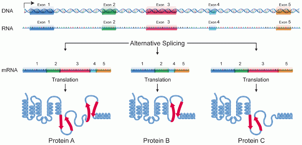
A protein isoform, or "protein variant", is a member of a set of highly similar proteins that originate from a single gene or gene family and are the result of genetic differences. While many perform the same or similar biological roles, some isoforms have unique functions. A set of protein isoforms may be formed from alternative splicings, variable promoter usage, or other post-transcriptional modifications of a single gene; post-translational modifications are generally not considered. (For that, see Proteoforms.) Through RNA splicing mechanisms, mRNA has the ability to select different protein-coding segments ( exons) of a gene, or even different parts of exons from RNA to form different mRNA sequences. Each unique sequence produces a specific form of a protein. The discovery of isoforms could explain the discrepancy between the small number of protein coding regions genes revealed by the human genome project and the large diversity of proteins seen in an organism: different ... [...More Info...] [...Related Items...] OR: [Wikipedia] [Google] [Baidu] |
Keratin 6
Keratin 6A is one of the 27 different type II keratins expressed in humans. Keratin 6A was the first type II keratin sequence determined. Analysis of the sequence of this keratin together with that of the first type I keratin led to the discovery of the four helical domains in the central rod of keratins. In humans Keratin 6A is encoded by the ''KRT6A'' gene. Keratins Keratins are the intermediate filament proteins that form a dense meshwork of filaments throughout the cytoplasm of epithelial cells. Keratins form heteropolymers consisting of a type I and a type II keratin. Keratins are generally expressed in particular pairs of type I and type II keratin proteins in a tissue-specific and cellular differentiation-specific manner. The keratin proteins of epithelial tissues are commonly known as "keratins" or are sometimes referred to as "epithelial keratins" or "cytokeratins". The specialized keratins of hair and nail are known as "hard keratins" or " trichocyte keratins". Tric ... [...More Info...] [...Related Items...] OR: [Wikipedia] [Google] [Baidu] |
Keratin 16
Keratin 16 is a protein that in humans is encoded by the ''KRT16'' gene. Keratin 16 is a type I cytokeratin. It is paired with keratin 6 in a number of epithelial tissues, including nail bed, esophagus, tongue, and hair follicles. Mutations in the gene encoding this protein are associated with the genetic skin disorders including pachyonychia congenita, non-epidermolytic palmoplantar keratoderma and unilateral palmoplantar verrucous nevus A Unilateral palmoplantar verrucous nevus is a cutaneous condition that has features of pachyonychia congenita. See also * Unilateral nevoid telangiectasia * List of cutaneous conditions Many skin conditions affect the human integumenta .... References External links GeneReviews/NCBI/NIH/UW entry on Pachyonychia Congenita Further reading * * * * * * * * * * * * * * * * * * Keratins {{Gene-17-stub ... [...More Info...] [...Related Items...] OR: [Wikipedia] [Google] [Baidu] |
Keratin 17
Keratin, type I cytoskeletal 17 is a protein that in humans is encoded by the ''KRT17'' gene. Keratin 17 is a type I cytokeratin. It is found in nail beds, hair follicles, sebaceous glands, and other epidermal appendages. Mutations in the gene encoding this protein lead to PC-K17 (previously known as Jackson-Lawler) type pachyonychia congenita and steatocystoma multiplex. Interactions Keratin 17 has been shown to interact with CCDC85B Coiled-coil domain-containing protein 85B is a protein that in humans is encoded by the ''CCDC85B'' gene. Function Hepatitis delta virus (HDV) is a pathogenic human virus whose RNA genome and replication cycle resemble those of plant viroids. .... References Further reading * * * * * * * * * * * * * * * * * External links GeneReviews/NCBI/NIH/UW entry on Pachyonychia Congenita Keratins {{gene-17-stub ... [...More Info...] [...Related Items...] OR: [Wikipedia] [Google] [Baidu] |
Hair Follicles
The hair follicle is an organ found in mammalian skin. It resides in the dermal layer of the skin and is made up of 20 different cell types, each with distinct functions. The hair follicle regulates hair growth via a complex interaction between hormones, neuropeptides, and immune cells. This complex interaction induces the hair follicle to produce different types of hair as seen on different parts of the body. For example, terminal hairs grow on the scalp and lanugo hairs are seen covering the bodies of fetuses in the uterus and in some newborn babies. The process of hair growth occurs in distinct sequential stages. The first stage is called ''anagen'' and is the active growth phase, ''telogen'' is the resting stage, ''catagen'' is the regression of the hair follicle phase, ''exogen'' is the active shedding of hair phase and lastly ''kenogen'' is the phase between the empty hair follicle and the growth of new hair. The function of hair in humans has long been a subject of interest ... [...More Info...] [...Related Items...] OR: [Wikipedia] [Google] [Baidu] |
Filiform Papillae
Lingual papillae (singular papilla) are small structures on the upper surface of the tongue that give it its characteristic rough texture. The four types of papillae on the human tongue have different structures and are accordingly classified as circumvallate (or vallate), fungiform, filiform, and foliate. All except the filiform papillae are associated with taste buds. Structure In living subjects, lingual papillae are more readily seen when the tongue is dry. There are four types of papillae present on the tongue: Filiform papillae Filiform papillae are the most numerous of the lingual papillae. They are fine, small, cone-shaped papillae covering most of the dorsum of the tongue. They are responsible for giving the tongue its texture and are responsible for the sensation of touch. Unlike the other kinds of papillae, filiform papillae do not contain taste buds. They cover most of the front two-thirds of the tongue's surface. They appear as very small, conical or cylindrical s ... [...More Info...] [...Related Items...] OR: [Wikipedia] [Google] [Baidu] |
Tongue
The tongue is a muscular organ (anatomy), organ in the mouth of a typical tetrapod. It manipulates food for mastication and swallowing as part of the digestive system, digestive process, and is the primary organ of taste. The tongue's upper surface (dorsum) is covered by taste buds housed in numerous lingual papillae. It is sensitive and kept moist by saliva and is richly supplied with nerves and blood vessels. The tongue also serves as a natural means of oral hygiene, cleaning the teeth. A major function of the tongue is the enabling of speech in humans and animal communication, vocalization in other animals. The human tongue is divided into two parts, an oral cavity, oral part at the front and a pharynx, pharyngeal part at the back. The left and right sides are also separated along most of its length by a vertical section of connective tissue, fibrous tissue (the lingual septum) that results in a groove, the median sulcus, on the tongue's surface. There are two groups of muscle ... [...More Info...] [...Related Items...] OR: [Wikipedia] [Google] [Baidu] |
Epithelial
Epithelium or epithelial tissue is one of the four basic types of animal tissue, along with connective tissue, muscle tissue and nervous tissue. It is a thin, continuous, protective layer of compactly packed cells with a little intercellular matrix. Epithelial tissues line the outer surfaces of organs and blood vessels throughout the body, as well as the inner surfaces of cavities in many internal organs. An example is the epidermis, the outermost layer of the skin. There are three principal shapes of epithelial cell: squamous (scaly), columnar, and cuboidal. These can be arranged in a singular layer of cells as simple epithelium, either squamous, columnar, or cuboidal, or in layers of two or more cells deep as stratified (layered), or ''compound'', either squamous, columnar or cuboidal. In some tissues, a layer of columnar cells may appear to be stratified due to the placement of the nuclei. This sort of tissue is called pseudostratified. All glands are made up of epitheli ... [...More Info...] [...Related Items...] OR: [Wikipedia] [Google] [Baidu] |
Mucosa
A mucous membrane or mucosa is a membrane that lines various cavities in the body of an organism and covers the surface of internal organs. It consists of one or more layers of epithelial cells overlying a layer of loose connective tissue. It is mostly of endodermal origin and is continuous with the skin at body openings such as the eyes, eyelids, ears, inside the nose, inside the mouth, lips, the genital areas, the urethral opening and the anus. Some mucous membranes secrete mucus, a thick protective fluid. The function of the membrane is to stop pathogens and dirt from entering the body and to prevent bodily tissues from becoming dehydrated. Structure The mucosa is composed of one or more layers of epithelial cells that secrete mucus, and an underlying lamina propria of loose connective tissue. The type of cells and type of mucus secreted vary from organ to organ and each can differ along a given tract. Mucous membranes line the digestive, respiratory and reproductive trac ... [...More Info...] [...Related Items...] OR: [Wikipedia] [Google] [Baidu] |
Esophagus
The esophagus (American English) or oesophagus (British English; both ), non-technically known also as the food pipe or gullet, is an organ in vertebrates through which food passes, aided by peristaltic contractions, from the pharynx to the stomach. The esophagus is a fibromuscular tube, about long in adults, that travels behind the trachea and heart, passes through the diaphragm, and empties into the uppermost region of the stomach. During swallowing, the epiglottis tilts backwards to prevent food from going down the larynx and lungs. The word ''oesophagus'' is from Ancient Greek οἰσοφάγος (oisophágos), from οἴσω (oísō), future form of φέρω (phérō, “I carry”) + ἔφαγον (éphagon, “I ate”). The wall of the esophagus from the lumen outwards consists of mucosa, submucosa (connective tissue), layers of muscle fibers between layers of fibrous tissue, and an outer layer of connective tissue. The mucosa is a stratified squamous epithel ... [...More Info...] [...Related Items...] OR: [Wikipedia] [Google] [Baidu] |
Keratin 6A
Keratin 6A is one of the 27 different type II keratins expressed in humans. Keratin 6A was the first type II keratin sequence determined. Analysis of the sequence of this keratin together with that of the first type I keratin led to the discovery of the four helical domains in the central rod of keratins. In humans Keratin 6A is encoded by the ''KRT6A'' gene. Keratins Keratins are the intermediate filament proteins that form a dense meshwork of filaments throughout the cytoplasm of epithelial cells. Keratins form heteropolymers consisting of a type I and a type II keratin. Keratins are generally expressed in particular pairs of type I and type II keratin proteins in a tissue-specific and cellular differentiation-specific manner. The keratin proteins of epithelial tissues are commonly known as "keratins" or are sometimes referred to as "epithelial keratins" or "cytokeratins". The specialized keratins of hair and nail are known as "hard keratins" or " trichocyte keratins". Tric ... [...More Info...] [...Related Items...] OR: [Wikipedia] [Google] [Baidu] |


