|
Kocher Maneuver
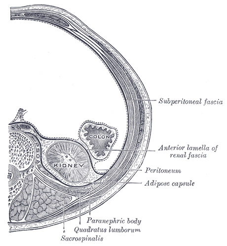
Kocher manoeuvre is a surgical manoeuvre to expose structures in the retroperitoneum behind the duodenum and pancreas. In vascular surgery, it is described as a method to expose the abdominal aorta. It usually has been in contrast with MLRRD (midline Laparotomy and right retroperitoneal space dissection). The Kocher manoeuvre and MLRRD have been used for diverse cases, but they have approximately equivalent outcomes. The Kocher manoeuvre may also refer to a procedure used to reduce anterior shoulder dislocations by externally rotating the shoulder, before adducting and internally rotating it. Uses The Kocher manoeuvre may be used to control bleeding from the inferior vena cava or aorta. It may also be used in pancreaticoduodenectomy, such as to facilitate removal of a pancreatic tumour. Technique The peritoneum is incised at the right edge of the duodenum, and the duodenum and the head of pancreas are reflected to the opposite direction; that is, to the left. History The K ... [...More Info...] [...Related Items...] OR: [Wikipedia] [Google] [Baidu] |
Surgery
Surgery ''cheirourgikē'' (composed of χείρ, "hand", and ἔργον, "work"), via la, chirurgiae, meaning "hand work". is a medical specialty that uses operative manual and instrumental techniques on a person to investigate or treat a pathological condition such as a disease or injury, to help improve bodily function, appearance, or to repair unwanted ruptured areas. The act of performing surgery may be called a surgical procedure, operation, or simply "surgery". In this context, the verb "operate" means to perform surgery. The adjective surgical means pertaining to surgery; e.g. surgical instruments or surgical nurse. The person or subject on which the surgery is performed can be a person or an animal. A surgeon is a person who practices surgery and a surgeon's assistant is a person who practices surgical assistance. A surgical team is made up of the surgeon, the surgeon's assistant, an anaesthetist, a circulating nurse and a surgical technologist. Surgery usually spa ... [...More Info...] [...Related Items...] OR: [Wikipedia] [Google] [Baidu] |
Retroperitoneum
The retroperitoneal space (retroperitoneum) is the anatomical space (sometimes a potential space) behind (''retro'') the peritoneum. It has no specific delineating anatomical structures. Organs are retroperitoneal if they have peritoneum on their anterior side only. Structures that are not suspended by mesentery in the abdominal cavity and that lie between the parietal peritoneum and abdominal wall are classified as retroperitoneal. This is different from organs that are not retroperitoneal, which have peritoneum on their posterior side and are suspended by mesentery in the abdominal cavity. The retroperitoneum can be further subdivided into the following: *Perirenal (or perinephric) space *Anterior pararenal (or paranephric) space *Posterior pararenal (or paranephric) space Retroperitoneal structures Structures that lie behind the peritoneum are termed "retroperitoneal". Organs that were once suspended within the abdominal cavity by mesentery but migrated posterior to the p ... [...More Info...] [...Related Items...] OR: [Wikipedia] [Google] [Baidu] |
Duodenum
The duodenum is the first section of the small intestine in most higher vertebrates, including mammals, reptiles, and birds. In fish, the divisions of the small intestine are not as clear, and the terms anterior intestine or proximal intestine may be used instead of duodenum. In mammals the duodenum may be the principal site for iron absorption. The duodenum precedes the jejunum and ileum and is the shortest part of the small intestine. In humans, the duodenum is a hollow jointed tube about 25–38 cm (10–15 inches) long connecting the stomach to the middle part of the small intestine. It begins with the duodenal bulb and ends at the suspensory muscle of duodenum. Duodenum can be divided into four parts: the first (superior), the second (descending), the third (horizontal) and the fourth (ascending) parts. Structure The duodenum is a C-shaped structure lying adjacent to the stomach. It is divided anatomically into four sections. The first part of the duodenum lies ... [...More Info...] [...Related Items...] OR: [Wikipedia] [Google] [Baidu] |
Pancreas
The pancreas is an organ of the digestive system and endocrine system of vertebrates. In humans, it is located in the abdomen behind the stomach and functions as a gland. The pancreas is a mixed or heterocrine gland, i.e. it has both an endocrine and a digestive exocrine function. 99% of the pancreas is exocrine and 1% is endocrine. As an endocrine gland, it functions mostly to regulate blood sugar levels, secreting the hormones insulin, glucagon, somatostatin, and pancreatic polypeptide. As a part of the digestive system, it functions as an exocrine gland secreting pancreatic juice into the duodenum through the pancreatic duct. This juice contains bicarbonate, which neutralizes acid entering the duodenum from the stomach; and digestive enzymes, which break down carbohydrates, proteins, and fats in food entering the duodenum from the stomach. Inflammation of the pancreas is known as pancreatitis, with common causes including chronic alcohol use and gallstones. Becaus ... [...More Info...] [...Related Items...] OR: [Wikipedia] [Google] [Baidu] |
Vascular Surgery
Vascular surgery is a surgical subspecialty in which diseases of the vascular system, or arteries, veins and lymphatic circulation, are managed by medical therapy, minimally-invasive catheter procedures and surgical reconstruction. The specialty evolved from general and cardiac surgery and includes treatment of the body's other major and essential veins and arteries. Open surgery techniques, as well as endovascular techniques are used to treat vascular diseases. The vascular surgeon is trained in the diagnosis and management of diseases affecting all parts of the vascular system excluding the coronaries and intracranial vasculature. Vascular surgeons often assist other physicians to address traumatic vascular injury, hemorrhage control, and safe exposure of vascular structures. History Early leaders of the field included Russian surgeon Nikolai Korotkov, noted for developing early surgical techniques, American interventional radiologist Charles Theodore Dotter who is credited wit ... [...More Info...] [...Related Items...] OR: [Wikipedia] [Google] [Baidu] |
Abdominal Aorta
In human anatomy, the abdominal aorta is the largest artery in the abdominal cavity. As part of the aorta, it is a direct continuation of the descending aorta (of the thorax). Structure The abdominal aorta begins at the level of the thoracic diaphragm, diaphragm, crossing it via the aortic hiatus, technically behind the diaphragm, at the vertebral level of T12. It travels down the posterior wall of the abdomen, anterior to the vertebral column. It thus follows the curvature of the lumbar vertebrae, that is, convex anteriorly. The peak of this convexity is at the level of the third lumbar vertebra (L3). It runs parallel to the inferior vena cava, which is located just to the right of the abdominal aorta, and becomes smaller in diameter as it gives off branches. This is thought to be due to the large size of its principal branches. At the 11th rib, the diameter is 122mm long and 55mm wide and this is because of the constant pressure. The abdominal aorta is clinically divided int ... [...More Info...] [...Related Items...] OR: [Wikipedia] [Google] [Baidu] |
Retroperitoneal Space
The retroperitoneal space (retroperitoneum) is the anatomical space (sometimes a potential space) behind (''retro'') the peritoneum. It has no specific delineating anatomical structures. Organs are retroperitoneal if they have peritoneum on their anterior side only. Structures that are not suspended by mesentery in the abdominal cavity and that lie between the parietal peritoneum and abdominal wall are classified as retroperitoneal. This is different from organs that are not retroperitoneal, which have peritoneum on their posterior side and are suspended by mesentery in the abdominal cavity. The retroperitoneum can be further subdivided into the following: *Perirenal (or perinephric) space *Anterior pararenal (or paranephric) space *Posterior pararenal (or paranephric) space Retroperitoneal structures Structures that lie behind the peritoneum are termed "retroperitoneal". Organs that were once suspended within the abdominal cavity by mesentery but migrated posterior to the p ... [...More Info...] [...Related Items...] OR: [Wikipedia] [Google] [Baidu] |
Bleeding
Bleeding, hemorrhage, haemorrhage or blood loss, is blood escaping from the circulatory system from damaged blood vessels. Bleeding can occur internally, or externally either through a natural opening such as the mouth, nose, ear, urethra, vagina or anus, or through a puncture in the skin. Hypovolemia is a massive decrease in blood volume, and death by excessive loss of blood is referred to as exsanguination. Typically, a healthy person can endure a loss of 10–15% of the total blood volume without serious medical difficulties (by comparison, blood donation typically takes 8–10% of the donor's blood volume). The stopping or controlling of bleeding is called hemostasis and is an important part of both first aid and surgery. Types * Upper head ** Intracranial hemorrhage – bleeding in the skull. ** Cerebral hemorrhage – a type of intracranial hemorrhage, bleeding within the brain tissue itself. ** Intracerebral hemorrhage – bleeding in the brain caused by the ruptur ... [...More Info...] [...Related Items...] OR: [Wikipedia] [Google] [Baidu] |
Venae Cavae
In anatomy, the venae cavae (; singular: vena cava ; ) are two large veins (great vessels) that return deoxygenated blood from the body into the heart. In humans they are the superior vena cava and the inferior vena cava, and both empty into the right atrium. They are located slightly off-center, toward the right side of the body. The right atrium receives deoxygenated blood through coronary sinus and two large veins called venae cavae. The inferior vena cava (or caudal vena cava in some animals) travels up alongside the abdominal aorta with blood from the lower part of the body. It is the largest vein in the human body. MadSci Network: Anatomy. Retrieved 19 September 2013. The superior vena cava (or cranial vena cava in animals) is above the heart, and ... [...More Info...] [...Related Items...] OR: [Wikipedia] [Google] [Baidu] |
Aorta
The aorta ( ) is the main and largest artery in the human body, originating from the left ventricle of the heart and extending down to the abdomen, where it splits into two smaller arteries (the common iliac arteries). The aorta distributes oxygenated blood to all parts of the body through the systemic circulation. Structure Sections In anatomical sources, the aorta is usually divided into sections. One way of classifying a part of the aorta is by anatomical compartment, where the thoracic aorta (or thoracic portion of the aorta) runs from the heart to the diaphragm. The aorta then continues downward as the abdominal aorta (or abdominal portion of the aorta) from the diaphragm to the aortic bifurcation. Another system divides the aorta with respect to its course and the direction of blood flow. In this system, the aorta starts as the ascending aorta, travels superiorly from the heart, and then makes a hairpin turn known as the aortic arch. Following the aortic arch ... [...More Info...] [...Related Items...] OR: [Wikipedia] [Google] [Baidu] |
Pancreaticoduodenectomy
A pancreaticoduodenectomy, also known as a Whipple procedure, is a major surgical operation most often performed to remove cancerous tumours from the head of the pancreas. It is also used for the treatment of pancreatic or duodenal trauma, or chronic pancreatitis. Due to the shared blood supply of organs in the proximal gastrointestinal system, surgical removal of the head of the pancreas also necessitates removal of the duodenum, proximal jejunum, gallbladder, and, occasionally, part of the stomach. Anatomy involved in the procedure The most common technique of a pancreaticoduodenectomy consists of the en bloc removal of the distal segment (antrum) of the stomach, the first and second portions of the duodenum, the head of the pancreas, the common bile duct, and the gallbladder. Lymph nodes in the area are often removed during the operation as well (lymphadenectomy). However, not all lymph nodes are removed in the most common type of pancreaticoduodenectomy because studies sho ... [...More Info...] [...Related Items...] OR: [Wikipedia] [Google] [Baidu] |
Pancreatic Tumor
Pancreatic tumors (Pancreatic cancer Pancreatic cancer arises when cell (biology), cells in the pancreas, a glandular organ behind the stomach, begin to multiply out of control and form a Neoplasm, mass. These cancerous cells have the malignant, ability to invade other parts of t ...) are often broken down into exocrine or endocrine tumors. Each different type of pancreatic tumor has a different appearance when examined under a microscope. These unique microscopic features and genetic markers are what allow for proper diagnosis and treatments in patients with pancreatic cancers. Types When talking about pancreatic cancers, the most common type is pancreatic adenocarcinoma, which accounts for greater than 85% of all pancreas cancers. Adenocarcinomas are exocrine tumors of the pancreas, which implies that they begin within the part of the pancreas responsible for creating digestive enzymes. Different subtypes of exocrine adenocarcinomas exist, with the most common being pancrea ... [...More Info...] [...Related Items...] OR: [Wikipedia] [Google] [Baidu] |
.jpg)


.gif)


