|
KNDy Neuron
Kisspeptin, neurokinin B, and dynorphin (KNDy) neurons are neurons in the hypothalamus of the brain that are central to the hormonal control of reproduction. KNDy neurons in the hypothalamus coexpress kisspeptin, neurokinin B (NKB) and dynorphin. They are involved in the negative feedback of gonadotropin-releasing hormone (GnRH) release in the hypothalamic–pituitary–gonadal (HPG) axis. Sex steroids released from the gonads act on KNDy neurons as inhibitors of kisspeptin release. This inhibition provides negative feedback control on the HPG axis. KNDy peptide colocalization was first discovered in 2007 in sheep and was later confirmed to be present in mice, rats, cows and nonhuman primates. KNDy neurons are thought to be located in the hypothalamus region of human brains due to conservation across most mammalian species. Other roles of KNDy neurons include influences on prolactin production; puberty; stress' effects on reproduction; and the control of thermoregulation. ... [...More Info...] [...Related Items...] OR: [Wikipedia] [Google] [Baidu] |
Reproductive System
The reproductive system of an organism, also known as the genital system, is the biological system made up of all the anatomical organs involved in sexual reproduction. Many non-living substances such as fluids, hormones, and pheromones are also important accessories to the reproductive system. Unlike most organ systems, the sexes of differentiated species often have significant differences. These differences allow for a combination of genetic material between two individuals, which allows for the possibility of greater genetic fitness of the offspring. Reproductive System 2001 Body Guide powered by Adam Animals In mammals, the major organs of the reproductive system include the external |
Thermoregulation
Thermoregulation is the ability of an organism to keep its body temperature within certain boundaries, even when the surrounding temperature is very different. A thermoconforming organism, by contrast, simply adopts the surrounding temperature as its own body temperature, thus avoiding the need for internal thermoregulation. The internal thermoregulation process is one aspect of homeostasis: a state of dynamic stability in an organism's internal conditions, maintained far from thermal equilibrium with its environment (the study of such processes in zoology has been called physiological ecology). If the body is unable to maintain a normal temperature and it increases significantly above normal, a condition known as hyperthermia occurs. Humans may also experience lethal hyperthermia when the wet bulb temperature is sustained above for six hours. The opposite condition, when body temperature decreases below normal levels, is known as hypothermia. It results when the homeostatic c ... [...More Info...] [...Related Items...] OR: [Wikipedia] [Google] [Baidu] |
Immunohistochemistry
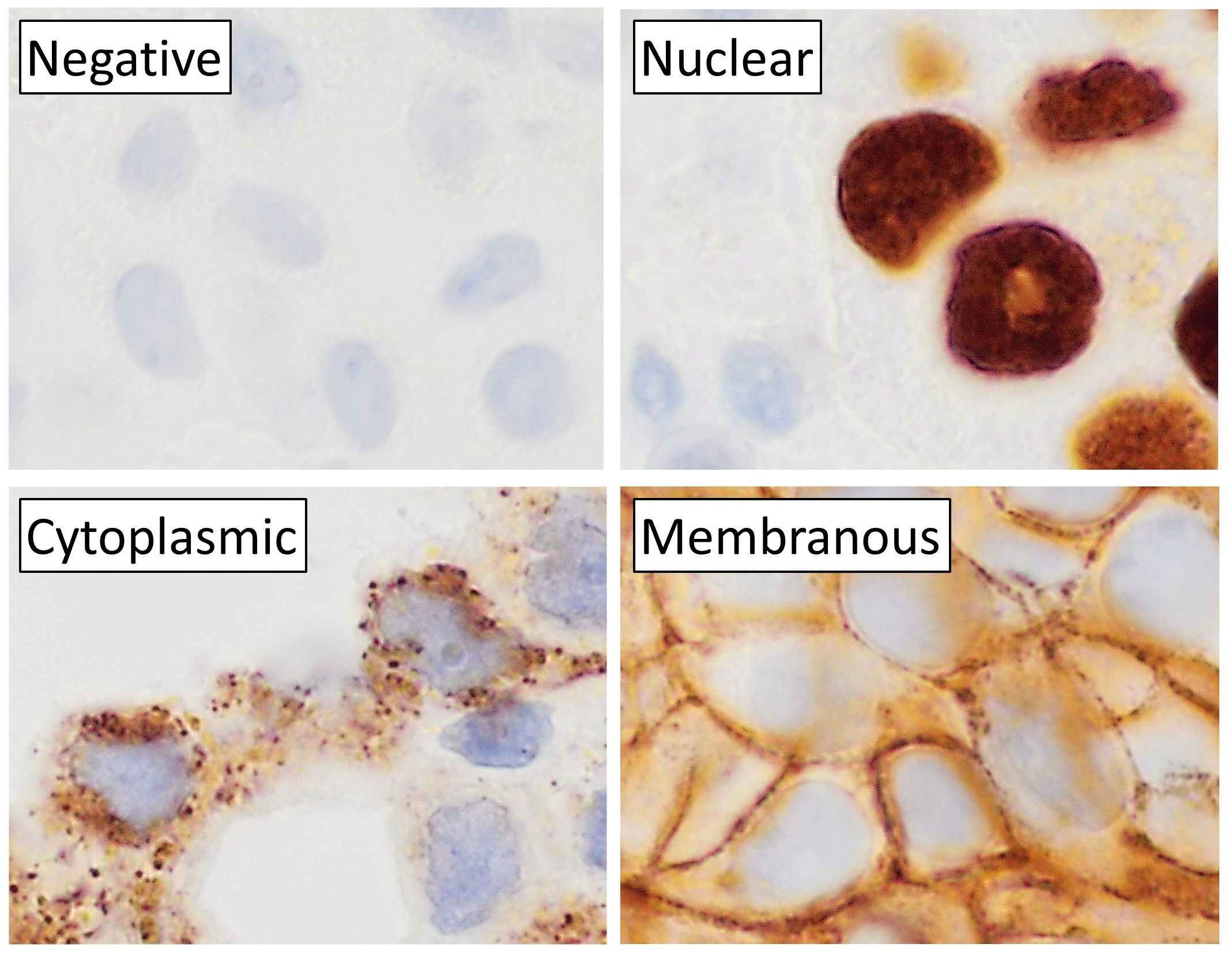
Immunohistochemistry (IHC) is the most common application of immunostaining. It involves the process of selectively identifying antigens (proteins) in cells of a tissue section by exploiting the principle of antibodies binding specifically to antigens in biological tissues. IHC takes its name from the roots "immuno", in reference to antibodies used in the procedure, and "histo", meaning tissue (compare to immunocytochemistry). Albert Coons conceptualized and first implemented the procedure in 1941. Visualising an antibody-antigen interaction can be accomplished in a number of ways, mainly either of the following: * ''Chromogenic immunohistochemistry'' (CIH), wherein an antibody is conjugated to an enzyme, such as peroxidase (the combination being termed immunoperoxidase), that can catalyse a colour-producing reaction. * '' Immunofluorescence'', where the antibody is tagged to a fluorophore, such as fluorescein or rhodamine. Immunohistochemical staining is widely used in the dia ... [...More Info...] [...Related Items...] OR: [Wikipedia] [Google] [Baidu] |
Steroid Hormone
A steroid hormone is a steroid that acts as a hormone. Steroid hormones can be grouped into two classes: corticosteroids (typically made in the adrenal cortex, hence ''cortico-'') and sex steroids (typically made in the gonads or placenta). Within those two classes are five types according to the receptors to which they bind: glucocorticoids and mineralocorticoids (both corticosteroids) and androgens, estrogens, and progestogens (sex steroids). Vitamin D derivatives are a sixth closely related hormone system with homologous receptors. They have some of the characteristics of true steroids as receptor ligands. Steroid hormones help control metabolism, inflammation, immune functions, salt and water balance, development of sexual characteristics, and the ability to withstand injury and illness. The term steroid describes both hormones produced by the body and artificially produced medications that duplicate the action for the naturally occurring steroids. Synthesis The nat ... [...More Info...] [...Related Items...] OR: [Wikipedia] [Google] [Baidu] |
Species
In biology, a species is the basic unit of classification and a taxonomic rank of an organism, as well as a unit of biodiversity. A species is often defined as the largest group of organisms in which any two individuals of the appropriate sexes or mating types can produce fertile offspring, typically by sexual reproduction. Other ways of defining species include their karyotype, DNA sequence, morphology, behaviour or ecological niche. In addition, paleontologists use the concept of the chronospecies since fossil reproduction cannot be examined. The most recent rigorous estimate for the total number of species of eukaryotes is between 8 and 8.7 million. However, only about 14% of these had been described by 2011. All species (except viruses) are given a two-part name, a "binomial". The first part of a binomial is the genus to which the species belongs. The second part is called the specific name or the specific epithet (in botanical nomenclature, also sometimes i ... [...More Info...] [...Related Items...] OR: [Wikipedia] [Google] [Baidu] |
Preoptic Area
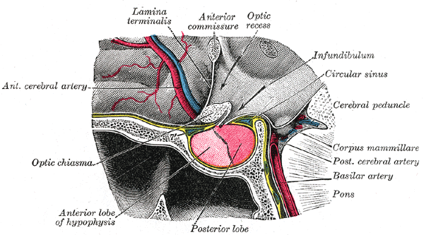
The preoptic area is a region of the hypothalamus. MeSH classifies it as part of the anterior hypothalamus. TA lists four nuclei in this region, (medial, median, lateral, and periventricular). Functions The preoptic area is responsible for thermoregulation and receives nervous stimulation from thermoreceptors in the skin, mucous membranes, and hypothalamus itself. Nuclei Median preoptic nucleus The median preoptic nucleus is located along the midline in a position significantly dorsal to the other three preoptic nuclei, at least in the crab-eating macaque brain. It wraps around the top (dorsal), front, and bottom (ventral) surfaces of the anterior commissure. The median preoptic nucleus generates thirst. Drinking decreases noradrenaline release in the median preoptic nucleus. Medial preoptic nucleus The medial preoptic nucleus is bounded laterally by the lateral preoptic nucleus, and medially by the preoptic periventricular nucleus. It releases gonadotropin-releasing hormon ... [...More Info...] [...Related Items...] OR: [Wikipedia] [Google] [Baidu] |
Third Ventricle
The third ventricle is one of the four connected ventricles of the ventricular system within the mammalian brain. It is a slit-like cavity formed in the diencephalon between the two thalami, in the midline between the right and left lateral ventricles, and is filled with cerebrospinal fluid (CSF). Running through the third ventricle is the interthalamic adhesion, which contains thalamic neurons and fibers that may connect the two thalami. Structure The third ventricle is a narrow, laterally flattened, vaguely rectangular region, filled with cerebrospinal fluid, and lined by ependyma. It is connected at the superior anterior corner to the lateral ventricles, by the interventricular foramina, and becomes the cerebral aqueduct (''aqueduct of Sylvius'') at the posterior caudal corner. Since the interventricular foramina are on the lateral edge, the corner of the third ventricle itself forms a bulb, known as the ''anterior recess'' (it is also known as the ''bulb of the ventricl ... [...More Info...] [...Related Items...] OR: [Wikipedia] [Google] [Baidu] |
Arcuate Nucleus
The arcuate nucleus of the hypothalamus (also known as ARH, ARC, or infundibular nucleus) is an aggregation of neurons in the mediobasal hypothalamus, adjacent to the third ventricle and the median eminence. The arcuate nucleus includes several important and diverse populations of neurons that help mediate different neuroendocrine and physiological functions, including neuroendocrine neurons, centrally projecting neurons, and astrocytes. The populations of neurons found in the arcuate nucleus are based on the hormones they secrete or interact with and are responsible for hypothalamic function, such as regulating hormones released from the pituitary gland or secreting their own hormones. Neurons in this region are also responsible for integrating information and providing inputs to other nuclei in the hypothalamus or inputs to areas outside this region of the brain. These neurons, generated from the ventral part of the periventricular epithelium during embryonic development, locat ... [...More Info...] [...Related Items...] OR: [Wikipedia] [Google] [Baidu] |
κ-opioid Receptor
The κ-opioid receptor or kappa opioid receptor, abbreviated KOR or KOP, is a G protein-coupled receptor that in humans is encoded by the ''OPRK1'' gene. The KOR is coupled to the G protein Gi/G0 and is one of four related receptors that bind opioid-like compounds in the brain and are responsible for mediating the effects of these compounds. These effects include altering nociception, consciousness, motor control, and mood. Dysregulation of this receptor system has been implicated in alcohol and drug addiction. The KOR is a type of opioid receptor that binds the opioid peptide dynorphin as the primary endogenous ligand (substrate naturally occurring in the body). In addition to dynorphin, a variety of natural alkaloids, terpenes and synthetic ligands bind to the receptor. The KOR may provide a natural addiction control mechanism, and therefore, drugs that target this receptor may have therapeutic potential in the treatment of addiction. There is evidence that distribution ... [...More Info...] [...Related Items...] OR: [Wikipedia] [Google] [Baidu] |
KiSS1-derived Peptide Receptor
The KiSS1-derived peptide receptor (also known as GPR54 or the Kisspeptin receptor) is a G protein-coupled receptor which binds the peptide hormone kisspeptin (metastin). Kisspeptin is encoded by the metastasis suppressor gene KISS1, which is expressed in a variety of endocrine and gonadal tissues. Activation of the kisspeptin receptor is linked to the phospholipase C and inositol trisphosphate second messenger cascades inside the cell. Function Kisspeptin is involved in the regulation of endocrine function and the onset of puberty, with activation of the kisspeptin receptor triggering release of gonadotropin-releasing hormone (GnRH), and release of kisspeptin itself being inhibited by oestradiol but enhanced by GnRH. Reductions in kisspeptin levels with age may conversely be one of the reasons behind age-related declines in levels of other endocrine hormones such as luteinizing hormone. Ligands No non-peptide ligands for this receptor have yet been discovered, but as of 2009 b ... [...More Info...] [...Related Items...] OR: [Wikipedia] [Google] [Baidu] |
Autocrine Signaling
Autocrine signaling is a form of cell signaling in which a cell secretes a hormone or chemical messenger (called the autocrine agent) that binds to autocrine receptors on that same cell, leading to changes in the cell. This can be contrasted with paracrine signaling, intracrine signaling, or classical endocrine signaling. Examples An example of an autocrine agent is the cytokine interleukin-1 in monocytes. When interleukin-1 is produced in response to external stimuli, it can bind to cell-surface receptors on the same cell that produced it. Another example occurs in activated T cell lymphocytes, i.e., when a T cell is induced to mature by binding to a peptide: MHC complex on a professional antigen-presenting cell and by the B7: CD28 costimulatory signal. Upon activation, "low-affinity" IL-2 receptors are replaced by "high-affinity" IL-2 receptors consisting of α, β, and γ chains. The cell then releases IL-2, which binds to its own new IL-2 receptors, causing self-stimulation ... [...More Info...] [...Related Items...] OR: [Wikipedia] [Google] [Baidu] |


