|
KIAA1211L
KIAA1211L is a protein that in humans is encoded by the KIAA1211L gene. It is highly expressed in the brain (Cerebral cortex, Cerebral Cortex). Furthermore, it is localized to the microtubules and the centrosomes and is subcellularly located in the Cell nucleus, nucleus. Finally, KIAA1211L is associated with certain mental disorders and various cancers. Gene KIAA1211L is a protein-coding gene. The table above presents the gene's alias, location, size and accession number. mRNA There are 11 splice isoforms of the gene KIAA1211L. The validated isoform has 10 exons. Protein The table above presents the protein's alias, size, and accession number. The KIAA1211L protein is proline rich and asparagine, isoleucine, phenylalanine, and tyrosine poor. Domains and motifs The KIAA1211L protein has one domain called the DUF4592 motif and spans amino acids 131–239. This domain is highly conserved among the KIAA1211L Homology (biology), orthologs. The DUF4592 motif is depicted ... [...More Info...] [...Related Items...] OR: [Wikipedia] [Google] [Baidu] |
Microtubule
Microtubules are polymers of tubulin that form part of the cytoskeleton and provide structure and shape to eukaryotic cells. Microtubules can be as long as 50 micrometres, as wide as 23 to 27 nm and have an inner diameter between 11 and 15 nm. They are formed by the polymerization of a dimer of two globular proteins, alpha and beta tubulin into protofilaments that can then associate laterally to form a hollow tube, the microtubule. The most common form of a microtubule consists of 13 protofilaments in the tubular arrangement. Microtubules play an important role in a number of cellular processes. They are involved in maintaining the structure of the cell and, together with microfilaments and intermediate filaments, they form the cytoskeleton. They also make up the internal structure of cilia and flagella. They provide platforms for intracellular transport and are involved in a variety of cellular processes, including the movement of secretory vesicles, organell ... [...More Info...] [...Related Items...] OR: [Wikipedia] [Google] [Baidu] |
GSK3B
Glycogen synthase kinase-3 beta, (GSK-3 beta), is an enzyme that in humans is encoded by the ''GSK3B'' gene. In mice, the enzyme is encoded by the Gsk3b gene. Abnormal regulation and expression of GSK-3 beta is associated with an increased susceptibility towards bipolar disorder. Function Glycogen synthase kinase-3 (GSK-3) is a proline-directed serine-threonine kinase that was initially identified as a phosphorylating and an inactivating agent of glycogen synthase. Two isoforms, alpha (GSK3A) and beta, show a high degree of amino acid homology. GSK3B is involved in energy metabolism, neuronal cell development, and body pattern formation. It might be a new therapeutic target for ischemic stroke. Disease relevance Homozygous disruption of the Gsk3b locus in mice results in embryonic lethality during mid-gestation. This lethality phenotype could be rescued by inhibition of tumor necrosis factor. Two SNPs at this gene, rs334558 (-50T/C) and rs3755557 (-1727A/T), are associat ... [...More Info...] [...Related Items...] OR: [Wikipedia] [Google] [Baidu] |
MiR-132
In molecular biology miR-132 microRNA is a short non-coding RNA molecule. MicroRNAs function to regulate the expression levels of other genes by several mechanisms, generally reducing protein levels through the cleavage of mRNAs or the repression of their translation. Several targets for miR-132 have been described, including mediators of neurological development, synaptic transmission, inflammation and angiogenesis. Expression miR-132 arises from the miR-212/132 cluster located in the intron of a non-coding gene on mouse chromosome 11. The transcription of this cluster was found to be enhanced by the transcription factor CREB (cAMP-response element binding protein). In neuronal cells BDNF (brain derived neurotrophic factor) is known to induce the transcription of this cluster; the pathway is thought to involve the BDNF-mediated activation of ERK1/2, which in turn activates MSK, another kinase enzyme. MSK-mediated phosphorylation of a serine residue on CREB may then enhan ... [...More Info...] [...Related Items...] OR: [Wikipedia] [Google] [Baidu] |
Cerebral Cortex
The cerebral cortex, also known as the cerebral mantle, is the outer layer of neural tissue of the cerebrum of the brain in humans and other mammals. The cerebral cortex mostly consists of the six-layered neocortex, with just 10% consisting of allocortex. It is separated into two cortices, by the longitudinal fissure that divides the cerebrum into the left and right cerebral hemispheres. The two hemispheres are joined beneath the cortex by the corpus callosum. The cerebral cortex is the largest site of neural integration in the central nervous system. It plays a key role in attention, perception, awareness, thought, memory, language, and consciousness. The cerebral cortex is part of the brain responsible for cognition. In most mammals, apart from small mammals that have small brains, the cerebral cortex is folded, providing a greater surface area in the confined volume of the cranium. Apart from minimising brain and cranial volume, cortical folding is crucial for the brain ... [...More Info...] [...Related Items...] OR: [Wikipedia] [Google] [Baidu] |
PAK1
Serine/threonine-protein kinase PAK 1 is an enzyme that in humans is encoded by the ''PAK1'' gene. PAK1 is one of six members of the PAK family of serine/threonine kinases which are broadly divided into group I (PAK1, PAK2 and PAK3) and group II (PAK4, PAK6 and PAK5/7). The PAKs are evolutionarily conserved. PAK1 localizes in distinct sub-cellular domains in the cytoplasm and nucleus. PAK1 regulates cytoskeleton remodeling, phenotypic signaling and gene expression, and affects a wide variety of cellular processes such as directional motility, invasion, metastasis, growth, cell cycle progression, angiogenesis. PAK1-signaling dependent cellular functions regulate both physiologic and disease processes, including cancer, as PAK1 is widely overexpressed and hyperstimulated in human cancer, at-large. Discovery PAK1 was first discovered as an effector of the Rho GTPases in rat brain by Manser and colleagues in 1994. The human PAK1 was identified as a GTP-dependent interacting partner ... [...More Info...] [...Related Items...] OR: [Wikipedia] [Google] [Baidu] |
Alpha-synuclein
Alpha-synuclein is a protein that, in humans, is encoded by the ''SNCA'' gene. Alpha-synuclein is a neuronal protein that regulates synaptic vesicle trafficking and subsequent neurotransmitter release. It is abundant in the brain, while smaller amounts are found in the heart, muscle and other tissues. In the brain, alpha-synuclein is found mainly in the axon terminals of presynaptic neurons. Within these terminals, alpha-synuclein interacts with phospholipids and proteins. Presynaptic terminals release chemical messengers, called neurotransmitters, from compartments known as synaptic vesicles. The release of neurotransmitters relays signals between neurons and is critical for normal brain function. The human alpha-synuclein protein is made of 140 amino acids. An alpha-synuclein fragment, known as the non- Abeta component (NAC) of Alzheimer's disease amyloid, originally found in an amyloid-enriched fraction, was shown to be a fragment of its precursor protein, NACP. It was later de ... [...More Info...] [...Related Items...] OR: [Wikipedia] [Google] [Baidu] |
Estrogen-related Receptor Alpha
Estrogen-related receptor alpha (ERRα), also known as NR3B1 (nuclear receptor subfamily 3, group B, member 1), is a nuclear receptor that in humans is encoded by the ''ESRRA'' (Estrogen Related Receptor Alpha) gene. ERRα was originally cloned by DNA sequence homology to the estrogen receptor alpha (ERα, NR3A1), but subsequent ligand binding and reporter-gene transfection experiments demonstrated that estrogens did not regulate ERRα. Currently, ERRα is considered an orphan nuclear receptor. Tissue distribution ERRα has wide tissue distribution but it is most highly expressed in tissues that preferentially use fatty acids as energy sources such as kidney, heart, brown adipose tissue, cerebellum, intestine, and skeletal muscle. Recently, ERRα has been detected in normal adrenal cortex tissues, in which its expression is possibly related to adrenal development, with a possible role in fetal adrenal function, in DHEAS production in adrenarche, and also in steroid production of p ... [...More Info...] [...Related Items...] OR: [Wikipedia] [Google] [Baidu] |
Oligodendrocyte
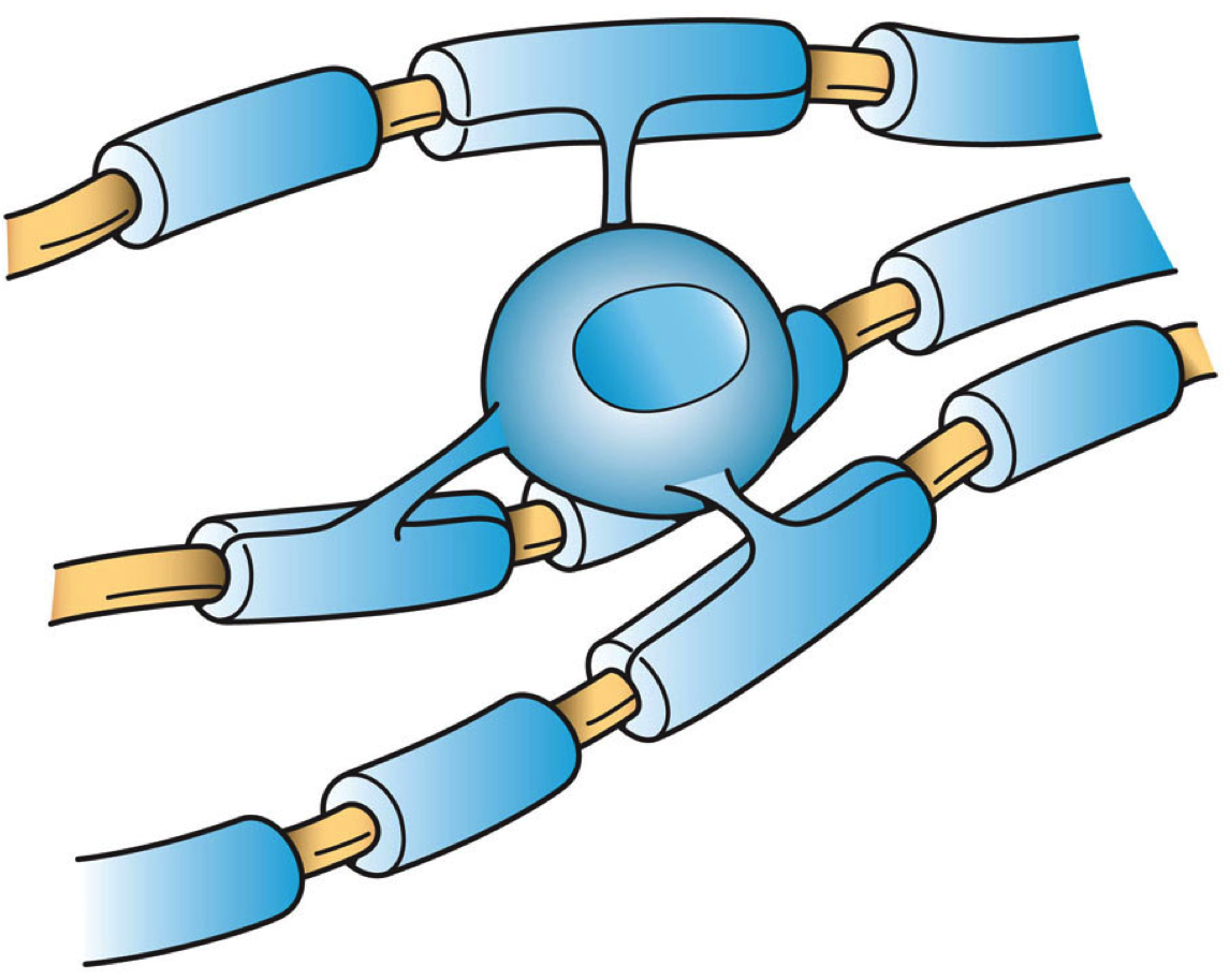
Oligodendrocytes (), or oligodendroglia, are a type of neuroglia whose main functions are to provide support and insulation to axons in the central nervous system of jawed vertebrates, equivalent to the function performed by Schwann cells in the peripheral nervous system. Oligodendrocytes do this by creating the myelin sheath. A single oligodendrocyte can extend its processes to 50 axons, wrapping approximately 1 μm of myelin sheath around each axon; Schwann cells, on the other hand, can wrap around only one axon. Each oligodendrocyte forms one segment of myelin for several adjacent axons. Oligodendrocytes are found only in the central nervous system, which comprises the brain and spinal cord. These cells were originally thought to have been produced in the ventral neural tube; however, research now shows oligodendrocytes originate from the ventral ventricular zone of the embryonic spinal cord and possibly have some concentrations in the forebrain. They are the last cell ... [...More Info...] [...Related Items...] OR: [Wikipedia] [Google] [Baidu] |
Neuroglia
Glia, also called glial cells (gliocytes) or neuroglia, are non-neuronal cells in the central nervous system (brain and spinal cord) and the peripheral nervous system that do not produce electrical impulses. They maintain homeostasis, form myelin in the peripheral nervous system, and provide support and protection for neurons. In the central nervous system, glial cells include oligodendrocytes, astrocytes, ependymal cells, and microglia, and in the peripheral nervous system they include Schwann cells and satellite cells. Function They have four main functions: *to surround neurons and hold them in place *to supply nutrients and oxygen to neurons *to insulate one neuron from another *to destroy pathogens and remove dead neurons. They also play a role in neurotransmission and synaptic connections, and in physiological processes such as breathing. While glia were thought to outnumber neurons by a ratio of 10:1, recent studies using newer methods and reappraisal of historical qua ... [...More Info...] [...Related Items...] OR: [Wikipedia] [Google] [Baidu] |
Transcription Factor
In molecular biology, a transcription factor (TF) (or sequence-specific DNA-binding factor) is a protein that controls the rate of transcription of genetic information from DNA to messenger RNA, by binding to a specific DNA sequence. The function of TFs is to regulate—turn on and off—genes in order to make sure that they are expressed in the desired cells at the right time and in the right amount throughout the life of the cell and the organism. Groups of TFs function in a coordinated fashion to direct cell division, cell growth, and cell death throughout life; cell migration and organization (body plan) during embryonic development; and intermittently in response to signals from outside the cell, such as a hormone. There are up to 1600 TFs in the human genome. Transcription factors are members of the proteome as well as regulome. TFs work alone or with other proteins in a complex, by promoting (as an activator), or blocking (as a repressor) the recruitment of RNA ... [...More Info...] [...Related Items...] OR: [Wikipedia] [Google] [Baidu] |
Promoter (genetics)
In genetics, a promoter is a sequence of DNA to which proteins bind to initiate transcription of a single RNA transcript from the DNA downstream of the promoter. The RNA transcript may encode a protein (mRNA), or can have a function in and of itself, such as tRNA or rRNA. Promoters are located near the transcription start sites of genes, upstream on the DNA (towards the 5' region of the sense strand). Promoters can be about 100–1000 base pairs long, the sequence of which is highly dependent on the gene and product of transcription, type or class of RNA polymerase recruited to the site, and species of organism. Promoters control gene expression in bacteria and eukaryotes. RNA polymerase must attach to DNA near a gene for transcription to occur. Promoter DNA sequences provide an enzyme binding site. The -10 sequence is TATAAT. -35 sequences are conserved on average, but not in most promoters. Artificial promoters with conserved -10 and -35 elements transcribe more slowly. All D ... [...More Info...] [...Related Items...] OR: [Wikipedia] [Google] [Baidu] |


