|
KCNK4
Potassium channel subfamily K member 4 is a protein that in humans is encoded by the ''KCNK4'' gene. KCNK4 protein channels are also called TRAAK channels. Function ''KNCK4'' is a gene segment that encodes for the TRAAK (TWIK-related Arachidonic Acid-Stimulated K+) subfamily of mechanosensitive potassium channels. Potassium channels play a role in many cellular processes including action potential depolarization, muscle contraction, hormone secretion, osmotic regulation, and ion flow. The K2P4.1 protein is a lipid-gated ion channel that belongs to the superfamily of potassium channel proteins containing two pore-forming P domains (K2P). K2P4.1 homodimerizes and functions as an outwardly rectifying channel. It is expressed primarily in neural tissues and is stimulated by membrane stretch and polyunsaturated fatty acids. TRAAK channels are found in mammalian neurons and are part of a protein family of weakly inward rectifying potassium channels. This subfamily of potassium chan ... [...More Info...] [...Related Items...] OR: [Wikipedia] [Google] [Baidu] |
Tandem Pore Domain Potassium Channel
The two-pore-domain or tandem pore domain potassium channels are a family of 15 members that form what is known as leak channels which possess Goldman-Hodgkin-Katz (open) rectification. These channels are regulated by several mechanisms including signaling lipids, oxygen tension, pH, mechanical stretch, and G-proteins . Their name is derived from the fact that the α subunits consist of four transmembrane segments, and each pair of transmembrane segments contains a pore loop between the two transmembrane segments. Thus, each subunit has two pore loops. As such, they structurally correspond to two inward-rectifier α subunits and thus form dimers in the membrane (whereas inward-rectifier α subunits form tetramers). Each single channel does ''not'' have two pores; the name of the channel comes from the fact that ''each subunit'' has two P (pore) domains in its primary sequence. To quote Rang and Dale (2015), "The nomenclature is misleading, especially when they are incorrectly ... [...More Info...] [...Related Items...] OR: [Wikipedia] [Google] [Baidu] |
Potassium Channel
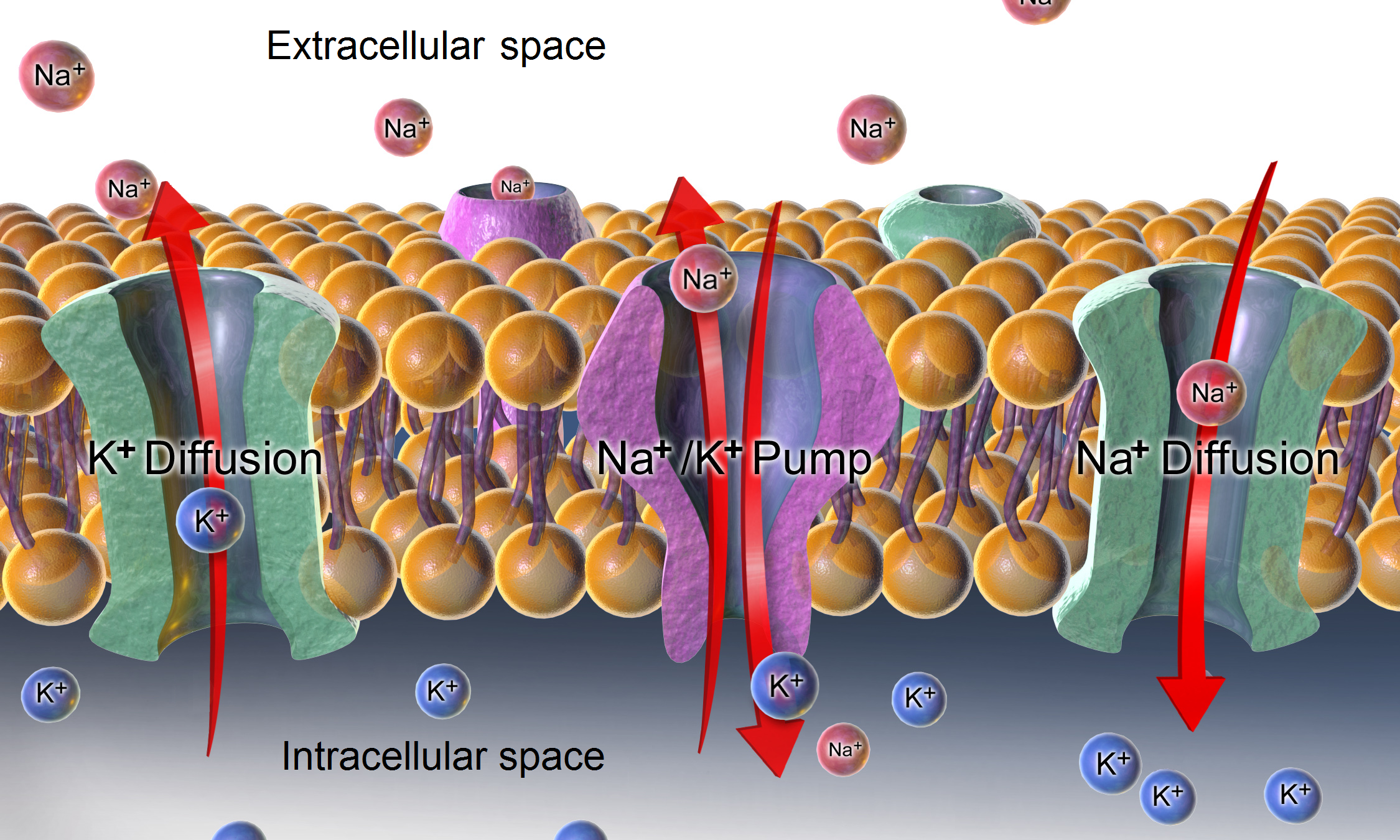
Potassium channels are the most widely distributed type of ion channel found in virtually all organisms. They form potassium-selective pores that span cell membranes. Potassium channels are found in most cell types and control a wide variety of cell functions. Function Potassium channels function to conduct potassium ions down their electrochemical gradient, doing so both rapidly (up to the diffusion rate of K+ ions in bulk water) and selectively (excluding, most notably, sodium despite the sub-angstrom difference in ionic radius). Biologically, these channels act to set or reset the resting potential in many cells. In excitable cells, such as neurons, the delayed counterflow of potassium ions shapes the action potential. By contributing to the regulation of the cardiac action potential duration in cardiac muscle, malfunction of potassium channels may cause life-threatening arrhythmias. Potassium channels may also be involved in maintaining vascular tone. They also regulate ce ... [...More Info...] [...Related Items...] OR: [Wikipedia] [Google] [Baidu] |
Habenula
In neuroanatomy, habenula (diminutive of Latin ''habena'' meaning rein) originally denoted the stalk of the pineal gland (pineal habenula; pedunculus of pineal body), but gradually came to refer to a neighboring group of nerve cells with which the pineal gland was believed to be associated, the habenular nucleus. The habenular nucleus is a set of well-conserved structures in all vertebrate animals. Currently, this term refers to this separate cell mass in the caudal portion of the dorsal diencephalon, known as the epithalamus, found in all vertebrates on both sides of the third ventricle. It connects the forebrain and midbrain within the epithalamus. It is embedded in the posterior end of the stria medullaris from which it receives most of its afferent fibers. By way of the fasciculus retroflexus (habenulointerpeduncular tract) it projects to the interpeduncular nucleus and other paramedian cell groups of the midbrain tegmentum. Although they were predominantly studied for their ... [...More Info...] [...Related Items...] OR: [Wikipedia] [Google] [Baidu] |
Hirschsprung's Disease
Hirschsprung's disease (HD or HSCR) is a birth defect in which nerves are missing from parts of the intestine. The most prominent symptom is constipation. Other symptoms may include vomiting, abdominal pain, diarrhea and slow growth. Symptoms usually become apparent in the first two months of life. Complications may include enterocolitis, megacolon, bowel obstruction and intestinal perforation. The disorder may occur by itself or in association with other genetic disorders such as Down syndrome or Waardenburg syndrome. About half of isolated cases are linked to a specific genetic mutation, and about 20% occur within families. Some of these occur in an autosomal dominant manner. The cause of the remaining cases is unclear. If otherwise normal parents have one child with the condition, the next child has a 4% risk of being affected. The condition is divided into two main types, short-segment and long-segment, depending on how much of the bowel is affected. Rarely, the small bowel m ... [...More Info...] [...Related Items...] OR: [Wikipedia] [Google] [Baidu] |
Saltatory Conduction
In neuroscience, saltatory conduction () is the propagation of action potentials along myelinated axons from one node of Ranvier to the next node, increasing the conduction velocity of action potentials. The uninsulated nodes of Ranvier are the only places along the axon where ions are exchanged across the axon membrane, regenerating the action potential between regions of the axon that are insulated by myelin, unlike electrical conduction in a simple circuit. Mechanism Myelinated axons only allow action potentials to occur at the unmyelinated nodes of Ranvier that occur between the myelinated internodes. It is by this restriction that saltatory conduction propagates an action potential along the axon of a neuron at rates significantly higher than would be possible in unmyelinated axons (150 m/s compared to 0.5 to 10 m/s). As sodium rushes into the node it creates an electrical force which pushes on the ions already inside the axon. This rapid conduction of electrical ... [...More Info...] [...Related Items...] OR: [Wikipedia] [Google] [Baidu] |
Node Of Ranvier
In neuroscience and anatomy, nodes of Ranvier ( ), also known as myelin-sheath gaps, occur along a myelinated axon where the axolemma is exposed to the extracellular space. Nodes of Ranvier are uninsulated and highly enriched in ion channels, allowing them to participate in the exchange of ions required to regenerate the action potential. Nerve conduction in myelinated axons is referred to as saltatory conduction () due to the manner in which the action potential seems to "jump" from one node to the next along the axon. This results in faster conduction of the action potential. Overview Many vertebrate axons are surrounded by a myelin sheath, allowing rapid and efficient saltatory ("jumping") propagation of action potentials. The contacts between neurons and glial cells display a very high level of spatial and temporal organization in myelinated fibers. The myelinating glial cells - oligodendrocytes in the central nervous system (CNS), and Schwann cells in the peripheral nervou ... [...More Info...] [...Related Items...] OR: [Wikipedia] [Google] [Baidu] |
Signal Transduction
Signal transduction is the process by which a chemical or physical signal is transmitted through a cell as a series of molecular events, most commonly protein phosphorylation catalyzed by protein kinases, which ultimately results in a cellular response. Proteins responsible for detecting stimuli are generally termed receptors, although in some cases the term sensor is used. The changes elicited by ligand binding (or signal sensing) in a receptor give rise to a biochemical cascade, which is a chain of biochemical events known as a signaling pathway. When signaling pathways interact with one another they form networks, which allow cellular responses to be coordinated, often by combinatorial signaling events. At the molecular level, such responses include changes in the transcription or translation of genes, and post-translational and conformational changes in proteins, as well as changes in their location. These molecular events are the basic mechanisms controlling cell growth, ... [...More Info...] [...Related Items...] OR: [Wikipedia] [Google] [Baidu] |
Resting Potential
A relatively static membrane potential which is usually referred to as the ground value for trans-membrane voltage. The relatively static membrane potential of quiescent cells is called the resting membrane potential (or resting voltage), as opposed to the specific dynamic electrochemical phenomena called action potential and graded membrane potential. Apart from the latter two, which occur in excitable cells (neurons, muscles, and some secretory cells in glands), membrane voltage in the majority of non-excitable cells can also undergo changes in response to environmental or intracellular stimuli. The resting potential exists due to the differences in membrane permeabilities for potassium, sodium, calcium, and chloride ions, which in turn result from functional activity of various ion channels, ion transporters, and exchangers. Conventionally, resting membrane potential can be defined as a relatively stable, ground value of transmembrane voltage in animal and plant cells. Beca ... [...More Info...] [...Related Items...] OR: [Wikipedia] [Google] [Baidu] |
Neurite
A neurite or neuronal process refers to any projection from the cell body of a neuron. This projection can be either an axon or a dendrite. The term is frequently used when speaking of immature or developing neurons, especially of cells in culture, because it can be difficult to tell axons from dendrites before differentiation is complete. Neurite development The development of a neurite requires a complex interplay of both extracellular and intracellular signals. At every given point along a developing neurite, there are receptors detecting both positive and negative growth cues from every direction in the surrounding space. The developing neurite sums together all of these growth signals in order to determine which direction the neurite will ultimately grow towards. While not all of the growth signals are known, several have been identified and characterized. Among the known extracellular growth signals are netrin, a midline chemoattractant, and semaphorin, ephrin and collaps ... [...More Info...] [...Related Items...] OR: [Wikipedia] [Google] [Baidu] |
Growth Cone
A growth cone is a large actin-supported extension of a developing or regenerating neurite seeking its synaptic target. It is the growth cone that drives axon growth. Their existence was originally proposed by Spanish histologist Santiago Ramón y Cajal based upon stationary images he observed under the microscope. He first described the growth cone based on fixed cells as "a concentration of protoplasm of conical form, endowed with amoeboid movements" (Cajal, 1890). Growth cones are situated on the tips of neurites, either dendrites or axons, of the nerve cell. The sensory, motor, integrative, and adaptive functions of growing axons and dendrites are all contained within this specialized structure. Structure The morphology of the growth cone can be easily described by using the hand as an analogy. The fine extensions of the growth cone are pointed filopodia known as microspikes. The filopodia are like the "fingers" of the growth cone; they contain bundles of actin filaments ... [...More Info...] [...Related Items...] OR: [Wikipedia] [Google] [Baidu] |
Cerebellum
The cerebellum (Latin for "little brain") is a major feature of the hindbrain of all vertebrates. Although usually smaller than the cerebrum, in some animals such as the mormyrid fishes it may be as large as or even larger. In humans, the cerebellum plays an important role in motor control. It may also be involved in some cognition, cognitive functions such as attention and language as well as emotion, emotional control such as regulating fear and pleasure responses, but its movement-related functions are the most solidly established. The human cerebellum does not initiate movement, but contributes to Motor coordination, coordination, precision, and accurate timing: it receives input from sensory systems of the spinal cord and from other parts of the brain, and integrates these inputs to fine-tune motor activity. Cerebellar damage produces disorders in Fine motor skill, fine movement, Equilibrioception, equilibrium, Human positions, posture, and motor learning in humans. Anatomica ... [...More Info...] [...Related Items...] OR: [Wikipedia] [Google] [Baidu] |
Basal Ganglia
The basal ganglia (BG), or basal nuclei, are a group of subcortical nuclei, of varied origin, in the brains of vertebrates. In humans, and some primates, there are some differences, mainly in the division of the globus pallidus into an external and internal region, and in the division of the striatum. The basal ganglia are situated at the base of the forebrain and top of the midbrain. Basal ganglia are strongly interconnected with the cerebral cortex, thalamus, and brainstem, as well as several other brain areas. The basal ganglia are associated with a variety of functions, including control of voluntary motor movements, procedural learning, habit learning, conditional learning, eye movements, cognition, and emotion. The main components of the basal ganglia – as defined functionally – are the striatum, consisting of both the dorsal striatum (caudate nucleus and putamen) and the ventral striatum (nucleus accumbens and olfactory tubercle), the globus pallidus, ... [...More Info...] [...Related Items...] OR: [Wikipedia] [Google] [Baidu] |


