|
Irvine–Gass Syndrome
Irvine–Gass syndrome, pseudophakic cystoid macular edema or postcataract CME is one of the most common causes of visual loss after cataract surgery. The syndrome is named in honor of S. Rodman Irvine and J. Donald M. Gass. The incidence is more common in older types of cataract surgery, where postcataract CME could occur in 20–60% of patients, but with modern cataract surgery, incidence of Irvine–Gass syndrome has reduced significantly. Replacement of the lens as treatment for cataract can cause pseudophakic macular edema (‘ pseudophakia’ means ‘replacement lens’). This could occur as the surgery involved sometimes irritates the retina (and other parts of the eye) causing the capillaries in the retina to dilate and leak fluid into the retina. This is less common today with modern lens replacement techniques. Signs and symptoms Most patients have decreased or fuzzy vision. Complications Foveolar photoreceptor damage and permanent vision impairment can arise from ... [...More Info...] [...Related Items...] OR: [Wikipedia] [Google] [Baidu] |
Ophthalmology
Ophthalmology (, ) is the branch of medicine that deals with the diagnosis, treatment, and surgery of eye diseases and disorders. An ophthalmologist is a physician who undergoes subspecialty training in medical and surgical eye care. Following a medical degree, a doctor specialising in ophthalmology must pursue additional postgraduate residency training specific to that field. In the United States, following graduation from medical school, one must complete a four-year residency in ophthalmology to become an ophthalmologist. Following residency, additional specialty training (or fellowship) may be sought in a particular aspect of eye pathology. Ophthalmologists prescribe medications to treat ailments, such as eye diseases, implement laser therapy, and perform surgery when needed. Ophthalmologists provide both primary and specialty eye care—medical and surgical. Most ophthalmologists participate in academic research on eye diseases at some point in their training and many inc ... [...More Info...] [...Related Items...] OR: [Wikipedia] [Google] [Baidu] |
Retinal Vein Occlusion
Central retinal vein occlusion, also CRVO, is when the central retinal vein becomes occluded, usually through thrombosis. The central retinal vein is the venous equivalent of the central retinal artery and both may become occluded. Since the central retinal artery and vein are the sole source of blood supply and drainage for the retina, such occlusion can lead to severe damage to the retina and blindness, due to ischemia (restriction in blood supply) and edema (swelling). CRVO can cause ocular ischemic syndrome. Nonischemic CRVO is the milder form of the disease. It may progress to the more severe ischemic type. CRVO can also cause glaucoma. Diagnosis Despite the role of thrombosis in the development of CRVO, a systematic review found no increased prevalence of thrombophilia (an inherent propensity to thrombosis) in patients with retinal vascular occlusion. Treatment Treatment consists of Anti-VEGF drugs like Lucentis or intravitreal steroid implant (Ozurdex) and Pan-Retinal L ... [...More Info...] [...Related Items...] OR: [Wikipedia] [Google] [Baidu] |
Anti Vascular Endothelial Growth Factor Therapy
Anti–vascular endothelial growth factor therapy, also known as anti-VEGF () therapy or medication, is the use of medications that block vascular endothelial growth factor. This is done in the treatment of certain cancers and in age-related macular degeneration. They can involve monoclonal antibodies such as bevacizumab, antibody derivatives such as ranibizumab (Lucentis), or orally-available small molecules that inhibit the tyrosine kinases stimulated by VEGF: sunitinib, sorafenib, axitinib, and pazopanib (some of these therapies target VEGF receptors rather than the VEGFs). Both antibody-based compounds and the first three orally available compounds are commercialized. The latter two, axitinib and pazopanib, are in clinical trials. Bergers and Hanahan concluded in 2008 that anti-VEGF drugs can show therapeutic efficacy in mouse models of cancer and in an increasing number of human cancers. But, "the benefits are at best transitory and are followed by a restoration of tumour g ... [...More Info...] [...Related Items...] OR: [Wikipedia] [Google] [Baidu] |
Nonsteroidal Anti-inflammatory Drug
Non-steroidal anti-inflammatory drugs (NSAID) are members of a Indication (medicine), therapeutic drug class which Analgesic, reduces pain, Anti-inflammatory, decreases inflammation, Antipyretic, decreases fever, and Antithrombotic, prevents blood clots. Side effects depend on the specific drug, its dose and duration of use, but largely include an increased risk of Stomach ulcers, gastrointestinal ulcers and bleeds, heart attack, and kidney disease. The term ''non-steroidal'', common from around 1960, distinguishes these drugs from corticosteroids, another class of anti-inflammatory drugs, which during the 1950s had acquired a bad reputation due to overuse and side-effect problems after their introduction in 1948. NSAIDs work by inhibiting the activity of cyclooxygenase enzymes (the COX-1 and COX-2 isozyme, isoenzymes). In cells, these enzymes are involved in the synthesis of key biological mediators, namely prostaglandins, which are involved in inflammation, and thromboxanes, ... [...More Info...] [...Related Items...] OR: [Wikipedia] [Google] [Baidu] |
Corticosteroid
Corticosteroids are a class of steroid hormones that are produced in the adrenal cortex of vertebrates, as well as the synthetic analogues of these hormones. Two main classes of corticosteroids, glucocorticoids and mineralocorticoids, are involved in a wide range of physiological processes, including stress response, immune response, and regulation of inflammation, carbohydrate metabolism, protein catabolism, blood electrolyte levels, and behavior. Some common naturally occurring steroid hormones are cortisol (), corticosterone (), cortisone () and aldosterone () (cortisone and aldosterone are isomers). The main corticosteroids produced by the adrenal cortex are cortisol and aldosterone. The etymology of the '' cortico-'' part of the name refers to the adrenal cortex, which makes these steroid hormones. Thus a corticosteroid is a "cortex steroid". Classes * Glucocorticoids such as cortisol affect carbohydrate, fat, and protein metabolism, and have anti ... [...More Info...] [...Related Items...] OR: [Wikipedia] [Google] [Baidu] |
Macular Hole
A macular hole is a small break in the macula, located in the center of the eye's light-sensitive tissue called the retina. Symptoms If the vitreous is firmly attached to the retina when it pulls away, it can tear the retina and create a macular hole. Also, once the vitreous has pulled away from the surface of the retina, some of the fibers can remain on the retinal surface and can contract. This increases tension (physics), tension on the retina and can lead to a macular hole. In either case, the fluid that has replaced the shrunken vitreous can then seep through the hole onto the macula, blurring and distorting central vision. Causes The eye contains a Gelatin dessert, jelly-like substance called the vitreous. Shrinking of the vitreous usually causes the hole. As a person ages, the vitreous becomes watery and begins to pull away from the retina. If the vitreous is firmly attached to the retina when it pulls away, a hole can result. Diagnosis Macular degeneration is ... [...More Info...] [...Related Items...] OR: [Wikipedia] [Google] [Baidu] |
Uveitis
Uveitis () is inflammation of the uvea, the pigmented layer of the eye between the inner retina and the outer fibrous layer composed of the sclera and cornea. The uvea consists of the middle layer of pigmented vascular structures of the eye and includes the iris, ciliary body, and choroid. Uveitis is described anatomically, by the part of the eye affected, as anterior, intermediate or posterior, or panuveitic if all parts are involved. Anterior uveitis ( iridocyclitis) is the most common, with the incidence of uveitis overall affecting approximately 1:4500, most commonly those between the ages of 20–60. Symptoms include eye pain, eye redness, floaters and blurred vision, and ophthalmic examination may show dilated ciliary blood vessels and the presence of cells in the anterior chamber. Uveitis may arise spontaneously, have a genetic component, or be associated with an autoimmune disease or infection. While the eye is a relatively protected environment, its immune mecha ... [...More Info...] [...Related Items...] OR: [Wikipedia] [Google] [Baidu] |
Epiretinal Membrane
Epiretinal membrane or macular pucker is a disease of the eye in response to changes in the vitreous humor or more rarely, diabetes. Sometimes, as a result of immune system response to protect the retina, cells converge in the macular area as the vitreous ages and pulls away in posterior vitreous detachment (PVD). PVD can create minor damage to the retina, stimulating exudate, inflammation, and leucocyte response. These cells can form a transparent layer gradually and, like all scar tissue, tighten to create tension on the retina which may bulge and pucker, or even cause swelling or macular edema. Often this results in distortions of vision that are clearly visible as bowing and blurring when looking at lines on chart paper (or an Amsler grid) within the macular area, or central 1.0 degree of visual arc. Usually it occurs in one eye first, and may cause binocular diplopia or double vision if the image from one eye is too different from the image of the other eye. The dis ... [...More Info...] [...Related Items...] OR: [Wikipedia] [Google] [Baidu] |
Anterior Chamber Flare
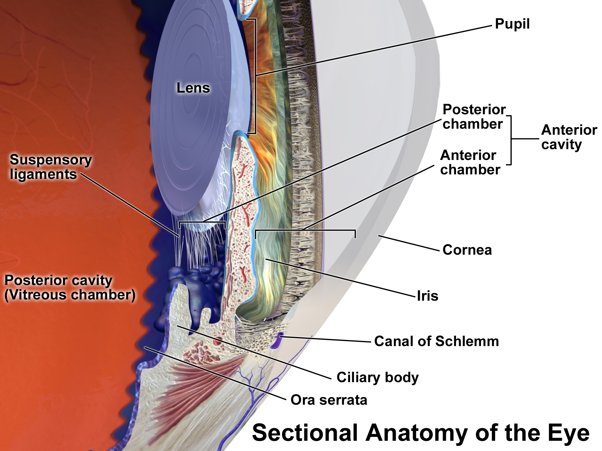
In ophthalmology and optometry, a slit lamp is an instrument consisting of a high-intensity light source that can be focused to shine a thin sheet of light into the eye. It is used in conjunction with a biomicroscope. The lamp facilitates an examination of the anterior segment and posterior segment of the human eye, which includes the eyelid, sclera, conjunctiva, iris, natural crystalline lens, and cornea. The binocular slit-lamp examination provides a stereoscopic magnified view of the eye structures in detail, enabling anatomical diagnoses to be made for a variety of eye conditions. A second, hand-held lens is used to examine the retina. History Two conflicting trends emerged in the development of the slit lamp. One trend originated from clinical research and aimed to apply the increasingly complex and advanced technology of the time. [...More Info...] [...Related Items...] OR: [Wikipedia] [Google] [Baidu] |
Prostaglandin Analogue
Prostaglandin analogues are a class of drugs that bind to a prostaglandin receptor. Wider use of prostaglandin analogues is limited by unwanted side effects and their abortive potential. Uses Prostaglandin analogues such as misoprostol are used in treatment of duodenal and gastric ulcers. Misoprostol and other prostaglandin analogues protect the lining of the gastrointestinal tract from harmful stomach acid and are especially indicated for the elderly on continuous doses of NSAIDs. In the field of ophthalmology, drugs of this class are used to lower intraocular pressure (IOP) in people with glaucoma. Up until the late 1970s prostaglandins were thought to raise IOP, but a paper published in 1977 showed that prostaglandin F2α lowered it, and subsequent studies found that this was due to increasing the outflow of aqueous humor, mainly by relaxing the ciliary muscle, and possibly also due to changes in extracellular matrix and to widening of spaces within the trabecular meshwo ... [...More Info...] [...Related Items...] OR: [Wikipedia] [Google] [Baidu] |
Prostaglandin
Prostaglandins (PG) are a group of physiology, physiologically active lipid compounds called eicosanoids that have diverse hormone-like effects in animals. Prostaglandins have been found in almost every Tissue (biology), tissue in humans and other animals. They are derived enzymatically from the fatty acid arachidonic acid. Every prostaglandin contains 20 carbon atoms, including a carbon ring, 5-carbon ring. They are a subclass of eicosanoids and of the prostanoid class of fatty acid derivatives. The structural differences between prostaglandins account for their different biological activities. A given prostaglandin may have different and even opposite effects in different tissues in some cases. The ability of the same prostaglandin to stimulate a reaction in one tissue and inhibit the same reaction in another tissue is determined by the type of receptor (biochemistry), receptor to which the prostaglandin binds. They act as autocrine or paracrine factors with their target cells ... [...More Info...] [...Related Items...] OR: [Wikipedia] [Google] [Baidu] |
Diabetes Mellitus
Diabetes mellitus, commonly known as diabetes, is a group of common endocrine diseases characterized by sustained hyperglycemia, high blood sugar levels. Diabetes is due to either the pancreas not producing enough of the hormone insulin, or the cells of the body becoming unresponsive to insulin's effects. Classic symptoms include polydipsia (excessive thirst), polyuria (excessive urination), polyphagia (excessive hunger), Weight loss#Unintentional, weight loss, and blurred vision. If left untreated, the disease can lead to various health complications, including disorders of the Cardiovascular disease, cardiovascular system, Diabetic retinopathy, eye, Diabetic nephropathy, kidney, and Diabetic neuropathy, nerves. Diabetes accounts for approximately 4.2 million deaths every year, with an estimated 1.5 million caused by either untreated or poorly treated diabetes. The major types of diabetes are Type 1 diabetes, type 1 and Type 2 diabetes, type 2. The most common treatment for ty ... [...More Info...] [...Related Items...] OR: [Wikipedia] [Google] [Baidu] |


