|
Introducer Sheath
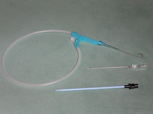
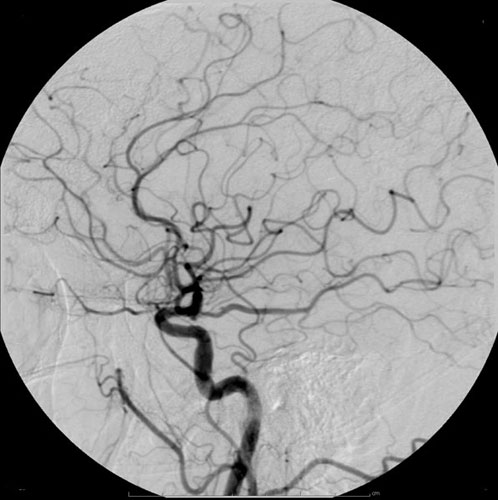
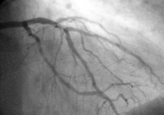
The Seldinger technique, also known as Seldinger wire technique, is a medical procedure to obtain safe access to blood vessels and other hollow organs. It is named after Sven Ivar Seldinger (1921–1998), a Swedish radiologist who introduced the procedure in 1953. Uses The Seldinger technique is used for angiography, insertion of chest drains and central venous catheters, insertion of PEG tubes using the push technique, insertion of the leads for an artificial pacemaker or implantable cardioverter-defibrillator, and numerous other interventional medical procedures. Complications The initial puncture is with a sharp instrument, and this may lead to hemorrhage or perforation of the organ in question. Infection is a possible complication, and hence asepsis is practiced during most Seldinger procedures. Loss of the guidewire into the cavity or blood vessel is a significant and generally preventable complication. Description The desired vessel or cavity is punctured with a sharp ... [...More Info...] [...Related Items...] OR: [Wikipedia] [Google] [Baidu] |
Seldinger Set
The Seldinger technique, also known as Seldinger wire technique, is a medical procedure to obtain safe access to blood vessels and other hollow organs. It is named after Sven Ivar Seldinger (1921–1998), a Swedish radiologist who introduced the procedure in 1953. Uses The Seldinger technique is used for angiography, insertion of chest drains and central venous catheters, insertion of PEG tubes using the push technique, insertion of the leads for an artificial pacemaker or implantable cardioverter-defibrillator, and numerous other interventional medical procedures. Complications The initial puncture is with a sharp instrument, and this may lead to hemorrhage or perforation of the organ in question. Infection is a possible complication, and hence asepsis is practiced during most Seldinger procedures. Loss of the guidewire into the cavity or blood vessel is a significant and generally preventable complication. Description The desired vessel or cavity is punctured with a shar ... [...More Info...] [...Related Items...] OR: [Wikipedia] [Google] [Baidu] |
Asepsis
Asepsis is the state of being free from disease-causing micro-organisms (such as pathogenic bacteria, viruses, pathogenic fungi, and parasites). There are two categories of asepsis: medical and surgical. The modern day notion of asepsis is derived from the older antiseptic techniques, a shift initiated by different individuals in the 19th century who introduced practices such as the sterilizing of surgical tools and the wearing of surgical gloves during operations. The goal of asepsis is to eliminate infection, not to achieve sterility. Ideally, a surgical field is sterile, meaning it is free of all biological contaminants (e.g. fungi, bacteria, viruses), not just those that can cause disease, putrefaction, or fermentation. Even in an aseptic state, a condition of sterile inflammation may develop. The term often refers to those practices used to promote or induce asepsis in an operative field of surgery or medicine to prevent infection. History The modern concept of asepsi ... [...More Info...] [...Related Items...] OR: [Wikipedia] [Google] [Baidu] |
Charles Dotter
Charles Theodore Dotter (14 June 1920 – 15 February 1985) was a pioneering US vascular radiologist who is credited with developing interventional radiology. Dotter, with his trainee Dr Melvin P. Judkins, described angioplasty in 1964. Dotter received a bachelor of arts degree in 1941 from Duke University. He went to medical school at Cornell, where he met his future wife, Pamela Beattie, a head nurse at New York Hospital. They married in 1944. He completed his internship at the United States Naval Hospital in New York State, and his residency at New York Hospital. Dotter invented angioplasty and the catheter-delivered stent, which were first used to treat peripheral arterial disease. It was Dotter who, in 1950, developed an automatic X-Ray Roll-Film magazine capable of producing images at the rate of 2 per second. On January 16, 1964, at Oregon Health and Science University Dotter percutaneously dilated a tight, localized stenosis of the superficial femoral artery (SFA) in ... [...More Info...] [...Related Items...] OR: [Wikipedia] [Google] [Baidu] |
Interventional Radiology
Interventional radiology (IR) is a medical specialty that performs various minimally-invasive procedures using medical imaging guidance, such as x-ray fluoroscopy, computed tomography, magnetic resonance imaging, or ultrasound. IR performs both diagnostic and therapeutic procedures through very small incisions or body orifices. Diagnostic IR procedures are those intended to help make a diagnosis or guide further medical treatment, and include image-guided biopsy of a tumor or injection of an imaging contrast agent into a hollow structure, such as a blood vessel or a duct. By contrast, therapeutic IR procedures provide direct treatment—they include catheter-based medicine delivery, medical device placement (e.g., stents), and angioplasty of narrowed structures. The main benefits of interventional radiology techniques are that they can reach the deep structures of the body through a body orifice or tiny incision using small needles and wires. That decreases risks, pain, ... [...More Info...] [...Related Items...] OR: [Wikipedia] [Google] [Baidu] |
Trochar
A trocar (or trochar) is a medical or veterinary device that is made up of an awl (which may be a metal or plastic sharpened or non-bladed tip), a cannula (essentially a hollow tube), and a seal. Trocars are placed through the abdomen during laparoscopic surgery. The trocar functions as a portal for the subsequent placement of other instruments, such as graspers, scissors, staplers, etc. Trocars also allow the escape of gas or fluid from organs within the body. Etymology The word ''trocar'', less commonly ''trochar'', comes from French ''trocart'', ''trois-quarts'' (three-fourths), from ''trois'' 'three' and ''carre'' 'side, face of an instrument', first recorded in the ''Dictionnaire des Arts et des Sciences'', 1694, by Thomas Corneille, younger brother of Pierre Corneille. History Originally, doctors used trocars to relieve pressure build-up of fluids (edema) or gases ( bloating). Patents for trocars appeared early in the 19th century, although their use dated back possibl ... [...More Info...] [...Related Items...] OR: [Wikipedia] [Google] [Baidu] |
Biopsy
A biopsy is a medical test commonly performed by a surgeon, interventional radiologist, or an interventional cardiologist. The process involves extraction of sample cells or tissues for examination to determine the presence or extent of a disease. The tissue is then fixed, dehydrated, embedded, sectioned, stained and mounted before it is generally examined under a microscope by a pathologist; it may also be analyzed chemically. When an entire lump or suspicious area is removed, the procedure is called an excisional biopsy. An incisional biopsy or core biopsy samples a portion of the abnormal tissue without attempting to remove the entire lesion or tumor. When a sample of tissue or fluid is removed with a needle in such a way that cells are removed without preserving the histological architecture of the tissue cells, the procedure is called a needle aspiration biopsy. Biopsies are most commonly performed for insight into possible cancerous or inflammatory conditions. H ... [...More Info...] [...Related Items...] OR: [Wikipedia] [Google] [Baidu] |
Catheter Ablation
Catheter ablation is a procedure used to remove or terminate a faulty electrical pathway from sections of the heart of those who are prone to developing cardiac arrhythmias such as atrial fibrillation, atrial flutter and Wolff-Parkinson-White syndrome. If not controlled, such arrhythmias increase the risk of ventricular fibrillation and sudden cardiac arrest. The ablation procedure can be classified by energy source: radiofrequency ablation and cryoablation. Medical uses Catheter ablation may be recommended for a recurrent or persistent arrhythmia resulting in symptoms or other dysfunction. Typically, catheter ablation is used only when pharmacologic treatment has been ineffective. Effectiveness Catheter ablation of most arrhythmias has a high success rate. Success rates for WPW syndrome have been as high as 95% For SVT, single procedure success is 91% to 96% (95% Confidence Interval) and multiple procedure success is 92% to 97% (95% Confidence Interval). For atrial flutter, s ... [...More Info...] [...Related Items...] OR: [Wikipedia] [Google] [Baidu] |
Radiocontrast
Radiocontrast agents are substances used to enhance the visibility of internal structures in X-ray-based imaging techniques such as computed tomography (contrast CT), projectional radiography, and fluoroscopy. Radiocontrast agents are typically iodine, or more rarely barium sulfate. The contrast agents absorb external X-rays, resulting in decreased exposure on the X-ray detector. This is different from radiopharmaceuticals used in nuclear medicine which emit radiation. Magnetic resonance imaging (MRI) functions through different principles and thus MRI contrast agents have a different mode of action. These compounds work by altering the magnetic properties of nearby hydrogen nuclei. Types and uses Radiocontrast agents used in X-ray examinations can be grouped in positive (iodinated agents, barium sulfate), and negative agents (air, carbon dioxide, methylcellulose). Iodine (circulatory system) Iodinated contrast contains iodine. It is the main type of radiocontrast used fo ... [...More Info...] [...Related Items...] OR: [Wikipedia] [Google] [Baidu] |
Fluoroscopy
Fluoroscopy () is an imaging technique that uses X-rays to obtain real-time moving images of the interior of an object. In its primary application of medical imaging, a fluoroscope () allows a physician to see the internal structure and function of a patient, so that the pumping action of the heart or the motion of swallowing, for example, can be watched. This is useful for both diagnosis and therapy and occurs in general radiology, interventional radiology, and image-guided surgery. In its simplest form, a fluoroscope consists of an X-ray source and a fluorescent screen, between which a patient is placed. However, since the 1950s most fluoroscopes have included X-ray image intensifiers and cameras as well, to improve the image's visibility and make it available on a remote display screen. For many decades, fluoroscopy tended to produce live pictures that were not recorded, but since the 1960s, as technology improved, recording and playback became the norm. Fluoroscopy i ... [...More Info...] [...Related Items...] OR: [Wikipedia] [Google] [Baidu] |
Angioplasty
Angioplasty, is also known as balloon angioplasty and percutaneous transluminal angioplasty (PTA), is a minimally invasive endovascular procedure used to widen narrowed or obstructed arteries or veins, typically to treat arterial atherosclerosis. A deflated balloon attached to a catheter (a balloon catheter) is passed over a guide-wire into the narrowed vessel and then inflated to a fixed size. The balloon forces expansion of the blood vessel and the surrounding muscular wall, allowing an improved blood flow. A stent may be inserted at the time of ballooning to ensure the vessel remains open, and the balloon is then deflated and withdrawn. Angioplasty has come to include all manner of vascular interventions that are typically performed percutaneously. The word is composed of the combining forms of the Greek words ἀνγεῖον ' "vessel" or "cavity" (of the human body) and πλάσσω ' "form" or "mould". Uses and indications Coronary angioplasty A coronary an ... [...More Info...] [...Related Items...] OR: [Wikipedia] [Google] [Baidu] |
Catheter
In medicine, a catheter (/ˈkæθətər/) is a thin tube made from medical grade materials serving a broad range of functions. Catheters are medical devices that can be inserted in the body to treat diseases or perform a surgical procedure. Catheters are manufactured for specific applications, such as cardiovascular, urological, gastrointestinal, neurovascular and ophthalmic procedures. The process of inserting a catheter is ''catheterization''. In most uses, a catheter is a thin, flexible tube (''soft'' catheter) though catheters are available in varying levels of stiffness depending on the application. A catheter left inside the body, either temporarily or permanently, may be referred to as an "indwelling catheter" (for example, a peripherally inserted central catheter). A permanently inserted catheter may be referred to as a "permcath" (originally a trademark). Catheters can be inserted into a body cavity, duct, or vessel, brain, skin or adipose tissue. Functionally, they a ... [...More Info...] [...Related Items...] OR: [Wikipedia] [Google] [Baidu] |
Nephrostomy
A nephrostomy is an artificial opening created between the kidney and the skin which allows for the urinary diversion directly from the upper part of the urinary system (renal pelvis). An urostomy is a related procedure performed more distally along the urinary system to provide urinary diversion. Uses A nephrostomy is performed whenever a blockage keeps urine from passing from the kidneys, through the ureter and into the urinary bladder. Without another way for urine to drain, pressure would rise within the urinary system and the kidneys would be damaged. The most common cause of blockage necessitating a nephrostomy is cancer, especially ovarian cancer and colon cancer. Nephrostomies may also be required to treat pyonephrosis, hydronephrosis and kidney stones. Process Nephrostomies are created by surgeons or interventional radiologists and typically consist of a catheter which pierces the skin and rests in the urinary tract. It is performed under ultrasound guidance, CT f ... [...More Info...] [...Related Items...] OR: [Wikipedia] [Google] [Baidu] |


