|
Inferior Thyroid Vein
The inferior thyroid veins appear two, frequently three or four, in number, and arise in the venous plexus on the thyroid gland, communicating with the middle and superior thyroid veins. While the superior and middle thyroid veins serve as direct tributaries to the internal jugular vein, the inferior thyroid veins drain directly to the brachiocephalic veins. The inferior thyroid veins form a plexus in front of the trachea, behind the sternothyroid muscle. From this plexus, the left vein descends and joins the left brachiocephalic vein, and the right vein passes obliquely downward and to the right across the brachiocephalic artery to open into the right brachiocephalic vein, just at its junction with the superior vena cava; sometimes the right and left veins open by a common trunk in the latter situation. The inferior thyroid veins receive esophageal, tracheal, and inferior laryngeal veins, and are provided with valves at their terminations in the brachiocephalic veins The l ... [...More Info...] [...Related Items...] OR: [Wikipedia] [Google] [Baidu] |
Thyroid Gland
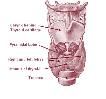
The thyroid, or thyroid gland, is an endocrine gland in vertebrates. In humans it is in the neck and consists of two connected lobe (anatomy), lobes. The lower two thirds of the lobes are connected by a thin band of Connective tissue, tissue called the thyroid isthmus. The thyroid is located at the front of the neck, below the Adam's apple. Microscopically, the functional unit of the thyroid gland is the spherical Thyroid follicular cell#Location, thyroid follicle, lined with thyroid follicular cell, follicular cells (thyrocytes), and occasional parafollicular cells that surround a follicular lumen, lumen containing colloid. The thyroid gland secretes three hormones: the two thyroid hormonestriiodothyronine, triiodothyronine (T3) and thyroid hormone, thyroxine (T4)and a peptide hormone, calcitonin. The thyroid hormones influence the basal metabolic rate, metabolic rate and protein biosynthesis, protein synthesis, and in children, growth and development. Calcitonin plays a role in ... [...More Info...] [...Related Items...] OR: [Wikipedia] [Google] [Baidu] |
Sternothyroid Muscle
The sternothyroid muscle, or sternothyroideus, is an infrahyoid muscle in the neck. It acts to depress the hyoid bone. It is below the sternohyoid muscle. It is shorter and wider than the sternohyoid. Structure The sternothyroid arises from the posterior surface of the manubrium of the sternum, below the origin of the sternohyoid. It also arises from the edge of the cartilage of the first rib. It is inserted into the oblique line on the lamina of the thyroid cartilage. It is in close contact with its fellow at the lower part of the neck, but diverges somewhat as it ascends. It is occasionally traversed by a transverse or oblique tendinous inscription. Innervation The sternothyroid muscle is innervated by the ansa cervicalis. Variations Doubling; absence; accessory slips to the thyrohyoid, inferior pharyngeal constrictor, or to the carotid sheath. Function The sternothyroid muscle depresses the hyoid bone, along with the other infrahyoid muscle. Clinical significance The u ... [...More Info...] [...Related Items...] OR: [Wikipedia] [Google] [Baidu] |
Inferior Laryngeal Veins
The inferior laryngeal vein is a vein which drains the larynx The larynx (), commonly called the voice box, is an organ in the top of the neck involved in breathing, producing sound and protecting the trachea against food aspiration. The opening of larynx into pharynx known as the laryngeal inlet is about .... It runs parallel to the laryngeal arteries. References Veins of the head and neck {{circulatory-stub ... [...More Info...] [...Related Items...] OR: [Wikipedia] [Google] [Baidu] |
Esophageal Veins
The esophageal veins drain blood from the esophagus to the azygos vein, in the thorax, and to the inferior thyroid vein in the neck. It also drains, although with less significance, to the hemiazygos vein, posterior intercostal vein and bronchial veins. In the abdomen, some drain to the left gastric vein which drains into the portal vein. See also * Esophageal varices Esophageal varices are extremely dilated sub-mucosal veins in the lower third of the esophagus. They are most often a consequence of portal hypertension, commonly due to cirrhosis. People with esophageal varices have a strong tendency to develop ... References External links * * () * https://web.archive.org/web/20080506103555/http://www.med.mun.ca/anatomyts/digest/avein6.htm Veins of the torso {{circulatory-stub ... [...More Info...] [...Related Items...] OR: [Wikipedia] [Google] [Baidu] |
Superior Vena Cava
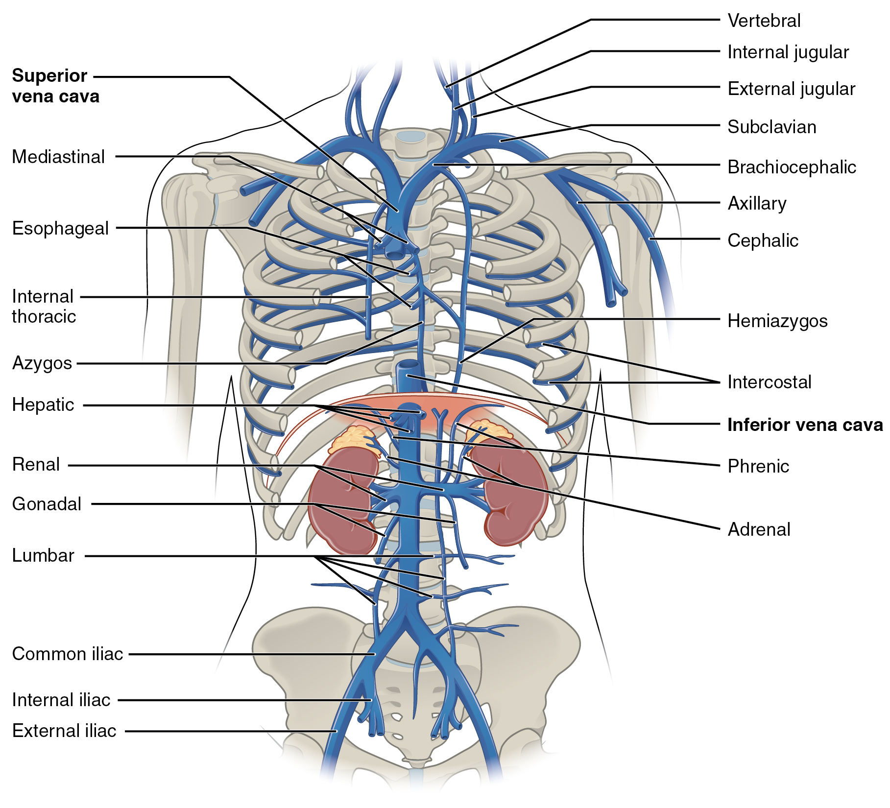
The superior vena cava (SVC) is the superior of the two venae cavae, the great venous trunks that return deoxygenated blood from the systemic circulation to the right atrium of the heart. It is a large-diameter (24 mm) short length vein that receives venous return from the upper half of the body, above the diaphragm. Venous return from the lower half, below the diaphragm, flows through the inferior vena cava. The SVC is located in the anterior right superior mediastinum. It is the typical site of central venous access via a central venous catheter or a peripherally inserted central catheter. Mentions of "the cava" without further specification usually refer to the SVC. Structure The superior vena cava is formed by the left and right brachiocephalic veins, which receive blood from the upper limbs, head and neck, behind the lower border of the first right costal cartilage. It passes vertically downwards behind first intercostal space and receives azygos vein just before it p ... [...More Info...] [...Related Items...] OR: [Wikipedia] [Google] [Baidu] |
Right Brachiocephalic Vein
The left and right brachiocephalic veins (previously called innominate veins) are major veins in the upper chest, formed by the union of each corresponding internal jugular vein and subclavian vein. This is at the level of the sternoclavicular joint. The left brachiocephalic vein is nearly always longer than the right. These veins merge to form the superior vena cava, a great vessel, posterior to the junction of the first costal cartilage with the manubrium of the sternum. The brachiocephalic veins are the major veins returning blood to the superior vena cava. Tributaries The brachiocephalic vein is formed by the confluence of the subclavian and internal jugular veins. In addition it receives drainage from: * Left and right internal thoracic vein (Also called internal mammary veins): drain into the inferior border of their corresponding vein * Left and right inferior thyroid veins: drain into the superior aspect of their corresponding veins near the confluence * Left and right ... [...More Info...] [...Related Items...] OR: [Wikipedia] [Google] [Baidu] |
Brachiocephalic Artery
The brachiocephalic artery (or brachiocephalic trunk or innominate artery) is an artery of the mediastinum that supplies blood to the right arm and the head and neck. It is the first branch of the aortic arch. Soon after it emerges, the brachiocephalic artery divides into the right common carotid artery and the right subclavian artery. There is no brachiocephalic artery for the left side of the body. The left common carotid, and the left subclavian artery, come directly off the aortic arch. However, there are two brachiocephalic veins. Structure The brachiocephalic artery arises, on a level with the upper border of the second right costal cartilage, from the start of the aortic arch, on a plane anterior to the origin of the left carotid artery. It ascends obliquely upward, backward, and to the right to the level of the upper border of the right sternoclavicular articulation, where it divides into the right common carotid artery and right subclavian arteries. The artery then cros ... [...More Info...] [...Related Items...] OR: [Wikipedia] [Google] [Baidu] |
Left Brachiocephalic Vein
The left and right brachiocephalic veins (previously called innominate veins) are major veins in the upper chest, formed by the union of each corresponding internal jugular vein and subclavian vein. This is at the level of the sternoclavicular joint. The left brachiocephalic vein is nearly always longer than the right. These veins merge to form the superior vena cava, a great vessel, posterior to the junction of the first costal cartilage with the manubrium of the sternum. The brachiocephalic veins are the major veins returning blood to the superior vena cava. Tributaries The brachiocephalic vein is formed by the confluence of the subclavian and internal jugular veins. In addition it receives drainage from: * Left and right internal thoracic vein (Also called internal mammary veins): drain into the inferior border of their corresponding vein * Left and right inferior thyroid veins: drain into the superior aspect of their corresponding veins near the confluence * Left and right ... [...More Info...] [...Related Items...] OR: [Wikipedia] [Google] [Baidu] |
Trachea
The trachea, also known as the windpipe, is a Cartilage, cartilaginous tube that connects the larynx to the bronchi of the lungs, allowing the passage of air, and so is present in almost all air-breathing animals with lungs. The trachea extends from the larynx and branches into the two primary bronchi. At the top of the trachea the cricoid cartilage attaches it to the larynx. The trachea is formed by a number of horseshoe-shaped rings, joined together vertically by overlying annular ligaments of trachea, ligaments, and by the trachealis muscle at their ends. The epiglottis closes the opening to the larynx during swallowing. The trachea begins to form in the second month of embryo development, becoming longer and more fixed in its position over time. It is epithelium lined with columnar epithelium, column-shaped cells that have hair-like extensions called cilia, with scattered goblet cells that produce protective mucins. The trachea can be affected by inflammation or infection, us ... [...More Info...] [...Related Items...] OR: [Wikipedia] [Google] [Baidu] |
Brachiocephalic Vein
The left and right brachiocephalic veins (previously called innominate veins) are major veins in the upper chest, formed by the union of each corresponding internal jugular vein and subclavian vein. This is at the level of the sternoclavicular joint. The left brachiocephalic vein is nearly always longer than the right. These veins merge to form the superior vena cava, a great vessel, posterior to the junction of the first costal cartilage with the manubrium of the sternum. The brachiocephalic veins are the major veins returning blood to the superior vena cava. Tributaries The brachiocephalic vein is formed by the confluence of the subclavian and internal jugular veins. In addition it receives drainage from: * Left and right internal thoracic vein (Also called internal mammary veins): drain into the inferior border of their corresponding vein * Left and right inferior thyroid veins: drain into the superior aspect of their corresponding veins near the confluence * Left and righ ... [...More Info...] [...Related Items...] OR: [Wikipedia] [Google] [Baidu] |
Brachiocephalic Veins
The left and right brachiocephalic veins (previously called innominate veins) are major veins in the upper chest, formed by the union of each corresponding internal jugular vein and subclavian vein. This is at the level of the sternoclavicular joint. The left brachiocephalic vein is nearly always longer than the right. These veins merge to form the superior vena cava, a great vessel, posterior to the junction of the first costal cartilage with the manubrium of the sternum. The brachiocephalic veins are the major veins returning blood to the superior vena cava. Tributaries The brachiocephalic vein is formed by the confluence of the subclavian and internal jugular veins. In addition it receives drainage from: * Left and right internal thoracic vein (Also called internal mammary veins): drain into the inferior border of their corresponding vein * Left and right inferior thyroid veins: drain into the superior aspect of their corresponding veins near the confluence * Left and right ... [...More Info...] [...Related Items...] OR: [Wikipedia] [Google] [Baidu] |
Internal Jugular Vein
The internal jugular vein is a paired jugular vein that collects blood from the brain and the superficial parts of the face and neck. This vein runs in the carotid sheath with the common carotid artery and vagus nerve. It begins in the posterior compartment of the jugular foramen, at the base of the skull. It is somewhat dilated at its origin, which is called the ''superior bulb''. This vein also has a common trunk into which drains the anterior branch of the retromandibular vein, the facial vein, and the lingual vein. It runs down the side of the neck in a vertical direction, being at one end lateral to the internal carotid artery, and then lateral to the common carotid artery, and at the root of the neck, it unites with the subclavian vein to form the brachiocephalic vein (innominate vein); a little above its termination is a second dilation, the ''inferior bulb''. Above, it lies upon the rectus capitis lateralis, behind the internal carotid artery and the nerves passing ... [...More Info...] [...Related Items...] OR: [Wikipedia] [Google] [Baidu] |


