|
Inferior Cardiac Nerve
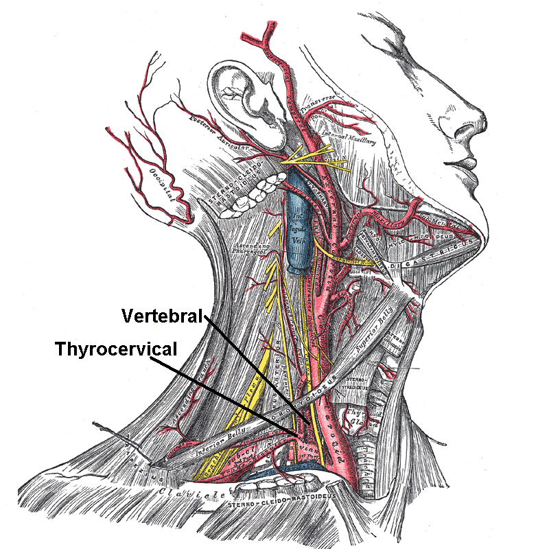
The inferior cardiac nerve arises from either the inferior cervical or the first thoracic ganglion. It descends behind the subclavian artery and along the front of the trachea, to join the deep part of the cardiac plexus. It communicates freely behind the subclavian artery with the recurrent nerve and the middle cardiac nerve. See also * inferior cervical ganglion * vagus nerve The vagus nerve, also known as the tenth cranial nerve, cranial nerve X, or simply CN X, is a cranial nerve that interfaces with the parasympathetic control of the heart, lungs, and digestive tract. It comprises two nerves—the left and right ... External links {{neuroanatomy-stub Sympathetic nervous system ... [...More Info...] [...Related Items...] OR: [Wikipedia] [Google] [Baidu] |
Heart
The heart is a muscular organ in most animals. This organ pumps blood through the blood vessels of the circulatory system. The pumped blood carries oxygen and nutrients to the body, while carrying metabolic waste such as carbon dioxide to the lungs. In humans, the heart is approximately the size of a closed fist and is located between the lungs, in the middle compartment of the chest. In humans, other mammals, and birds, the heart is divided into four chambers: upper left and right atria and lower left and right ventricles. Commonly the right atrium and ventricle are referred together as the right heart and their left counterparts as the left heart. Fish, in contrast, have two chambers, an atrium and a ventricle, while most reptiles have three chambers. In a healthy heart blood flows one way through the heart due to heart valves, which prevent backflow. The heart is enclosed in a protective sac, the pericardium, which also contains a small amount of fluid. The wall ... [...More Info...] [...Related Items...] OR: [Wikipedia] [Google] [Baidu] |
Cardiac Plexus
The cardiac plexus is a plexus of nerves situated at the base of the heart that innervates the heart. Structure The cardiac plexus is divided into a superficial part, which lies in the concavity of the aortic arch, and a deep part, between the aortic arch and the trachea. The two parts are, however, closely connected. The sympathetic component of the cardiac plexus comes from cardiac nerves, which originate from the sympathetic trunk. The parasympathetic component of the cardiac plexus originates from the cardiac branches of the vagus nerve. Superficial part The superficial part of the cardiac plexus lies beneath the arch of the aorta, in front of the right pulmonary artery. It is formed by the superior cervical cardiac branch of the left sympathetic trunk and the inferior cardiac branch of the left vagus nerve. A small ganglion, the ''cardiac ganglion of Wrisberg'', is occasionally found connected with these nerves at their point of junction. This ganglion, when present, is si ... [...More Info...] [...Related Items...] OR: [Wikipedia] [Google] [Baidu] |
Thoracic Ganglion
The thoracic ganglia are paravertebral ganglia. The thoracic portion of the sympathetic trunk typically has 12 thoracic ganglia. Emerging from the ganglia are thoracic splanchnic nerves (the cardiopulmonary, the greater, lesser, and least splanchnic nerves) that help provide sympathetic innervation to thoracic and abdominal structures. The thoracic part of sympathetic trunk lies posterior to the costovertebral pleura and is hence not a content of the posterior mediastinum Also, the ganglia of the thoracic sympathetic trunk have both white and gray rami communicantes. The white rami communicantes carry sympathetic fibers arising in the spinal cord into the sympathetic trunk, while the gray rami communicantes carry postganglionic nerve fibers of the sympathetic nervous system back to the spinal nerves A spinal nerve is a mixed nerve, which carries motor, sensory, and autonomic signals between the spinal cord and the body. In the human body there are 31 pairs of spinal nerves, on ... [...More Info...] [...Related Items...] OR: [Wikipedia] [Google] [Baidu] |
Subclavian Artery
In human anatomy, the subclavian arteries are paired major arteries of the upper thorax, below the clavicle. They receive blood from the aortic arch. The left subclavian artery supplies blood to the left arm and the right subclavian artery supplies blood to the right arm, with some branches supplying the head and thorax. On the left side of the body, the subclavian comes directly off the aortic arch, while on the right side it arises from the relatively short brachiocephalic artery when it bifurcates into the subclavian and the right common carotid artery. The usual branches of the subclavian on both sides of the body are the vertebral artery, the internal thoracic artery, the thyrocervical trunk, the costocervical trunk and the dorsal scapular artery, which may branch off the transverse cervical artery, which is a branch of the thyrocervical trunk. The subclavian becomes the axillary artery at the lateral border of the first rib. Structure From its origin, the subclavian artery t ... [...More Info...] [...Related Items...] OR: [Wikipedia] [Google] [Baidu] |
Vertebrate Trachea
The trachea, also known as the windpipe, is a cartilaginous tube that connects the larynx to the bronchi of the lungs, allowing the passage of air, and so is present in almost all air-breathing animals with lungs. The trachea extends from the larynx and branches into the two primary bronchi. At the top of the trachea the cricoid cartilage attaches it to the larynx. The trachea is formed by a number of horseshoe-shaped rings, joined together vertically by overlying ligaments, and by the trachealis muscle at their ends. The epiglottis closes the opening to the larynx during swallowing. The trachea begins to form in the second month of embryo development, becoming longer and more fixed in its position over time. It is epithelium lined with column-shaped cells that have hair-like extensions called cilia, with scattered goblet cells that produce protective mucins. The trachea can be affected by inflammation or infection, usually as a result of a viral illness affecting other parts ... [...More Info...] [...Related Items...] OR: [Wikipedia] [Google] [Baidu] |
Cardiac Plexus
The cardiac plexus is a plexus of nerves situated at the base of the heart that innervates the heart. Structure The cardiac plexus is divided into a superficial part, which lies in the concavity of the aortic arch, and a deep part, between the aortic arch and the trachea. The two parts are, however, closely connected. The sympathetic component of the cardiac plexus comes from cardiac nerves, which originate from the sympathetic trunk. The parasympathetic component of the cardiac plexus originates from the cardiac branches of the vagus nerve. Superficial part The superficial part of the cardiac plexus lies beneath the arch of the aorta, in front of the right pulmonary artery. It is formed by the superior cervical cardiac branch of the left sympathetic trunk and the inferior cardiac branch of the left vagus nerve. A small ganglion, the ''cardiac ganglion of Wrisberg'', is occasionally found connected with these nerves at their point of junction. This ganglion, when present, is si ... [...More Info...] [...Related Items...] OR: [Wikipedia] [Google] [Baidu] |
Recurrent Nerve
The recurrent laryngeal nerve (RLN) is a branch of the vagus nerve ( cranial nerve X) that supplies all the intrinsic muscles of the larynx, with the exception of the cricothyroid muscles. There are two recurrent laryngeal nerves, right and left. The right and left nerves are not symmetrical, with the left nerve looping under the aortic arch, and the right nerve looping under the right subclavian artery then traveling upwards. They both travel alongside the trachea. Additionally, the nerves are among the few nerves that follow a ''recurrent'' course, moving in the opposite direction to the nerve they branch from, a fact from which they gain their name. The recurrent laryngeal nerves supply sensation to the larynx below the vocal cords, give cardiac branches to the deep cardiac plexus, and branch to the trachea, esophagus and the inferior constrictor muscles. The posterior cricoarytenoid muscles, the only muscles that can open the vocal folds, are innervated by this nerve. ... [...More Info...] [...Related Items...] OR: [Wikipedia] [Google] [Baidu] |
Middle Cardiac Nerve
The middle cardiac nerve (''great cardiac nerve''), the largest of the three cardiac nerves, arises from the middle cervical ganglion, or from the trunk between the middle and inferior ganglia. On the right side it descends behind the common carotid artery, and at the root of the neck runs either in front of or behind the subclavian artery; it then descends on the trachea, receives a few filaments from the recurrent nerve, and joins the right half of the deep part of the cardiac plexus. In the neck, it communicates with the superior cardiac and recurrent nerves. On the left side, the middle cardiac nerve enters the chest between the left carotid and the left subclavian artery, and joins the left half of the deep part of the cardiac plexus. See also * Middle cervical ganglion The middle cervical ganglion is the smallest of the three cervical ganglia, and is occasionally absent. It is placed opposite the sixth cervical vertebra, usually in front of, or close to, the inferior t ... [...More Info...] [...Related Items...] OR: [Wikipedia] [Google] [Baidu] |
Inferior Cervical Ganglion
The inferior cervical ganglion is situated between the base of the transverse process of the last cervical vertebra and the neck of the first rib, on the medial side of the costocervical artery. Its form is irregular; it is larger in size than the middle cervical ganglion, and is frequently fused with the first thoracic ganglion, under which circumstances it is then called the "stellate ganglion." Structure It is connected to the middle cervical ganglion by two or more cords, one of which forms a loop around the subclavian artery and supplies offsets to it. This loop is named the ''ansa subclavia'' (Vieussenii). The ganglion sends gray rami communicantes to the seventh and eighth cervical nerves. Branches The inferior cervical ganglion gives off two branches: * The Inferior cardiac nerve * ''offsets to bloodvessels'' form plexuses on the subclavian artery and its branches. The plexus on the vertebral artery is continued on to the basilar, posterior cerebral, and cerebellar art ... [...More Info...] [...Related Items...] OR: [Wikipedia] [Google] [Baidu] |
Vagus Nerve
The vagus nerve, also known as the tenth cranial nerve, cranial nerve X, or simply CN X, is a cranial nerve that interfaces with the parasympathetic control of the heart, lungs, and digestive tract. It comprises two nerves—the left and right vagus nerves—but they are typically referred to collectively as a single subsystem. The vagus is the longest nerve of the autonomic nervous system in the human body and comprises both sensory and motor fibers. The sensory fibers originate from neurons of the nodose ganglion, whereas the motor fibers come from neurons of the dorsal motor nucleus of the vagus and the nucleus ambiguus. The vagus was also historically called the pneumogastric nerve. Structure Upon leaving the medulla oblongata between the olive and the inferior cerebellar peduncle, the vagus nerve extends through the jugular foramen, then passes into the carotid sheath between the internal carotid artery and the internal jugular vein down to the neck, chest, and abdom ... [...More Info...] [...Related Items...] OR: [Wikipedia] [Google] [Baidu] |


