|
Infantile Digital Fibromatosis
Infantile digital fibromatosis (IDF), also termed inclusion body fibromatosis, Reye tumor, or Reye's tumor, usually occurs as a single, small, asymptomatic, nodule in the dermis on a finger or toeFreedberg, et al. (2003). ''Fitzpatrick's Dermatology in General Medicine''. (6th ed.). Page 989. McGraw-Hill. . of infants and young children. IMF is a rare disorder with approximately 200 cases reported in the medical literature as of 2021. The World Health Organization, 2020, classified these nodules as a specific benign tumor type in the category of fibroblastic and myofibroblastic tumors. IDF was first described by the Australian pathologist, Douglas Reye, in 1965. IDF consists of an overgrowth of spindle-shaped cells in a collagen fiber-rich background located in the dermis (i.e. the layer of skin between the epidermis and subcutaneous tissue) but may extend into the subcutaneous tissue. These spindle-shaped cells contain distinctive inclusion bodies within their cytoplasm that grea ... [...More Info...] [...Related Items...] OR: [Wikipedia] [Google] [Baidu] |
Pediatrics
Pediatrics ( also spelled ''paediatrics'' or ''pædiatrics'') is the branch of medicine that involves the medical care of infants, children, adolescents, and young adults. In the United Kingdom, paediatrics covers many of their youth until the age of 18. The American Academy of Pediatrics recommends people seek pediatric care through the age of 21, but some pediatric subspecialists continue to care for adults up to 25. Worldwide age limits of pediatrics have been trending upward year after year. A medical doctor who specializes in this area is known as a pediatrician, or paediatrician. The word ''pediatrics'' and its cognates mean "healer of children," derived from the two Greek words: (''pais'' "child") and (''iatros'' "doctor, healer"). Pediatricians work in clinics, research centers, universities, general hospitals and children's hospitals, including those who practice pediatric subspecialties (e.g. neonatology requires resources available in a NICU). History The ear ... [...More Info...] [...Related Items...] OR: [Wikipedia] [Google] [Baidu] |
Protein Filament
In biology, a protein filament is a long chain of protein monomers, such as those found in hair, muscle, or in flagella. Protein filaments form together to make the cytoskeleton of the cell. They are often bundled together to provide support, strength, and rigidity to the cell. When the filaments are packed up together, they are able to form three different cellular parts. The three major classes of protein filaments that make up the cytoskeleton include: actin filaments, microtubules and intermediate filaments. Cellular types Microfilaments Compared to the other parts of the cytoskeletons, the microfilaments contain the thinnest filaments, with a diameter of approximately 7 nm. Microfilaments are part of the cytoskeleton that are composed of protein called actin. Two strands of actin intertwined together form a filamentous structure allowing for the movement of motor proteins. Microfilaments can either occur in the monomeric G-actin or filamentous F-actin. Microfilamen ... [...More Info...] [...Related Items...] OR: [Wikipedia] [Google] [Baidu] |
Calponin
Calponin is a calcium binding protein. Calponin tonically inhibits the ATPase activity of myosin in smooth muscle. Phosphorylation of calponin by a protein kinase, which is dependent upon calcium binding to calmodulin, releases the calponin's inhibition of the smooth muscle ATPase. Structure and function Calponin is mainly made up of α-helices with hydrogen bond turns. It is a binding protein and is made up of three domains. These domains in order of appearance are Calponin Homology (CH), regulatory domain (RD), and Click-23, domain that contains the calponin repeats. At the CH domain calponin binds to α-actin and filamin and binds to actin within the RD domain. Calmodulin Calmodulin (CaM) (an abbreviation for calcium-modulated protein) is a multifunctional intermediate calcium-binding messenger protein expressed in all eukaryotic cells. It is an intracellular target of the secondary messenger Ca2+, and the bind ..., when activated by calcium may bind weakly to the C ... [...More Info...] [...Related Items...] OR: [Wikipedia] [Google] [Baidu] |
Immunohistochemistry
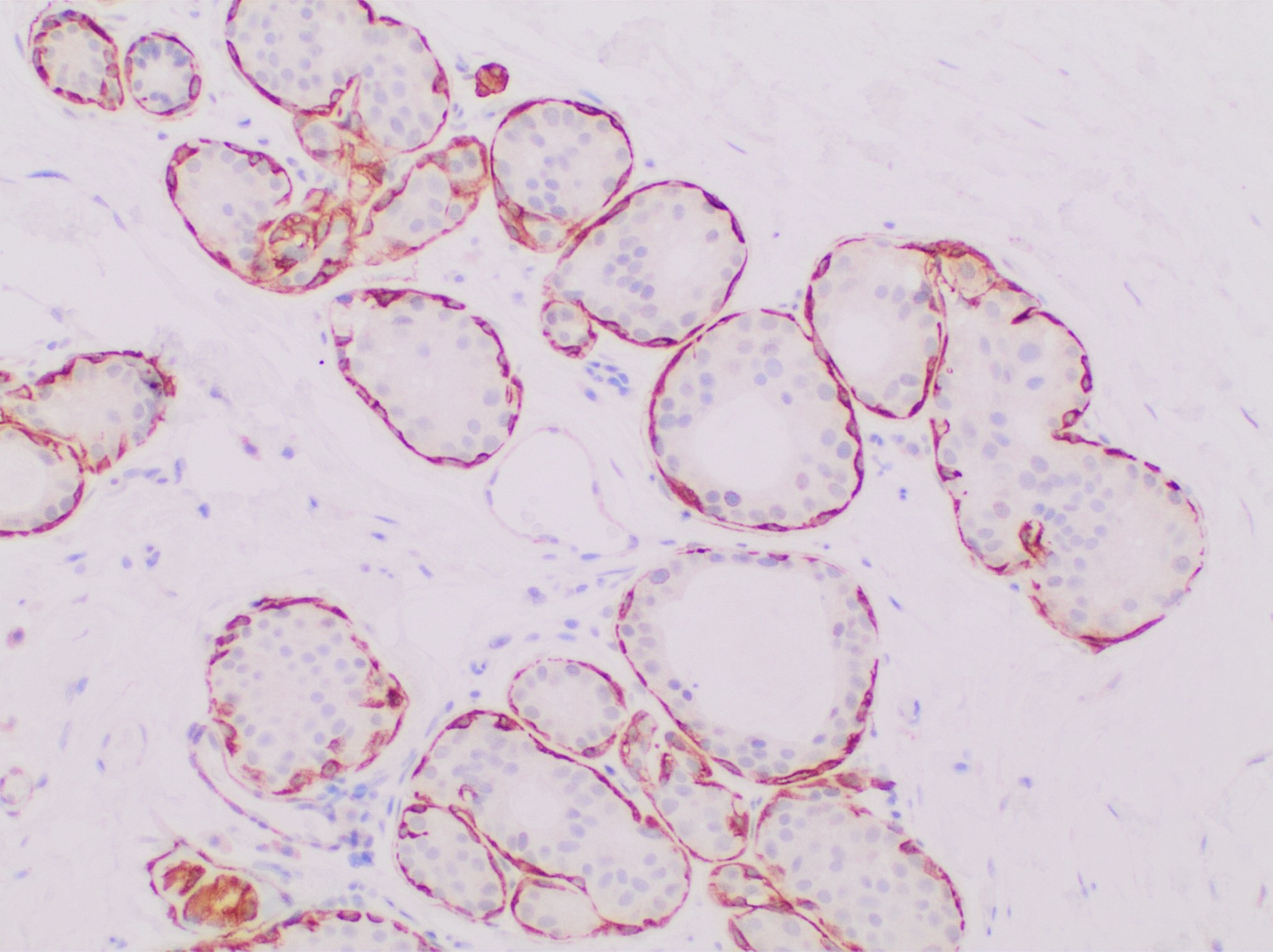
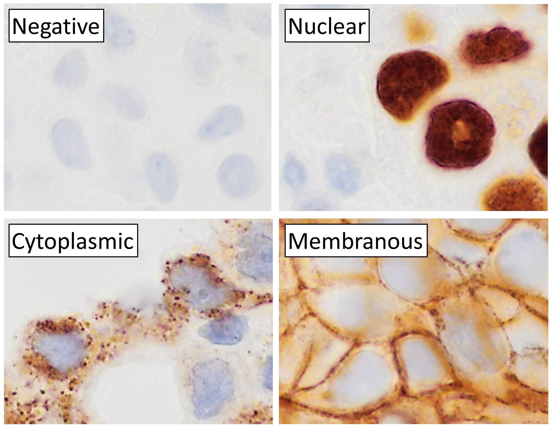
Immunohistochemistry (IHC) is the most common application of immunostaining. It involves the process of selectively identifying antigens (proteins) in cells of a tissue section by exploiting the principle of antibodies binding specifically to antigens in biological tissues. IHC takes its name from the roots "immuno", in reference to antibodies used in the procedure, and "histo", meaning tissue (compare to immunocytochemistry). Albert Coons conceptualized and first implemented the procedure in 1941. Visualising an antibody-antigen interaction can be accomplished in a number of ways, mainly either of the following: * ''Chromogenic immunohistochemistry'' (CIH), wherein an antibody is conjugated to an enzyme, such as peroxidase (the combination being termed immunoperoxidase), that can catalyse a colour-producing reaction. * '' Immunofluorescence'', where the antibody is tagged to a fluorophore, such as fluorescein or rhodamine. Immunohistochemical staining is widely used in the dia ... [...More Info...] [...Related Items...] OR: [Wikipedia] [Google] [Baidu] |
Cytoplasmic
In cell biology, the cytoplasm is all of the material within a eukaryotic cell, enclosed by the cell membrane, except for the cell nucleus. The material inside the nucleus and contained within the nuclear membrane is termed the nucleoplasm. The main components of the cytoplasm are cytosol (a gel-like substance), the organelles (the cell's internal sub-structures), and various cytoplasmic inclusions. The cytoplasm is about 80% water and is usually colorless. The submicroscopic ground cell substance or cytoplasmic matrix which remains after exclusion of the cell organelles and particles is groundplasm. It is the hyaloplasm of light microscopy, a highly complex, polyphasic system in which all resolvable cytoplasmic elements are suspended, including the larger organelles such as the ribosomes, mitochondria, the plant plastids, lipid droplets, and vacuoles. Most cellular activities take place within the cytoplasm, such as many metabolic pathways including glycolysis, and p ... [...More Info...] [...Related Items...] OR: [Wikipedia] [Google] [Baidu] |
Pathognomonic
Pathognomonic (rare synonym ''pathognomic'') is a term, often used in medicine, that means "characteristic for a particular disease". A pathognomonic sign is a particular sign whose presence means that a particular disease is present beyond any doubt. Labelling a sign or symptom "pathognomonic" represents a marked intensification of a "diagnostic" sign or symptom. The word is an adjective of Greek origin derived from πάθος ''pathos'' "disease" and γνώμων ''gnomon'' "indicator" (from γιγνώσκω ''gignosko'' "I know, I recognize"). Practical use While some findings may be classic, typical or highly suggestive in a certain condition, they may not occur ''uniquely'' in this condition and therefore may not directly imply a specific diagnosis. A pathognomonic sign or symptom has very high positive predictive value but does not need to have high sensitivity: for example it can sometimes be absent in a certain disease, since the term only implies that, when it is prese ... [...More Info...] [...Related Items...] OR: [Wikipedia] [Google] [Baidu] |
Chromatin
Chromatin is a complex of DNA and protein found in eukaryotic cells. The primary function is to package long DNA molecules into more compact, denser structures. This prevents the strands from becoming tangled and also plays important roles in reinforcing the DNA during cell division, preventing DNA damage, and regulating gene expression and DNA replication. During mitosis and meiosis, chromatin facilitates proper segregation of the chromosomes in anaphase; the characteristic shapes of chromosomes visible during this stage are the result of DNA being coiled into highly condensed chromatin. The primary protein components of chromatin are histones. An octamer of two sets of four histone cores (Histone H2A, Histone H2B, Histone H3, and Histone H4) bind to DNA and function as "anchors" around which the strands are wound.Maeshima, K., Ide, S., & Babokhov, M. (2019). Dynamic chromatin organization without the 30-nm fiber. ''Current opinion in cell biology, 58,'' 95–104. https://doi.o ... [...More Info...] [...Related Items...] OR: [Wikipedia] [Google] [Baidu] |
Cell Nucleus
The cell nucleus (pl. nuclei; from Latin or , meaning ''kernel'' or ''seed'') is a membrane-bound organelle found in eukaryotic cells. Eukaryotic cells usually have a single nucleus, but a few cell types, such as mammalian red blood cells, have no nuclei, and a few others including osteoclasts have many. The main structures making up the nucleus are the nuclear envelope, a double membrane that encloses the entire organelle and isolates its contents from the cellular cytoplasm; and the nuclear matrix, a network within the nucleus that adds mechanical support. The cell nucleus contains nearly all of the cell's genome. Nuclear DNA is often organized into multiple chromosomes – long stands of DNA dotted with various proteins, such as histones, that protect and organize the DNA. The genes within these chromosomes are structured in such a way to promote cell function. The nucleus maintains the integrity of genes and controls the activities of the cell by regulating gene expres ... [...More Info...] [...Related Items...] OR: [Wikipedia] [Google] [Baidu] |
H&E Stain
Hematoxylin and eosin stain ( or haematoxylin and eosin stain or hematoxylin-eosin stain; often abbreviated as H&E stain or HE stain) is one of the principal tissue stains used in histology. It is the most widely used stain in medical diagnosis and is often the gold standard. For example, when a pathologist looks at a biopsy of a suspected cancer, the histological section is likely to be stained with H&E. H&E is the combination of two histological stains: hematoxylin and eosin. The hematoxylin stains cell nuclei a purplish blue, and eosin stains the extracellular matrix and cytoplasm pink, with other structures taking on different shades, hues, and combinations of these colors. Hence a pathologist can easily differentiate between the nuclear and cytoplasmic parts of a cell, and additionally, the overall patterns of coloration from the stain show the general layout and distribution of cells and provides a general overview of a tissue sample's structure. Thus, pattern recogniti ... [...More Info...] [...Related Items...] OR: [Wikipedia] [Google] [Baidu] |
Smooth Muscle
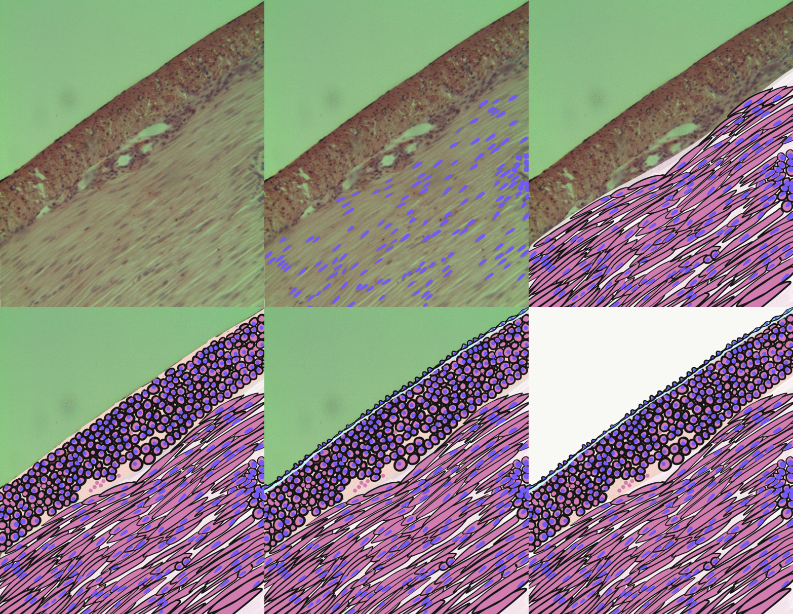
Smooth muscle is an involuntary non-striated muscle, so-called because it has no sarcomeres and therefore no striations (''bands'' or ''stripes''). It is divided into two subgroups, single-unit and multiunit smooth muscle. Within single-unit muscle, the whole bundle or sheet of smooth muscle cells contracts as a syncytium. Smooth muscle is found in the walls of hollow organs, including the stomach, intestines, bladder and uterus; in the walls of passageways, such as blood, and lymph vessels, and in the tracts of the respiratory, urinary, and reproductive systems. In the eyes, the ciliary muscles, a type of smooth muscle, dilate and contract the iris and alter the shape of the lens. In the skin, smooth muscle cells such as those of the arrector pili cause hair to stand erect in response to cold temperature or fear. Structure Gross anatomy Smooth muscle is grouped into two types: single-unit smooth muscle, also known as visceral smooth muscle, and multiunit smooth muscle. ... [...More Info...] [...Related Items...] OR: [Wikipedia] [Google] [Baidu] |
Myofibroblast
A myofibroblast is a cell phenotype that was first described as being in a state between a fibroblast and a smooth muscle cell. Structure Myofibroblasts are contractile web-like fusiform cells that are identifiable by their expression of α-smooth muscle actin within their cytoplasmic stress fibers. In the gastrointestinal and genitourinary tracts, myofibroblasts are found subepithelially in mucosal surfaces. Here they not only act as a regulator of the shape of the crypts and villi, but also act as stem-niche cells in the intestinal crypts and as parts of atypical antigen-presenting cells. They have both support as well as paracrine function in most places. Location Myofibroblasts were first identified in granulation tissue during skin wound healing. Typically, these cells are found in granulation tissue, scar tissue (fibrosis) and the stroma of tumours. They also line the gastrointestinal tract, wherein they regulate the shapes of crypts and villi. Markers Myofibroblasts ... [...More Info...] [...Related Items...] OR: [Wikipedia] [Google] [Baidu] |
Fibroblasts
A fibroblast is a type of biological cell that synthesizes the extracellular matrix and collagen, produces the structural framework ( stroma) for animal tissues, and plays a critical role in wound healing. Fibroblasts are the most common cells of connective tissue in animals. Structure Fibroblasts have a branched cytoplasm surrounding an elliptical, speckled nucleus having two or more nucleoli. Active fibroblasts can be recognized by their abundant rough endoplasmic reticulum. Inactive fibroblasts (called fibrocytes) are smaller, spindle-shaped, and have a reduced amount of rough endoplasmic reticulum. Although disjointed and scattered when they have to cover a large space, fibroblasts, when crowded, often locally align in parallel clusters. Unlike the epithelial cells lining the body structures, fibroblasts do not form flat monolayers and are not restricted by a polarizing attachment to a basal lamina on one side, although they may contribute to basal lamina components in s ... [...More Info...] [...Related Items...] OR: [Wikipedia] [Google] [Baidu] |


.jpg)