|
Image-guided Surgery
Image-guided surgery (IGS) is any surgical procedure where the surgeon uses tracked surgical instruments in conjunction with preoperative or intraoperative images in order to directly or indirectly guide the procedure. Image guided surgery systems use cameras, ultrasonic, electromagnetic or a combination or fields to capture and relay the patient's anatomy and the surgeon's precise movements in relation to the patient, to computer monitors in the operating room or to augmented reality headsets (augmented reality surgical navigation technology). This is generally performed in real-time though there may be delays of seconds or minutes depending on the modality and application. Image-guided surgery helps surgeons perform safer and less invasive procedures and has become a recognized standard of care in managing disorders including cranial, otorhinolaryngology, spine, orthopedic, and cardiovascular. Benefits The benefits of Image-guided surgery include greater control of the surgica ... [...More Info...] [...Related Items...] OR: [Wikipedia] [Google] [Baidu] |
Surgery
Surgery ''cheirourgikē'' (composed of χείρ, "hand", and ἔργον, "work"), via la, chirurgiae, meaning "hand work". is a medical specialty that uses operative manual and instrumental techniques on a person to investigate or treat a pathological condition such as a disease or injury, to help improve bodily function, appearance, or to repair unwanted ruptured areas. The act of performing surgery may be called a surgical procedure, operation, or simply "surgery". In this context, the verb "operate" means to perform surgery. The adjective surgical means pertaining to surgery; e.g. surgical instruments or surgical nurse. The person or subject on which the surgery is performed can be a person or an animal. A surgeon is a person who practices surgery and a surgeon's assistant is a person who practices surgical assistance. A surgical team is made up of the surgeon, the surgeon's assistant, an anaesthetist, a circulating nurse and a surgical technologist. Surgery usually span ... [...More Info...] [...Related Items...] OR: [Wikipedia] [Google] [Baidu] |
Neurosurgery
Neurosurgery or neurological surgery, known in common parlance as brain surgery, is the medical specialty concerned with the surgical treatment of disorders which affect any portion of the nervous system including the brain, spinal cord and peripheral nervous system. Education and context In different countries, there are different requirements for an individual to legally practice neurosurgery, and there are varying methods through which they must be educated. In most countries, neurosurgeon training requires a minimum period of seven years after graduating from medical school. United States In the United States, a neurosurgeon must generally complete four years of undergraduate education, four years of medical school, and seven years of residency (PGY-1-7). Most, but not all, residency programs have some component of basic science or clinical research. Neurosurgeons may pursue additional training in the form of a fellowship after residency, or, in some cases, as a senior ... [...More Info...] [...Related Items...] OR: [Wikipedia] [Google] [Baidu] |
Radiosurgery
Radiosurgery is surgery using radiation, that is, the destruction of precisely selected areas of tissue using ionizing radiation rather than excision with a blade. Like other forms of radiation therapy (also called radiotherapy), it is usually used to treat cancer. Radiosurgery was originally defined by the Swedish neurosurgeon Lars Leksell as "a single high dose fraction of radiation, stereotactically directed to an intracranial region of interest". In stereotactic radiosurgery (SRS), the word " stereotactic" refers to a three-dimensional coordinate system that enables accurate correlation of a virtual target seen in the patient's diagnostic images with the actual target position in the patient. Stereotactic radiosurgery may also be called stereotactic body radiation therapy (SBRT) or stereotactic ablative radiotherapy (SABR) when used outside the central nervous system (CNS). History Stereotactic radiosurgery was first developed in 1949 by the Swedish neurosurgeon Lars Lek ... [...More Info...] [...Related Items...] OR: [Wikipedia] [Google] [Baidu] |
Microsoft Hololens
Microsoft HoloLens is an augmented reality (AR)/ mixed reality (MR) headset developed and manufactured by Microsoft. HoloLens runs the Windows Mixed Reality platform under the Windows 10 operating system. Some of the positional tracking technology used in HoloLens can trace its lineage to the Microsoft Kinect, an accessory for Microsoft's Xbox game console that was introduced in 2010. The pre-production version of HoloLens, the Development Edition, shipped on March 30, 2016, and is targeted to developers in the United States and Canada for a list price of $3000 which allowed hobbyist, professionals, and corporations to participate in the pre-production version of HoloLens. Samsung and Asus have extended an offer to Microsoft to help produce their own mixed-reality products, in collaboration with Microsoft, based around the concept and hardware on HoloLens. On October 12, 2016, Microsoft announced global expansion of HoloLens and publicized that HoloLens would be available f ... [...More Info...] [...Related Items...] OR: [Wikipedia] [Google] [Baidu] |
Intraoperative MRI
Intraoperative magnetic resonance imaging (iMRI) refers to an operating room configuration that enables surgeons to image the patient via an MRI scanner while the patient is undergoing surgery, particularly brain surgery. iMRI reduces the risk of damaging critical parts of the brain and helps confirm that the surgery was successful or if additional resection is needed before the patient’s head is closed and the surgery completed.Chicoine MR, Lim CC, et al. 2011. Implementation and preliminary clinical experience with the use of ceiling mounted mobile high field intraoperative magnetic resonance imaging between two operating rooms. Acta Neurochir Suppl. 2011:109:97-102 Equipment and operating suite configuration Compared to other imaging types, high-field iMRI requires the additional cost of specialized operating suites, instrumentation and longer anesthesia and operating room time; however, published studies show use of iMRI increases physicians’ ability to detect residual tumo ... [...More Info...] [...Related Items...] OR: [Wikipedia] [Google] [Baidu] |
Interventional Radiology
Interventional radiology (IR) is a medical specialty that performs various minimally-invasive procedures using medical imaging guidance, such as x-ray fluoroscopy, computed tomography, magnetic resonance imaging, or ultrasound. IR performs both diagnostic and therapeutic procedures through very small incisions or body orifices. Diagnostic IR procedures are those intended to help make a diagnosis or guide further medical treatment, and include image-guided biopsy of a tumor or injection of an imaging contrast agent into a hollow structure, such as a blood vessel or a duct. By contrast, therapeutic IR procedures provide direct treatment—they include catheter-based medicine delivery, medical device placement (e.g., stents), and angioplasty of narrowed structures. The main benefits of interventional radiology techniques are that they can reach the deep structures of the body through a body orifice or tiny incision using small needles and wires. That decreases risks, pain, ... [...More Info...] [...Related Items...] OR: [Wikipedia] [Google] [Baidu] |
Computer Assisted Surgery
Computer-assisted surgery (CAS) represents a surgical concept and set of methods, that use computer technology for surgical planning, and for guiding or performing surgical interventions. CAS is also known as computer-aided surgery, computer-assisted intervention, image-guided surgery, digital surgery and surgical navigation, but these are terms that are more or less synonymous with CAS. CAS has been a leading factor in the development of robotic surgery. General principles Creating a virtual image of the patient The most important component for CAS is the development of an accurate model of the patient. This can be conducted through a number of medical imaging technologies including CT, MRI, x-rays, ultrasound plus many more. For the generation of this model, the anatomical region to be operated has to be scanned and uploaded into the computer system. It is possible to employ a number of scanning methods, with the datasets combined through data fusion techniques. ... [...More Info...] [...Related Items...] OR: [Wikipedia] [Google] [Baidu] |
N-localizer
The N-localizer is a device that enables guidance of stereotactic surgery or radiosurgery using tomographic images that are obtained via computed tomography (CT), magnetic resonance imaging (MRI), or positron emission tomography (PET). The N-localizer comprises a diagonal rod that spans two vertical rods to form an N-shape (Figure 1) and permits calculation of the point where a tomographic image plane intersects the diagonal rod. Attaching three N-localizers to a stereotactic instrument allows calculation of three points where a tomographic image plane intersects three diagonal rods (Figure 2). These points determine the spatial orientation of the tomographic image plane relative to the stereotactic frame. The N-localizer is integrated with the Brown-Roberts-Wells (BRW), Kelly-Goerss, Leksell, Cosman-Roberts-Wells (CRW), Micromar-ETM03B, FiMe-BlueFrame, Macom, and Adeor-Zeppelin stereotactic frames and with the Gamma Knife radiosurgery system. An alternative to the N-localizer is ... [...More Info...] [...Related Items...] OR: [Wikipedia] [Google] [Baidu] |
Positron Emission Tomography
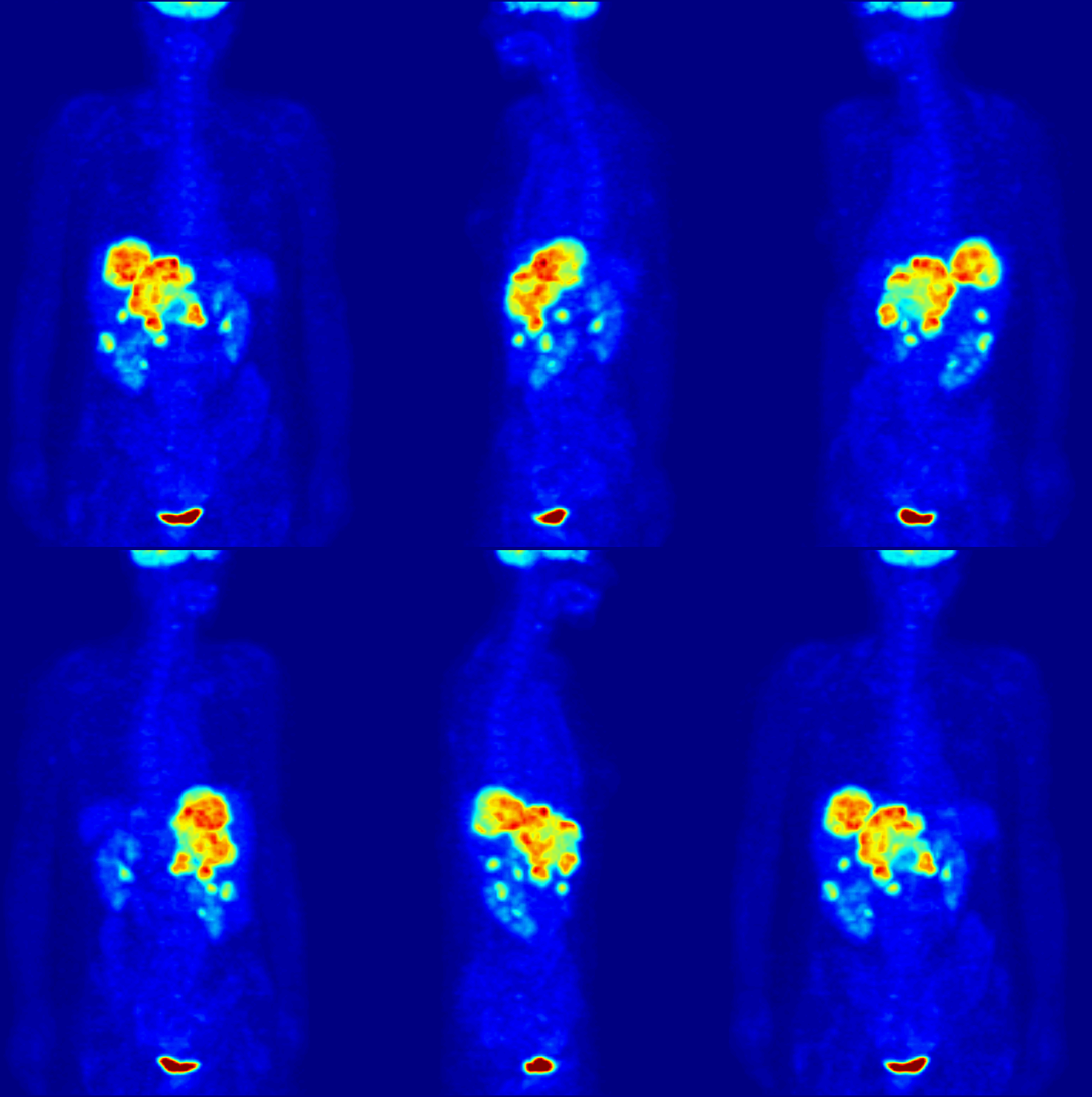
Positron emission tomography (PET) is a functional imaging technique that uses radioactive substances known as radiotracers to visualize and measure changes in metabolic processes, and in other physiological activities including blood flow, regional chemical composition, and absorption. Different tracers are used for various imaging purposes, depending on the target process within the body. For example: * Fluorodeoxyglucose ( 18F">sup>18FDG or FDG) is commonly used to detect cancer; * 18Fodium fluoride">sup>18Fodium fluoride (Na18F) is widely used for detecting bone formation; * Oxygen-15 (15O) is sometimes used to measure blood flow. PET is a common imaging technique, a medical scintillography technique used in nuclear medicine. A radiopharmaceutical – a radioisotope attached to a drug – is injected into the body as a radioactive tracer, tracer. When the radiopharmaceutical undergoes beta plus decay, a positron is emitted, and when the positron interacts with an or ... [...More Info...] [...Related Items...] OR: [Wikipedia] [Google] [Baidu] |
Magnetic Resonance Imaging
Magnetic resonance imaging (MRI) is a medical imaging technique used in radiology to form pictures of the anatomy and the physiological processes inside the body. MRI scanners use strong magnetic fields, magnetic field gradients, and radio waves to generate images of the organs in the body. MRI does not involve X-rays or the use of ionizing radiation, which distinguishes it from computed tomography (CT) and positron emission tomography (PET) scans. MRI is a medical application of nuclear magnetic resonance (NMR) which can also be used for imaging in other NMR applications, such as NMR spectroscopy. MRI is widely used in hospitals and clinics for medical diagnosis, staging and follow-up of disease. Compared to CT, MRI provides better contrast in images of soft tissues, e.g. in the brain or abdomen. However, it may be perceived as less comfortable by patients, due to the usually longer and louder measurements with the subject in a long, confining tube, although "open ... [...More Info...] [...Related Items...] OR: [Wikipedia] [Google] [Baidu] |
Computed Tomography
A computed tomography scan (CT scan; formerly called computed axial tomography scan or CAT scan) is a medical imaging technique used to obtain detailed internal images of the body. The personnel that perform CT scans are called radiographers or radiology technologists. CT scanners use a rotating X-ray tube and a row of detectors placed in a gantry to measure X-ray attenuations by different tissues inside the body. The multiple X-ray measurements taken from different angles are then processed on a computer using tomographic reconstruction algorithms to produce tomographic (cross-sectional) images (virtual "slices") of a body. CT scans can be used in patients with metallic implants or pacemakers, for whom magnetic resonance imaging (MRI) is contraindicated. Since its development in the 1970s, CT scanning has proven to be a versatile imaging technique. While CT is most prominently used in medical diagnosis, it can also be used to form images of non-living objects. The 1979 N ... [...More Info...] [...Related Items...] OR: [Wikipedia] [Google] [Baidu] |
Radiosurgery
Radiosurgery is surgery using radiation, that is, the destruction of precisely selected areas of tissue using ionizing radiation rather than excision with a blade. Like other forms of radiation therapy (also called radiotherapy), it is usually used to treat cancer. Radiosurgery was originally defined by the Swedish neurosurgeon Lars Leksell as "a single high dose fraction of radiation, stereotactically directed to an intracranial region of interest". In stereotactic radiosurgery (SRS), the word " stereotactic" refers to a three-dimensional coordinate system that enables accurate correlation of a virtual target seen in the patient's diagnostic images with the actual target position in the patient. Stereotactic radiosurgery may also be called stereotactic body radiation therapy (SBRT) or stereotactic ablative radiotherapy (SABR) when used outside the central nervous system (CNS). History Stereotactic radiosurgery was first developed in 1949 by the Swedish neurosurgeon Lars Lek ... [...More Info...] [...Related Items...] OR: [Wikipedia] [Google] [Baidu] |
.jpg)


