|
Idiopathic Orbital Inflammatory Disease
Idiopathic orbital inflammatory (IOI) disease refers to a marginated mass-like enhancing soft tissue involving any area of the orbit. It is the most common painful orbital mass in the adult population, and is associated with proptosis, cranial nerve palsy (Tolosa–Hunt syndrome), uveitis, and retinal detachment. Idiopathic orbital inflammatory syndrome, also known as orbital pseudotumor, was first described by GleasonGleason JE. Idipathic myositis involving the extraocular muscles. Ophthalmol Rec.12:471–478, 1903 in 1903 and by Busse and Hochheim.Busse O, Hochheim W. cited by Dunnington JH, Berke RN. Exophthalmos due to chronic orbital myositic. Arch Ophthal . 30:446–466, 1943 It was then characterized as a distinct entity in 1905 by Birch-Hirschfeld.Birch-Hirschfeld A. Zur diagnostic and pathologic der orbital tumoren. Ber Dtsch Ophthalmol Ges. 32: 127–135, 1905Birch-Hirschfeld A. Handbuch der gesamten augenheilkunde, vol. 9. Berlin: Julius Springer. p. 251–253, 1930 It ... [...More Info...] [...Related Items...] OR: [Wikipedia] [Google] [Baidu] |
Proptosis
Exophthalmos (also called exophthalmus, exophthalmia, proptosis, or exorbitism) is a bulging of the eye anteriorly out of the orbit. Exophthalmos can be either bilateral (as is often seen in Graves' disease) or unilateral (as is often seen in an orbital tumor). Complete or partial dislocation from the orbit is also possible from trauma or swelling of surrounding tissue resulting from trauma. In the case of Graves' disease, the displacement of the eye results from abnormal connective tissue deposition in the orbit and extraocular muscles, which can be visualized by CT or MRI. If left untreated, exophthalmos can cause the eyelids to fail to close during sleep, leading to corneal dryness and damage. Another possible complication is a form of redness or irritation called superior limbic keratoconjunctivitis, in which the area above the cornea becomes inflamed as a result of increased friction when blinking. The process that is causing the displacement of the eye may also compre ... [...More Info...] [...Related Items...] OR: [Wikipedia] [Google] [Baidu] |
Cranial Nerve
Cranial nerves are the nerves that emerge directly from the brain (including the brainstem), of which there are conventionally considered twelve pairs. Cranial nerves relay information between the brain and parts of the body, primarily to and from regions of the head and neck, including the special senses of vision, taste, smell, and hearing. The cranial nerves emerge from the central nervous system above the level of the first vertebra of the vertebral column. Each cranial nerve is paired and is present on both sides. There are conventionally twelve pairs of cranial nerves, which are described with Roman numerals I–XII. Some considered there to be thirteen pairs of cranial nerves, including cranial nerve zero. The numbering of the cranial nerves is based on the order in which they emerge from the brain and brainstem, from front to back. The terminal nerves (0), olfactory nerves (I) and optic nerves (II) emerge from the cerebrum, and the remaining ten pairs arise from t ... [...More Info...] [...Related Items...] OR: [Wikipedia] [Google] [Baidu] |
Tolosa–Hunt Syndrome
Tolosa–Hunt syndrome is a rare disorder characterized by severe and unilateral headaches with orbital pain, along with weakness and paralysis (ophthalmoplegia) of certain eye muscles ( extraocular palsies). In 2004, the International Headache Society provided a definition of the diagnostic criteria which included granuloma. Signs and symptoms Symptoms are usually limited to one side of the head, and in most cases the individual affected will experience intense, sharp pain and paralysis of muscles around the eye. Symptoms may subside without medical intervention, yet recur without a noticeable pattern. In addition, affected individuals may experience paralysis of various facial nerves and drooping of the upper eyelid ( ptosis). Other signs include double vision, fever, chronic fatigue, vertigo or arthralgia. Occasionally the patient may present with a feeling of protrusion of one or both eyeballs (exophthalmos). Causes The cause of Tolosa–Hunt syndrome is not known, but the d ... [...More Info...] [...Related Items...] OR: [Wikipedia] [Google] [Baidu] |
Uveitis
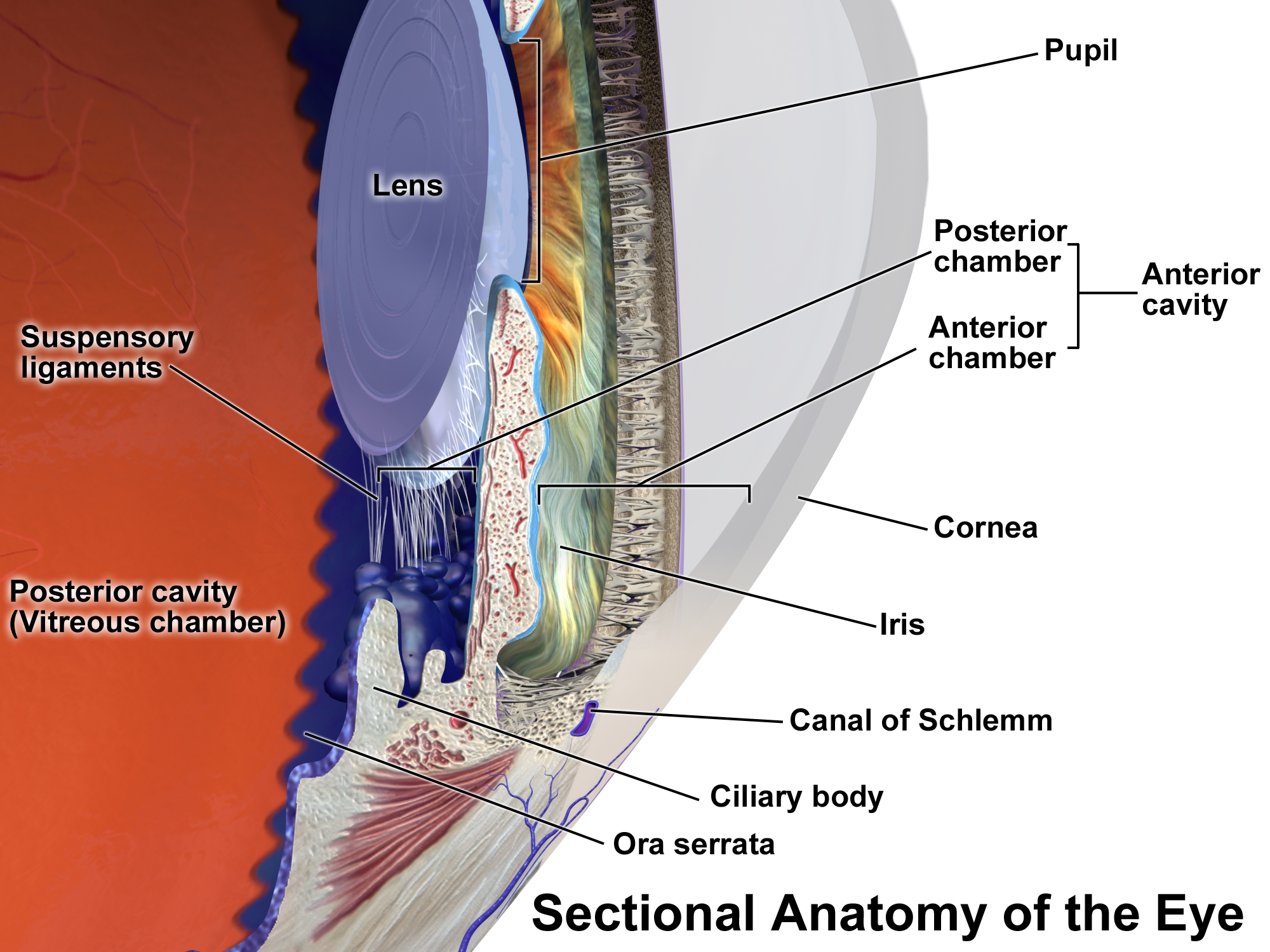
Uveitis () is inflammation of the uvea, the pigmented layer of the eye between the inner retina and the outer fibrous layer composed of the sclera and cornea. The uvea consists of the middle layer of pigmented vascular structures of the eye and includes the iris, ciliary body, and choroid. Uveitis is described anatomically, by the part of the eye affected, as anterior, intermediate or posterior, or panuveitic if all parts are involved. Anterior uveitis ( iridocyclytis) is the most common, with the incidence of uveitis overall affecting approximately 1:4500, most commonly those between the ages of 20-60. Symptoms include eye pain, eye redness, floaters and blurred vision, and ophthalmic examination may show dilated ciliary blood vessels and the presence of cells in the anterior chamber. Uveitis may arise spontaneously, have a genetic component, or be associated with an autoimmune disease or infection. While the eye is a relatively protected environment, its immune mechanisms ... [...More Info...] [...Related Items...] OR: [Wikipedia] [Google] [Baidu] |
Retinal Detachment
Retinal detachment is a disorder of the eye in which the retina peels away from its underlying layer of support tissue. Initial detachment may be localized, but without rapid treatment the entire retina may detach, leading to vision loss and blindness. It is a surgical emergency. The retina is a thin layer of light-sensitive tissue on the back wall of the eye. The optical system of the eye focuses light on the retina much like light is focused on the film in a camera. The retina translates that focused image into neural impulses and sends them to the brain via the optic nerve. Occasionally, posterior vitreous detachment, injury or trauma to the eye or head may cause a small tear in the retina. The tear allows vitreous fluid to seep through it under the retina, and peel it away like a bubble in wallpaper. Diagnosis Symptoms As the retina is responsible for vision, persons experiencing a retinal detachment have vision loss. This can be painful or painless. Imaging Ultraso ... [...More Info...] [...Related Items...] OR: [Wikipedia] [Google] [Baidu] |
Graves' Ophthalmopathy
Graves’ ophthalmopathy, also known as thyroid eye disease (TED), is an autoimmune inflammatory disorder of the orbit and periorbital tissues, characterized by upper eyelid retraction, lid lag, swelling, redness (erythema), conjunctivitis, and bulging eyes (exophthalmos). It occurs most commonly in individuals with Graves' disease, and less commonly in individuals with Hashimoto's thyroiditis, or in those who are euthyroid. It is part of a systemic process with variable expression in the eyes, thyroid, and skin, caused by autoantibodies that bind to tissues in those organs. The autoantibodies target the fibroblasts in the eye muscles, and those fibroblasts can differentiate into fat cells (adipocytes). Fat cells and muscles expand and become inflamed. Veins become compressed and are unable to drain fluid, causing edema. Annual incidence is 16/100,000 in women, 3/100,000 in men. About 3–5% have severe disease with intense pain, and sight-threatening corneal ulceration or compr ... [...More Info...] [...Related Items...] OR: [Wikipedia] [Google] [Baidu] |
IgG4-related Ophthalmic Disease
IgG4-related ophthalmic disease (IgG4-ROD) is the recommended term to describe orbital (eye socket) manifestations of the systemic condition IgG4-related disease, which is characterised by infiltration of lymphocytes and plasma cells and subsequent fibrosis in involved structures. It can involve one or more of the orbital structures. Frequently involved structures include the lacrimal glands, extraocular muscles, infraorbital nerve, supraorbital nerve and eyelids. It has also been speculated that ligneous conjunctivitis may be a manifestation of IgG4-related disease (IgG4-RD). As is the case with other manifestations of IgG4-related disease, a prompt response to steroid therapy is a characteristic feature of IgG4-ROD in most cases, unless significant fibrosis has already occurred. It basically can cause loss of vision and in other cases, cause skin irritation and loss of brain function Symptoms Symptoms, if any, can be mild even in the presence of sig ... [...More Info...] [...Related Items...] OR: [Wikipedia] [Google] [Baidu] |
Sarcoidosis
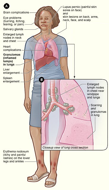
Sarcoidosis (also known as ''Besnier-Boeck-Schaumann disease'') is a disease involving abnormal collections of inflammatory cells that form lumps known as granulomata. The disease usually begins in the lungs, skin, or lymph nodes. Less commonly affected are the eyes, liver, heart, and brain. Any organ can be affected though. The signs and symptoms depend on the organ involved. Often, no, or only mild, symptoms are seen. When it affects the lungs, wheezing, coughing, shortness of breath, or chest pain may occur. Some may have Löfgren syndrome with fever, large lymph nodes, arthritis, and a rash known as erythema nodosum. The cause of sarcoidosis is unknown. Some believe it may be due to an immune reaction to a trigger such as an infection or chemicals in those who are genetically predisposed. Those with affected family members are at greater risk. Diagnosis is partly based on signs and symptoms, which may be supported by biopsy. Findings that make it likely include large lymph n ... [...More Info...] [...Related Items...] OR: [Wikipedia] [Google] [Baidu] |
Granulomatosis With Polyangiitis
Granulomatosis with polyangiitis (GPA), previously known as Wegener's granulomatosis (WG), is a rare long-term systemic disorder that involves the formation of granulomas and inflammation of blood vessels (vasculitis). It is a form of vasculitis that affects small- and medium-size vessels in many organs but most commonly affects the upper respiratory tract, lungs and kidneys. The signs and symptoms of GPA are highly varied and reflect which organs are supplied by the affected blood vessels. Typical signs and symptoms include nosebleeds, stuffy nose and crustiness of nasal secretions, and inflammation of the uveal layer of the eye. Damage to the heart, lungs and kidneys can be fatal. The cause of GPA is unknown. Genetics have been found to play a role in GPA though the risk of inheritance appears to be low. GPA treatment depends on the severity of the disease. Severe disease is typically treated with a combination of immunosuppressive medications such as rituximab or cyclopho ... [...More Info...] [...Related Items...] OR: [Wikipedia] [Google] [Baidu] |
Orbital Cellulitis
Orbital cellulitis is inflammation of eye tissues behind the orbital septum. It is most commonly caused by an acute spread of infection into the eye socket from either the adjacent sinuses or through the blood. It may also occur after trauma. When it affects the rear of the eye, it is known as retro-orbital cellulitis. It should not be confused with periorbital cellulitis, which refers to cellulitis anterior to the septum. Without proper treatment, orbital cellulitis may lead to serious consequences, including permanent loss of vision or even death. Signs and symptoms Orbital cellulitis commonly presents with painful eye movement, sudden vision loss, chemosis, bulging of the infected eye, and limited eye movement. Along with these symptoms, patients typically have redness and swelling of the eyelid, pain, discharge, inability to open the eye, occasional fever and lethargy. Complications Complications include hearing loss, blood infection, meningitis, cavernous sinus thromb ... [...More Info...] [...Related Items...] OR: [Wikipedia] [Google] [Baidu] |
Carotid-cavernous Fistula
A carotid-cavernous fistula results from an abnormal communication between the arterial and venous systems within the cavernous sinus in the skull. It is a type of arteriovenous fistula. As arterial blood under high pressure enters the cavernous sinus, the normal venous return to the cavernous sinus is impeded and this causes engorgement of the draining veins, manifesting most dramatically as a sudden engorgement and redness of the eye of the same side. Presentation CCF symptoms include bruit (a humming sound within the skull due to high blood flow through the arteriovenous fistula), progressive visual loss, and pulsatile proptosis Exophthalmos (also called exophthalmus, exophthalmia, proptosis, or exorbitism) is a bulging of the eye anteriorly out of the orbit. Exophthalmos can be either bilateral (as is often seen in Graves' disease) or unilateral (as is often seen i ... or progressive bulging of the eye due to dilatation of the veins draining the eye. Pain is the sympto ... [...More Info...] [...Related Items...] OR: [Wikipedia] [Google] [Baidu] |


