|
Head Of Ulna
The ulna (''pl''. ulnae or ulnas) is a long bone found in the forearm that stretches from the elbow to the smallest finger, and when in anatomical position, is found on the medial side of the forearm. That is, the ulna is on the same side of the forearm as the little finger. It runs parallel to the radius, the other long bone in the forearm. The ulna is usually slightly longer than the radius, but the radius is thicker. Therefore, the radius is considered to be the larger of the two. Structure The ulna is a long bone found in the forearm that stretches from the elbow to the smallest finger, and when in anatomical position, is found on the medial side of the forearm. It is broader close to the elbow, and narrows as it approaches the wrist. Close to the elbow, the ulna has a bony process, the olecranon process, a hook-like structure that fits into the olecranon fossa of the humerus. This prevents hyperextension and forms a hinge joint with the trochlea of the humerus. There is ... [...More Info...] [...Related Items...] OR: [Wikipedia] [Google] [Baidu] |
Standard Anatomical Position
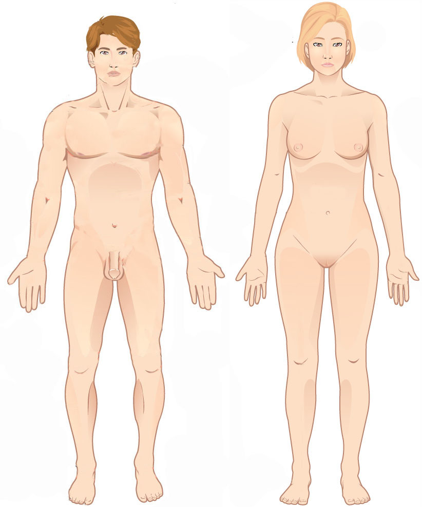
The standard anatomical position, or standard anatomical model, is the scientifically agreed upon reference position for anatomical location terms. Standard anatomical positions are used to standardise the position of appendages of animals with respect to the main body of the organism. In medical disciplines, all references to a location on or in the body are made based upon the standard anatomical position. A straight position is assumed when describing a proximo-distal axis (towards or away from a point of attachment). This helps avoid confusion in terminology when referring to the same organism in different postures. For example, if the elbow is flexed, the hand remains distal to the shoulder even if it approaches the shoulder. Human anatomy In standard anatomical position, the human body is standing erect and at rest. Unlike the situation in other vertebrates, the limbs are placed in positions reminiscent of the supine position imposed on cadavers during autopsy. Theref ... [...More Info...] [...Related Items...] OR: [Wikipedia] [Google] [Baidu] |
Gray212
Grey (more common in British English) or gray (more common in American English) is an intermediate color between black and white. It is a neutral or achromatic color, meaning literally that it is "without color", because it can be composed of black and white. It is the color of a cloud-covered sky, of ash and of lead. The first recorded use of ''grey'' as a color name in the English language was in 700 CE.Maerz and Paul ''A Dictionary of Color'' New York:1930 McGraw-Hill Page 196 ''Grey'' is the dominant spelling in European and Commonwealth English, while ''gray'' has been the preferred spelling in American English; both spellings are valid in both varieties of English. In Europe and North America, surveys show that grey is the color most commonly associated with neutrality, conformity, boredom, uncertainty, old age, indifference, and modesty. Only one percent of respondents chose it as their favorite color. Etymology ''Grey'' comes from the Middle English o ... [...More Info...] [...Related Items...] OR: [Wikipedia] [Google] [Baidu] |
Flexor Pollicis Longus
The flexor pollicis longus (; FPL, Latin ''flexor'', bender; ''pollicis'', of the thumb; ''longus'', long) is a muscle in the forearm and hand that flexes the thumb. It lies in the same plane as the flexor digitorum profundus. This muscle is unique to humans, being either rudimentary or absent in other primates. A meta-analysis indicated accessory flexor pollicis longus is present in around 48% of the population. Human anatomy Origin and insertion It arises from the grooved anterior (side of palm) surface of the body of the radius, extending from immediately below the radial tuberosity and oblique line to within a short distance of the pronator quadratus muscle.Gray 1918, ''Flexor Pollicis Longus'', paras 20, 25 An occasionally present accessory long head of the flexor pollicis longus muscle is called 'Gantzer's muscle'. It may cause compression of the anterior interosseous nerve. It arises also from the adjacent part of the interosseous membrane of the forearm, and genera ... [...More Info...] [...Related Items...] OR: [Wikipedia] [Google] [Baidu] |
Pronator Teres
The pronator teres is a muscle (located mainly in the forearm) that, along with the pronator quadratus, serves to pronate the forearm (turning it so that the palm faces posteriorly when from the anatomical position). It has two attachments, to the medial humeral supracondylar ridge and the ulnar tuberosity, and inserts near the middle of the radius. Structure The pronator teres has two heads—humeral and ulnar. * The humeral head, the larger and more superficial, arises from the medial supracondylar ridge immediately superior to the medial epicondyle of the humerus, and from the common flexor tendon (which arises from the medial epicondyle). * The ulnar head (or ulnar tuberosity) is a thin fasciculus, which arises from the medial side of the coronoid process of the ulna, and joins the preceding at an acute angle. The median nerve enters the forearm between the two heads of the muscle, and is separated from the ulnar artery by the ulnar head. The muscle passes obliquely acros ... [...More Info...] [...Related Items...] OR: [Wikipedia] [Google] [Baidu] |
Flexor Digitorum Profundus
The flexor digitorum profundus is a muscle in the forearm of humans that flexes the fingers (also known as digits). It is considered an extrinsic hand muscle because it acts on the hand while its muscle belly is located in the forearm. Together the flexor pollicis longus, pronator quadratus, and flexor digitorum profundus form the deep layer of ventral forearm muscles.Platzer 2004, p 162 The muscle is named . Structure Flexor digitorum profundus originates in the upper 3/4 of the anterior and medial surfaces of the ulna, interosseous membrane and deep fascia of the forearm. The muscle fans out into four tendons (one to each of the second to fifth fingers) to the palmar base of the distal phalanx. Along with the flexor digitorum superficialis, it has long tendons that run down the arm and through the carpal tunnel and attach to the palmar side of the phalanges of the fingers. Flexor digitorum profundus lies deep to the superficialis, but it attaches more distally. There ... [...More Info...] [...Related Items...] OR: [Wikipedia] [Google] [Baidu] |
Flexor Digitorum Superficialis Muscle
Flexor digitorum superficialis (''flexor digitorum sublimis'') is an extrinsic flexor muscle of the fingers at the proximal interphalangeal joints. It is in the anterior compartment of the forearm. It is sometimes considered to be the deepest part of the superficial layer of this compartment, and sometimes considered to be a distinct, "intermediate layer" of this compartment. It is relatively common for the Flexor digitorum superficialis to be missing from the little finger, bilaterally and unilaterally, which can cause problems when diagnosing a little finger injury. Structure The muscle has two classically described heads – the humeroulnar and radial – and it is between these heads that the median nerve and ulnar artery pass. The ulnar collateral ligament of elbow joint gives its origin to part of this muscle. Four long tendons come off this muscle near the wrist and travel through the carpal tunnel formed by the flexor retinaculum. These tendons, along with those of f ... [...More Info...] [...Related Items...] OR: [Wikipedia] [Google] [Baidu] |
Ulnar Collateral Ligament Of Elbow Joint
The ulnar collateral ligament (UCL) or internal lateral ligament is a thick triangular ligament at the medial aspect of the elbow uniting the distal aspect of the humerus to the proximal aspect of the ulna. Structure It consists of two portions, an anterior and posterior united by a thinner intermediate portion. Note that this ligament is also referred to as the medial collateral ligament and should not be confused with the lateral ulnar collateral ligament (LUCL). The ''anterior portion'', directed obliquely forward, is attached, above, by its apex, to the front part of the medial epicondyle of the humerus; and, below, by its broad base to the medial margin of the coronoid process of the ulna. The ''posterior portion'', also of triangular form, is attached, above, by its apex, to the lower and back part of the medial epicondyle; below, to the medial margin of the olecranon. Between these two bands a few intermediate fibers descend from the medial epicondyle to blend wi ... [...More Info...] [...Related Items...] OR: [Wikipedia] [Google] [Baidu] |
Brachialis
The brachialis (brachialis anticus), also known as the Teichmann muscle, is a muscle in the upper arm that flexes the elbow. It lies deeper than the biceps brachii, and makes up part of the floor of the region known as the cubital fossa (elbow pit). The brachialis is the prime mover of elbow flexion generating about 50% more power than the biceps.Saladin, Kenneth S, Stephen J. Sullivan, and Christina A. Gan. Anatomy & Physiology: The Unity of Form and Function. 2015. Print. Structure The brachialis originates from the anterior surface of the distal half of the humerus, near the insertion of the deltoid muscle, which it embraces by two angular processes. Its origin extends below to within 2.5 cm of the margin of the articular surface of the humerus at the elbow joint. Its fibers converge to a thick tendon, which is inserted into the tuberosity of the ulna and the rough depression on the anterior surface of the coronoid process of the ulna. Blood supply The brachialis ... [...More Info...] [...Related Items...] OR: [Wikipedia] [Google] [Baidu] |
Anconeus Muscle
The anconeus muscle (or anconaeus/anconæus) is a small muscle on the posterior aspect of the elbow joint. Some consider anconeus to be a continuation of the triceps brachii muscle. Some sources consider it to be part of the posterior compartment of the arm, while others consider it part of the posterior compartment of the forearm. The anconeus muscle can easily be palpated just lateral to the olecranon process of the ulna. Structure Anconeus originates on the posterior surface of the lateral epicondyle of the humerus and inserts distally on the superior posterior surface of the ulna and the lateral aspect of the olecranon. Innervation Anconeus is innervated by a branch of the radial nerve (cervical roots 7 and 8) from the posterior cord of the brachial plexus called the nerve to the anconeus. The somatomotor portion of radial nerve innervating anconeus bifurcates from the main branch in the radial groove of the humerus. This innervation pattern follows the rules of in ... [...More Info...] [...Related Items...] OR: [Wikipedia] [Google] [Baidu] |
Flexor Carpi Ulnaris
The flexor carpi ulnaris (FCU) is a muscle of the forearm that flexes and adducts at the wrist joint. Structure Origin The flexor carpi ulnaris has two heads; a humeral head and ulnar head. The humeral head originates from the medial epicondyle of the humerus via the common flexor tendon. The ulnar head originates from the medial margin of the olecranon of the ulnar and the upper two-thirds of the dorsal border of the ulnar by an aponeurosis. Between the two heads passes the ulnar nerve and ulnar artery. Insertion The flexor carpi ulnaris inserts onto the pisiform, hook of the hamate (via the pisohamate ligament) and the anterior surface of the base of the fifth metacarpal (via the pisometacarpal ligament). Action The flexor carpi ulnaris flexes and adducts at the wrist joint. Innervation The flexor carpi ulnaris is innervated by the ulnar nerve. The corresponding spinal nerves are C8 and T1. Tendon The tendon of flexor carpi ulnaris can be seen on the anterior surface of ... [...More Info...] [...Related Items...] OR: [Wikipedia] [Google] [Baidu] |
Triceps Brachii
The triceps, or triceps brachii (Latin for "three-headed muscle of the arm"), is a large muscle on the back of the upper limb of many vertebrates. It consists of 3 parts: the medial, lateral, and long head. It is the muscle principally responsible for extension of the elbow joint (straightening of the arm). Structure The long head arises from the infraglenoid tubercle of the scapula. It extends distally anterior to the teres minor and posterior to the teres major. The medial head arises proximally in the humerus, just inferior to the groove of the radial nerve; from the dorsal (back) surface of the humerus; from the medial intermuscular septum; and its distal part also arises from the lateral intermuscular septum. The medial head is mostly covered by the lateral and long heads, and is only visible distally on the humerus. The lateral head arises from the dorsal surface of the humerus, lateral and proximal to the groove of the radial nerve, from the greater tuber ... [...More Info...] [...Related Items...] OR: [Wikipedia] [Google] [Baidu] |
Olecranon Fossa
The olecranon fossa is a deep triangular depression on the posterior side of the humerus, superior to the trochlea. It provides space for the olecranon of the ulna during extension of the forearm. Structure The olecranon fossa is located on the posterior side of the distal humerus. The joint capsule of the elbow attaches to the humerus just proximal to the olecranon fossa. Function The olecranon fossa provides space for the olecranon of the ulna during extension of the forearm, from which it gets its name. Other animals The olecranon fossa is present in various mammals, including dogs The dog (''Canis familiaris'' or ''Canis lupus familiaris'') is a domesticated descendant of the wolf. Also called the domestic dog, it is derived from the extinct Pleistocene wolf, and the modern wolf is the dog's nearest living relative. .... Additional images File:Slide1bgbg.JPG, Elbow joint. Deep dissection. Posterior view. File:Slide2bgbg.JPG, Elbow joint. Deep dissec ... [...More Info...] [...Related Items...] OR: [Wikipedia] [Google] [Baidu] |


