|
Flexor Digitorum Profundus
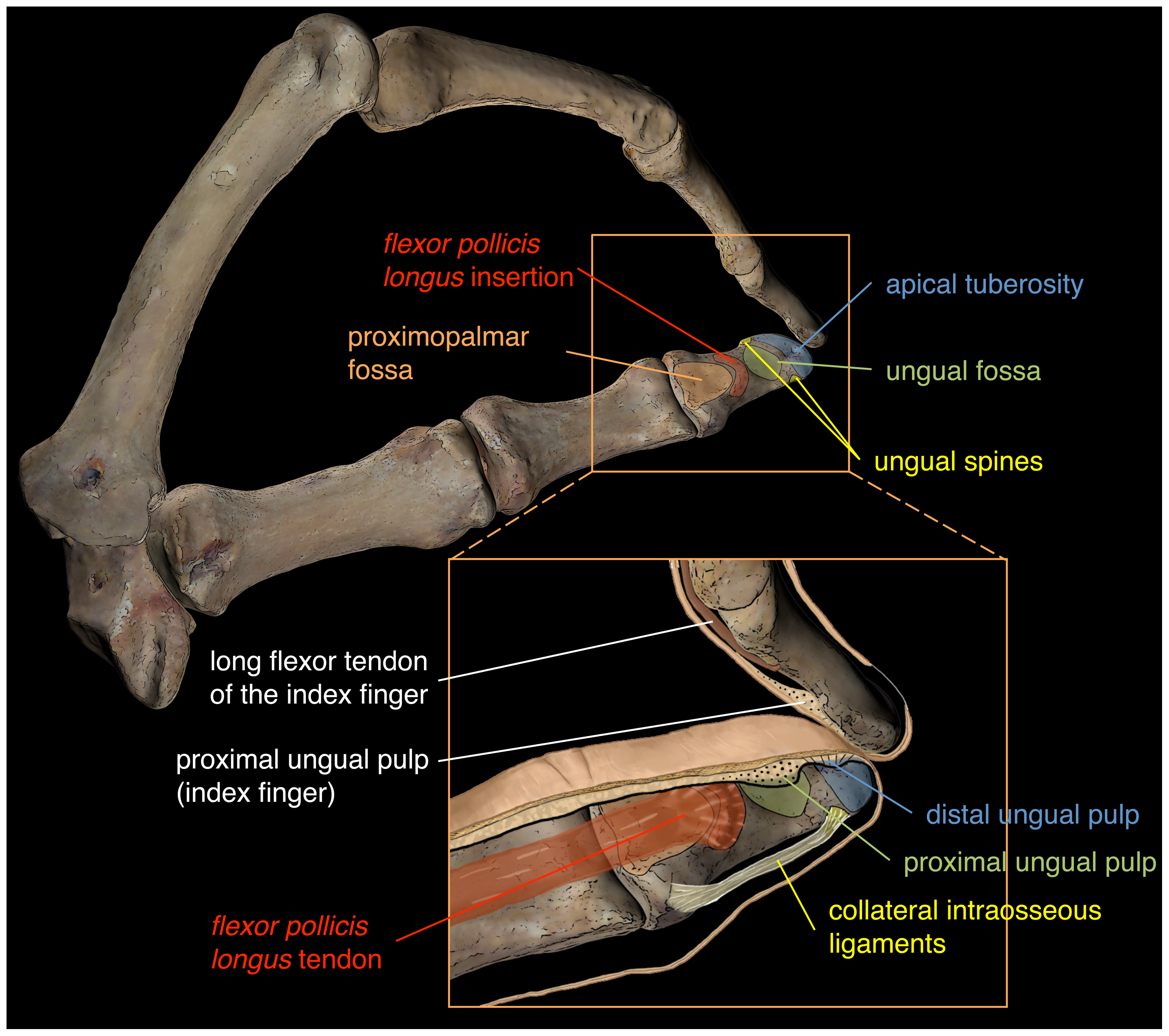
The flexor digitorum profundus is a muscle in the forearm of humans that flexes the fingers (also known as digits). It is considered an extrinsic hand muscle because it acts on the hand while its muscle belly is located in the forearm. Together the flexor pollicis longus, pronator quadratus, and flexor digitorum profundus form the deep layer of ventral forearm muscles.Platzer 2004, p 162 The muscle is named . Structure Flexor digitorum profundus originates in the upper 3/4 of the anterior and medial surfaces of the ulna, interosseous membrane and deep fascia of the forearm. The muscle fans out into four tendons (one to each of the second to fifth fingers) to the palmar base of the distal phalanx. Along with the flexor digitorum superficialis, it has long tendons that run down the arm and through the carpal tunnel and attach to the palmar side of the phalanges of the fingers. Flexor digitorum profundus lies deep to the superficialis, but it attaches more distally. Therefore, ... [...More Info...] [...Related Items...] OR: [Wikipedia] [Google] [Baidu] |
Ulna
The ulna (''pl''. ulnae or ulnas) is a long bone found in the forearm that stretches from the elbow to the smallest finger, and when in anatomical position, is found on the medial side of the forearm. That is, the ulna is on the same side of the forearm as the little finger. It runs parallel to the radius, the other long bone in the forearm. The ulna is usually slightly longer than the radius, but the radius is thicker. Therefore, the radius is considered to be the larger of the two. Structure The ulna is a long bone found in the forearm that stretches from the elbow to the smallest finger, and when in anatomical position, is found on the medial side of the forearm. It is broader close to the elbow, and narrows as it approaches the wrist. Close to the elbow, the ulna has a bony process, the olecranon process, a hook-like structure that fits into the olecranon fossa of the humerus. This prevents hyperextension and forms a hinge joint with the trochlea of the humerus. There is ... [...More Info...] [...Related Items...] OR: [Wikipedia] [Google] [Baidu] |
Interosseous Membrane
An interosseous membrane is a thick dense fibrous sheet of connective tissue that spans the space between two bones, forming a type of syndesmosis joint. Interosseous membranes in the human body: * Interosseous membrane of forearm * Interosseous membrane of leg The interosseous membrane of the leg (middle tibiofibular ligament) extends between the interosseous crests of the tibia and fibula, helps stabilize the Tib-Fib relationship and separates the muscles on the front from those on the back of the leg. ... Gallery File:5 ligaments of interosseous membrane of forearm.png, Five ligaments of interosseous membrane of forearm:* Central band (key portion to be reconstructed in case of injury)* Accessory band * Distal oblique bundle * Proximal oblique cord* Dorsal oblique accessory cord Notes External links * * {{Authority control Skeletal system ... [...More Info...] [...Related Items...] OR: [Wikipedia] [Google] [Baidu] |
Wrist
In human anatomy, the wrist is variously defined as (1) the Carpal bones, carpus or carpal bones, the complex of eight bones forming the proximal skeletal segment of the hand; "The wrist contains eight bones, roughly aligned in two rows, known as the carpal bones." (2) the wrist joint or radiocarpal joint, the joint between the radius (bone), radius and the Carpal bones, carpus and; (3) the anatomical region surrounding the carpus including the distal parts of the bones of the forearm and the proximal parts of the metacarpus or five metacarpal bones and the series of joints between these bones, thus referred to as ''wrist joints''. "With the large number of bones composing the wrist (ulna, radius, eight carpas, and five metacarpals), it makes sense that there are many, many joints that make up the structure known as the wrist." This region also includes the carpal tunnel, the anatomical snuff box, bracelet lines, the Flexor retinaculum of the hand, flexor retinaculum, and the ex ... [...More Info...] [...Related Items...] OR: [Wikipedia] [Google] [Baidu] |
Flexor Digitorum Superficialis Muscle
Flexor digitorum superficialis (''flexor digitorum sublimis'') is an extrinsic flexor muscle of the fingers at the proximal interphalangeal joints. It is in the anterior compartment of the forearm. It is sometimes considered to be the deepest part of the superficial layer of this compartment, and sometimes considered to be a distinct, "intermediate layer" of this compartment. It is relatively common for the Flexor digitorum superficialis to be missing from the little finger, bilaterally and unilaterally, which can cause problems when diagnosing a little finger injury. Structure The muscle has two classically described heads – the humeroulnar and radial – and it is between these heads that the median nerve and ulnar artery pass. The ulnar collateral ligament of elbow joint gives its origin to part of this muscle. Four long tendons come off this muscle near the wrist and travel through the carpal tunnel formed by the flexor retinaculum. These tendons, along with those of flex ... [...More Info...] [...Related Items...] OR: [Wikipedia] [Google] [Baidu] |
Flexor Carpi Ulnaris
The flexor carpi ulnaris (FCU) is a muscle of the forearm that flexes and adducts at the wrist joint. Structure Origin The flexor carpi ulnaris has two heads; a humeral head and ulnar head. The humeral head originates from the medial epicondyle of the humerus via the common flexor tendon. The ulnar head originates from the medial margin of the olecranon of the ulnar and the upper two-thirds of the dorsal border of the ulnar by an aponeurosis. Between the two heads passes the ulnar nerve and ulnar artery. Insertion The flexor carpi ulnaris inserts onto the pisiform, hook of the hamate (via the pisohamate ligament) and the anterior surface of the base of the fifth metacarpal (via the pisometacarpal ligament). Action The flexor carpi ulnaris flexes and adducts at the wrist joint. Innervation The flexor carpi ulnaris is innervated by the ulnar nerve. The corresponding spinal nerves are C8 and T1. Tendon The tendon of flexor carpi ulnaris can be seen on the anterior surface of th ... [...More Info...] [...Related Items...] OR: [Wikipedia] [Google] [Baidu] |
Anterior Interosseous Branch
The anterior interosseous nerve (volar interosseous nerve) is a branch of the median nerve that supplies the deep muscles on the anterior of the forearm, except the ulnar (medial) half of the flexor digitorum profundus. Its nerve roots come from C8 and T1. It accompanies the anterior interosseous artery along the anterior of the interosseous membrane of the forearm, in the interval between the flexor pollicis longus and flexor digitorum profundus, supplying the whole of the former and (most commonly) the radial half of the latter, and ending below in the pronator quadratus and wrist joint. Note that the median nerve supplies all flexor muscles of the forearm except for the ulnar half of flexor digitorum profundus and the flexor carpi ulnaris, which is a superficial muscle of the forearm. Innervation The anterior interosseous nerve classically innervates 2.5 muscles: which are deep muscles of the forearm * flexor pollicis longus * pronator quadratus * the radial (lateral) half of ... [...More Info...] [...Related Items...] OR: [Wikipedia] [Google] [Baidu] |
Ulnar Nerve
In human anatomy, the ulnar nerve is a nerve that runs near the ulna bone. The ulnar collateral ligament of elbow joint is in relation with the ulnar nerve. The nerve is the largest in the human body unprotected by muscle or bone, so injury is common. This nerve is directly connected to the little finger, and the adjacent half of the ring finger, innervating the palmar aspect of these fingers, including both front and back of the tips, perhaps as far back as the fingernail beds. This nerve can cause an electric shock-like sensation by striking the medial epicondyle of the humerus posteriorly, or inferiorly with the elbow flexed. The ulnar nerve is trapped between the bone and the overlying skin at this point. This is commonly referred to as bumping one's "funny bone". This name is thought to be a pun, based on the sound resemblance between the name of the bone of the upper arm, the humerus, and the word "humorous". Alternatively, according to the Oxford English Dictionary, i ... [...More Info...] [...Related Items...] OR: [Wikipedia] [Google] [Baidu] |
Composite Muscle
Composite or hybrid muscles are those muscles which have more than one set of fibers but perform the same function and are usually supplied by different nerves for different set of fibers. Examples * Brachialis: Musculocutaneous nerve is motor and radial nerve is proprioceptive. * Adductor magnus : Its adductor part by posterior division of obturator nerve and hamstring part by tibial part of sciatic nerve. * Biceps femoris: Its long head is supplied by the tibial part of sciatic nerve, whereas the short head is supplied by the common peroneal nerve. This reflects the composite derivation from the flexor and extensor musculature. * Pectineus: Its anterior set of fibers are supplied by the femoral nerve, whereas posterior set of fibers are supplied by the obturator nerve. * Flexor digitorum profundus: Its radial half of is supplied by the median nerve and the ulnar half is supplied by the ulnar nerve. * Flexor policis brevis: Supplied by recurrent branch of the median nerve and the ... [...More Info...] [...Related Items...] OR: [Wikipedia] [Google] [Baidu] |
Lumbricals Of The Hand
The lumbricals are intrinsic muscles of the hand that flex the metacarpophalangeal joints, and extend the interphalangeal joints. p. 97 The lumbrical muscles of the foot also have a similar action, though they are of less clinical concern. Structure The lumbricals are four, small, worm-like muscles on each hand. These muscles are unusual in that they do not attach to bone. Instead, they attach proximally to the tendons of flexor digitorum profundus, and distally to the extensor expansions. The first and second lumbricals are unipennate, while the third and fourth lumbricals are bipennate. Nerve supply The first and second lumbricals (the most radial two) are innervated by the median nerve. The third and fourth lumbricals (most ulnar two) are innervated by the deep branch of ulnar nerve. This is the usual innervation of the lumbricals (occurring in 60% of individuals). However 1:3 (median:ulnar - 20% of individuals) and 3:1 (median:ulnar - 20% of individuals) also exist ... [...More Info...] [...Related Items...] OR: [Wikipedia] [Google] [Baidu] |
Distal Phalanx
The phalanges (singular: ''phalanx'' ) are digital bones in the hands and feet of most vertebrates. In primates, the thumbs and big toes have two phalanges while the other digits have three phalanges. The phalanges are classed as long bones. Structure The phalanges are the bones that make up the fingers of the hand and the toes of the foot. There are 56 phalanges in the human body, with fourteen on each hand and foot. Three phalanges are present on each finger and toe, with the exception of the thumb and large toe, which possess only two. The middle and far phalanges of the fifth toes are often fused together (symphalangism). The phalanges of the hand are commonly known as the finger bones. The phalanges of the foot differ from the hand in that they are often shorter and more compressed, especially in the proximal phalanges, those closest to the torso. A phalanx is named according to whether it is proximal, middle, or distal and its associated finger or toe. The proximal ... [...More Info...] [...Related Items...] OR: [Wikipedia] [Google] [Baidu] |
Phalanx Bones
The phalanges (singular: ''phalanx'' ) are digital bones in the hands and feet of most vertebrates. In primates, the thumbs and big toes have two phalanges while the other digits have three phalanges. The phalanges are classed as long bones. Structure The phalanges are the bones that make up the fingers of the hand and the toes of the foot. There are 56 phalanges in the human body, with fourteen on each hand and foot. Three phalanges are present on each finger and toe, with the exception of the thumb and large toe, which possess only two. The middle and far phalanges of the fifth toes are often fused together (symphalangism). The phalanges of the hand are commonly known as the finger bones. The phalanges of the foot differ from the hand in that they are often shorter and more compressed, especially in the proximal phalanges, those closest to the torso. A phalanx is named according to whether it is proximal, middle, or distal and its associated finger or toe. The proximal ... [...More Info...] [...Related Items...] OR: [Wikipedia] [Google] [Baidu] |
Carpal Tunnel
In the human body, the carpal tunnel or carpal canal is the passageway on the palmar side of the wrist that connects the forearm to the hand. The tunnel is bounded by the bones of the wrist and flexor retinaculum from connective tissue. Normally several tendons from the flexor group of forearm muscles and the median nerve pass through it. There are described cases of variable median artery occurrence. When any of the nine long flexor tendons passing through the narrow carpal canal swell or degenerate, the narrowing of the canal may result in the median nerve becoming entrapped or compressed, a common medical condition known as carpal tunnel syndrome (CTS). Structure The carpal bones that make up the wrist form an arch which is convex on the dorsal side of the hand and concave on the palmar side. The groove on the palmar side, the ''sulcus carpi'', is covered by the flexor retinaculum, a sheath of tough connective tissue, thus forming the carpal tunnel. On the side of the ... [...More Info...] [...Related Items...] OR: [Wikipedia] [Google] [Baidu] |


