|
Hyperkeratotic
Hyperkeratosis is thickening of the stratum corneum (the outermost layer of the epidermis, or skin), often associated with the presence of an abnormal quantity of keratin,Kumar, Vinay; Fausto, Nelso; Abbas, Abul (2004) ''Robbins & Cotran Pathologic Basis of Disease'' (7th ed.). Saunders. Page 1230. . and also usually accompanied by an increase in the granular layer. As the corneum layer normally varies greatly in thickness in different sites, some experience is needed to assess minor degrees of hyperkeratosis. It can be caused by vitamin A deficiency or chronic exposure to arsenic. Hyperkeratosis can also be caused by B-Raf inhibitor drugs such as Vemurafenib and Dabrafenib.Niezgoda, Anna; Niezgoda, Piotr; Czajkowski, Rafal (2015) ''Novel Approaches to Treatment of Advanced Melanoma: A Review of Targeted Therapy and Immunotherapy'' BioMed Research International It can be treated with urea-containing creams, which dissolve the intercellular matrix of the cells of the stratum corn ... [...More Info...] [...Related Items...] OR: [Wikipedia] [Google] [Baidu] |
Epidermolytic Hyperkeratosis
Epidermolytic ichthyosis (EI), also known as bullous epidermis ichthyosis (BEI), epidermolytic hyperkeratosis (EHK), bullous congenital ichthyosiform erythroderma (BCIE), bullous ichthyosiform erythrodermaFreedberg, et al. (2003). ''Fitzpatrick's Dermatology in General Medicine''. (6th ed.). McGraw-Hill. . or bullous congenital ichthyosiform erythroderma Brocq, is a rare and severe form of ichthyosis that affects around 1 in 300,000 people. It is caused by a genetic mutation, and thus cannot be completely cured without some form of gene therapy. While some research has been done into possible gene therapy treatments, the work hasn't yet been successfully developed to the stage where it can be routinely given to patients. The condition involves the clumping of keratin filaments.James, William; Berger, Timothy; Elston, Dirk (2005). ''Andrews' Diseases of the Skin: Clinical Dermatology''. (10th ed.). Saunders. . Presentation Epidermolytic hyperkeratosis is a skin disorder that is ... [...More Info...] [...Related Items...] OR: [Wikipedia] [Google] [Baidu] |
Keratoderma
Keratoderma is a hornlike skin condition. Classification The keratodermas are classified into the following subgroups:Freedberg, et al. (2003). ''Fitzpatrick's Dermatology in General Medicine''. (6th ed.). McGraw-Hill. . Congenital * Simple keratodermas ** Diffuse palmoplantar keratodermas *** Diffuse epidermolytic palmoplantar keratoderma *** Diffuse nonepidermolytic palmoplantar keratoderma *** mal de Meleda ** Focal palmoplantar keratoderma *** Striate palmoplantar keratoderma ** Punctate palmoplantar keratoderma *** Keratosis punctata palmaris et plantaris *** Spiny keratoderma *** Focal acral hyperkeratosis * Complex keratodermas ** Diffuse palmoplantar keratoderma *** Erythrokeratodermia variabilis *** Palmoplantar keratoderma of Sybert *** Olmsted syndrome *** Naegeli–Franceschetti–Jadassohn syndrome ** Focal palmoplantar keratoderma *** Papillon–Lefèvre syndrome *** Pachyonychia congenita type I *** Pachyonychia congenita type II *** Focal palmoplantar ... [...More Info...] [...Related Items...] OR: [Wikipedia] [Google] [Baidu] |
Focal Acral Hyperkeratosis
Palmoplantar keratodermas are a heterogeneous group of disorders characterized by abnormal thickening of the stratum corneum of the palms and soles. Autosomal recessive, dominant, X-linked, and acquired forms have all been described. Types Clinically, three distinct patterns of palmoplantar keratoderma may be identified: diffuse, focal, and punctate. Diffuse Diffuse palmoplantar keratoderma is a type of palmoplantar keratoderma that is characterized by an even, thick, symmetric hyperkeratosis over the whole of the palm and sole, usually evident at birth or in the first few months of life. Restated, diffuse palmoplantar keratoderma is an autosomal dominant disorder in which hyperkeratosis is confined to the palms and soles. The two major types can have a similar clinical appearance: *''Diffuse epidermolytic palmoplantar keratoderma'' (also known as "Palmoplantar keratoderma cum degeneratione granulosa Vörner," "Vörner's epidermolytic palmoplantar keratoderma", and "Vörn ... [...More Info...] [...Related Items...] OR: [Wikipedia] [Google] [Baidu] |
Urea-containing Cream
Urea, also known as carbamide-containing cream, is used as a medication and applied to the skin to treat dryness and itching such as may occur in psoriasis, dermatitis, or ichthyosis. It may also be used to soften nails. In adults side effects are generally few. It may occasionally cause skin irritation. Urea works in part by loosening dried skin. Preparations generally contain 5 to 50% urea. Urea containing creams have been used since the 1940s. It is on the World Health Organization's List of Essential Medicines. It is available over the counter. Medical uses Urea cream is indicated for debridement and promotion of normal healing of skin areas with hyperkeratosis, particularly where healing is inhibited by local skin infection, skin necrosis, fibrinous or itching debris or eschar. Specific condition with hyperkeratosis where urea cream is useful include: * Dry skin and rough skin * Dermatitis * Psoriasis * Ichthyosis * Eczema * Keratosis * Keratoderma * Corns * Calluses * ... [...More Info...] [...Related Items...] OR: [Wikipedia] [Google] [Baidu] |
Sebum
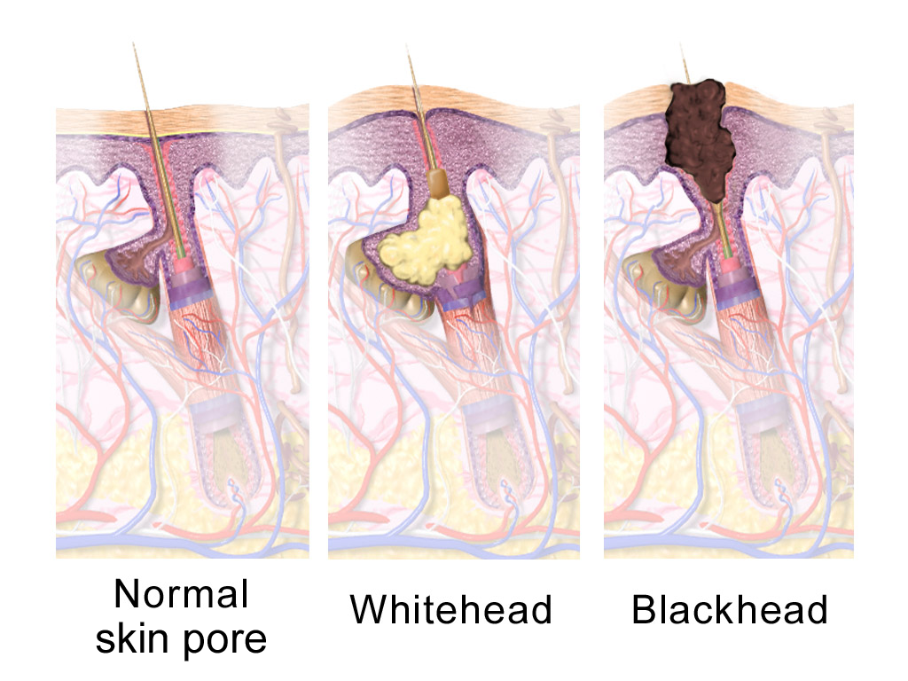
A sebaceous gland is a microscopic exocrine gland in the skin that opens into a hair follicle to secrete an oily or waxy matter, called sebum, which lubricates the hair and skin of mammals. In humans, sebaceous glands occur in the greatest number on the face and scalp, but also on all parts of the skin except the palms of the hands and soles of the feet. In the eyelids, meibomian glands, also called tarsal glands, are a type of sebaceous gland that secrete a special type of sebum into tears. Surrounding the female nipple, areolar glands are specialized sebaceous glands for lubricating the nipple. Fordyce spots are benign, visible, sebaceous glands found usually on the lips, gums and inner cheeks, and genitals. Structure Location Sebaceous glands are found throughout all areas of the skin, except the palms of the hands and soles of the feet. There are two types of sebaceous glands, those connected to hair follicles and those that exist independently. Sebaceous glands ... [...More Info...] [...Related Items...] OR: [Wikipedia] [Google] [Baidu] |
Hair Follicle
The hair follicle is an organ found in mammalian skin. It resides in the dermal layer of the skin and is made up of 20 different cell types, each with distinct functions. The hair follicle regulates hair growth via a complex interaction between hormones, neuropeptides, and immune cells. This complex interaction induces the hair follicle to produce different types of hair as seen on different parts of the body. For example, terminal hairs grow on the scalp and lanugo hairs are seen covering the bodies of fetuses in the uterus and in some newborn babies. The process of hair growth occurs in distinct sequential stages. The first stage is called ''anagen'' and is the active growth phase, ''telogen'' is the resting stage, ''catagen'' is the regression of the hair follicle phase, ''exogen'' is the active shedding of hair phase and lastly ''kenogen'' is the phase between the empty hair follicle and the growth of new hair. The function of hair in humans has long been a subject of interest ... [...More Info...] [...Related Items...] OR: [Wikipedia] [Google] [Baidu] |
Multiple Minute Digitate Hyperkeratosis
Multiple minute digitate hyperkeratosis (also known as "Digitate keratoses," "Disseminated spiked hyperkeratosis," "Familial disseminated piliform hyperkeratosis," and "Minute aggregate keratosis") is a rare cutaneous condition, with about half of cases being familial, inherited in an autosomal dominant fashion, while the other half are sporadic. This disease has a unique histology, so a biopsy and further tests should be done to confirm the diagnosis and rule out other disorders and malignancy. See also * Epidermis (skin), Epidermis * Skin lesion References Epidermal nevi, neoplasms, and cysts {{Epidermal-growth-stub ... [...More Info...] [...Related Items...] OR: [Wikipedia] [Google] [Baidu] |
Ichthyosis
Ichthyosis is a family of genetic skin disorders characterized by dry, thickened, scaly skin. The more than 20 types of ichthyosis range in severity of symptoms, outward appearance, underlying genetic cause and mode of inheritance (e.g., dominant, recessive, autosomal or X-linked). Ichthyosis comes from the Greek ἰχθύς ''ichthys'', literally "fish", since dry, scaly skin is the defining feature of all forms of ichthyosis. The severity of symptoms can vary enormously, from the mildest, most common, types such as ichthyosis vulgaris, which may be mistaken for normal dry skin, up to life-threatening conditions such as harlequin-type ichthyosis. Ichthyosis vulgaris accounts for more than 95% of cases. Types Many types of ichthyoses exist, and an exact diagnosis may be difficult. Types of ichthyoses are classified by their appearance, if they are syndromic or not, and by mode of inheritance. For example, non-syndromic ichthyoses that are inherited recessively come under the um ... [...More Info...] [...Related Items...] OR: [Wikipedia] [Google] [Baidu] |
Louis-Anne-Jean Brocq
Louis-Anne-Jean Brocq (1 February 1856 – 18 December 1928) was a French dermatologist born in Laroque-Timbaut, a village in the department of Lot-et-Garonne. He practiced medicine in Paris at the Hospice la Rochefoucauld, the Hôpital Broca, and from 1906 to 1921, the Hôpital Saint-Louis. As a young physician he studied and worked with Jean Alfred Fournier (1832–1915), Jean Baptiste Emile Vidal (1825–1893) and Ernest Henri Besnier (1831–1909). Brocq provided early, comprehensive descriptions of numerous skin disorders, including keratosis pilaris, parapsoriasis and a form of dermatitis called "Duhring-Brocq disease" (named with Louis Adolphus Duhring and sometimes referred to as dermatitis herpetiformis). Other eponymous skin diseases named after him are " Brocq's pseudopelade", a condition involving progressive scarring of the scalp, and "Brocq-Pautrier angiolupoid", a specific type of sarcoidosis of the skin named in conjunction with Dr. Lucien-Marie Pautrier (1876 ... [...More Info...] [...Related Items...] OR: [Wikipedia] [Google] [Baidu] |
Foot
The foot ( : feet) is an anatomical structure found in many vertebrates. It is the terminal portion of a limb which bears weight and allows locomotion. In many animals with feet, the foot is a separate organ at the terminal part of the leg made up of one or more segments or bones, generally including claws or nails. Etymology The word "foot", in the sense of meaning the "terminal part of the leg of a vertebrate animal" comes from "Old English fot "foot," from Proto-Germanic *fot (source also of Old Frisian fot, Old Saxon fot, Old Norse fotr, Danish fod, Swedish fot, Dutch voet, Old High German fuoz, German Fuß, Gothic fotus "foot"), from PIE root *ped- "foot". The "plural form feet is an instance of i-mutation." Structure The human foot is a strong and complex mechanical structure containing 26 bones, 33 joints (20 of which are actively articulated), and more than a hundred muscles, tendons, and ligaments.Podiatry Channel, ''Anatomy of the foot and ankle'' The joints of the ... [...More Info...] [...Related Items...] OR: [Wikipedia] [Google] [Baidu] |
Sole (foot)
The sole is the bottom of the foot. In humans the sole of the foot is anatomically referred to as the plantar aspect. Structure The glabrous skin on the sole of the foot lacks the hair and pigmentation found elsewhere on the body, and it has a high concentration of sweat pores. The sole contains the thickest layers of skin on the body due to the weight that is continually placed on it. It is crossed by a set of creases that form during the early stages of embryonic development. Like those of the palm, the sweat pores of the sole lack sebaceous glands. The sole is a sensory organ by which we can perceive the ground while standing and walking. The subcutaneous tissue in the sole has adapted to deal with the high local compressive forces on the heel and the ball (between the toes and the arch) by developing a system of "pressure chambers." Each chamber is composed of internal fibrofatty tissue covered by external collagen connective tissue. The septa (internal walls) ... [...More Info...] [...Related Items...] OR: [Wikipedia] [Google] [Baidu] |
B Vitamins
B vitamins are a class of water-soluble vitamins that play important roles in cell metabolism and synthesis of red blood cells. Though these vitamins share similar names (B1, B2, B3, etc.), they are chemically distinct compounds that often coexist in the same foods. In general, dietary supplements containing all eight are referred to as a vitamin B complex. Individual B vitamin supplements are referred to by the specific number or name of each vitamin, such as B1 for thiamine, B2 for riboflavin, and B3 for niacin. Some are more commonly recognized by name than by number, for example pantothenic acid, biotin, and folate. Each B vitamin is either a cofactor (generally a coenzyme) for key metabolic processes or is a precursor needed to make one and is thus an essential nutrient. List of B vitamins Note: other substances once thought to be vitamins were given numbers in the B-vitamin numbering scheme, but were subsequently discovered to be either not essential for life or manufact ... [...More Info...] [...Related Items...] OR: [Wikipedia] [Google] [Baidu] |


