|
Hyalectan
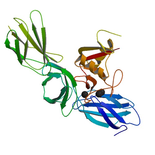
Lecticans, also known as hyalectans, are a family of proteoglycans (a type protein that is attached to chains of negatively charged polysaccharides) that are components of the extracellular matrix. There are four members of the lectican family: aggrecan, brevican, neurocan, and versican. Lecticans interact with hyaluronic acid and tenascin-R to form a ternary complex. Tissue distribution Aggrecan is a major component of extracellular matrix in cartilage whereas versican is widely expressed in a number of connective tissues including those in vascular smooth muscle, skin epithelial cells, and the cells of central and peripheral nervous system. The expression of neurocan and brevican is largely restricted to neural tissues. Structure All four lecticans contain an N-terminal globular domain (G1 domain) that in turn contains an immunoglobulin V-set domain and a Link domain that binds hyaluronic acid; a long extended central domain (CS) that is modified with covalently attached s ... [...More Info...] [...Related Items...] OR: [Wikipedia] [Google] [Baidu] |
Proteoglycan
Proteoglycans are proteins that are heavily glycosylated. The basic proteoglycan unit consists of a "core protein" with one or more covalently attached glycosaminoglycan (GAG) chain(s). The point of attachment is a serine (Ser) residue to which the glycosaminoglycan is joined through a tetrasaccharide bridge (e.g. chondroitin sulfate- GlcA- Gal-Gal- Xyl-PROTEIN). The Ser residue is generally in the sequence -Ser-Gly-X-Gly- (where X can be any amino acid residue but proline), although not every protein with this sequence has an attached glycosaminoglycan. The chains are long, linear carbohydrate polymers that are negatively charged under physiological conditions due to the occurrence of sulfate and uronic acid groups. Proteoglycans occur in connective tissue. Types Proteoglycans are categorized by their relative size (large and small) and the nature of their glycosaminoglycan chains. Types include: Certain members are considered members of the "small leucine-rich proteoglyc ... [...More Info...] [...Related Items...] OR: [Wikipedia] [Google] [Baidu] |
Epithelial
Epithelium or epithelial tissue is one of the four basic types of animal tissue, along with connective tissue, muscle tissue and nervous tissue. It is a thin, continuous, protective layer of compactly packed cells with a little intercellular matrix. Epithelial tissues line the outer surfaces of organs and blood vessels throughout the body, as well as the inner surfaces of cavities in many internal organs. An example is the epidermis, the outermost layer of the skin. There are three principal shapes of epithelial cell: squamous (scaly), columnar, and cuboidal. These can be arranged in a singular layer of cells as simple epithelium, either squamous, columnar, or cuboidal, or in layers of two or more cells deep as stratified (layered), or ''compound'', either squamous, columnar or cuboidal. In some tissues, a layer of columnar cells may appear to be stratified due to the placement of the nuclei. This sort of tissue is called pseudostratified. All glands are made up of epitheli ... [...More Info...] [...Related Items...] OR: [Wikipedia] [Google] [Baidu] |
Hyaladherin
Hyaladherins, also known as hyaluronan-binding proteins, are proteins capable of binding to hyaluronic acid. Most hyaladherins belong to the Link module superfamily, including its main receptor CD44, hyalectans and TSG-6. In addition there is a diverse group of hyaladherins lacking a Link module; these include the receptor RHAMM, C1QBP (HABP1) and HABP2. The primary roles of hyaladherins are cell adhesion, structural support of the extracellular matrix (ECM) and cell signalling. Due to the role of aberrant hyaluronic acid synthesis and degradation in various cancers, hyaladherins, as well as hyaluronic acid, are considered a promising target for cancer therapy. See also *Hyaluronan synthase *Hyaluronidase Hyaluronidases are a family of enzymes that catalyse the degradation of hyaluronic acid (HA). Karl Meyer classified these enzymes in 1971, into three distinct groups, a scheme based on the enzyme reaction products. The three main types of hyal ... References {{reflist ... [...More Info...] [...Related Items...] OR: [Wikipedia] [Google] [Baidu] |
C-type Lectin
A C-type lectin (CLEC) is a type of carbohydrate-binding protein known as a lectin. The C-type designation is from their requirement for calcium for binding. Proteins that contain C-type lectin domains have a diverse range of functions including cell-cell adhesion, immune response to pathogens and apoptosis. Classification Drickamer ''et al.'' classified C-type lectins into 7 subgroups (I to VII) based on the order of the various protein domains in each protein. This classification was subsequently updated in 2002, leading to seven additional groups (VIII to XIV). Most recently, three further subgroups were added (XV to XVII). CLECs include: * CLEC1A, CLEC1B * CLEC2A, CLEC2B, CD69 (CLEC2C), CLEC2D, CLEC2L * CLEC3A, CLEC3B * CLEC4A, CLEC4C, CLEC4D, CLEC4E, CLEC4F, CLEC4G, ASGR1 (CLEC4H1), ASGR2 (CLEC4H2), FCER2 (CLEC4J), CD207 (CLEC4K), CD209 (CLEC4L), CLEC4M * CLEC5A * CLEC6A * CLEC7A * OLR1 (CLEC8A) * CLEC9A * CLEC10A * CLEC11A * CLEC12A, CLEC12B * CD302 (C ... [...More Info...] [...Related Items...] OR: [Wikipedia] [Google] [Baidu] |
EGF-like Domain
The EGF-like domain is an evolutionary conserved protein domain, which derives its name from the epidermal growth factor where it was first described. It comprises about 30 to 40 amino-acid residues and has been found in a large number of mostly animal proteins. Most occurrences of the EGF-like domain are found in the extracellular domain of membrane-bound proteins or in proteins known to be secreted. An exception to this is the prostaglandin-endoperoxide synthase. The EGF-like domain includes 6 cysteine residues which in the epidermal growth factor have been shown to form 3 disulfide bonds. The structures of 4-disulfide EGF-domains have been solved from the laminin and integrin proteins. The main structure of EGF-like domains is a two-stranded β-sheet followed by a loop to a short C-terminal, two-stranded β-sheet. These two β-sheets are usually denoted as the major (N-terminal) and minor (C-terminal) sheets. EGF-like domains frequently occur in numerous tandem copies in pro ... [...More Info...] [...Related Items...] OR: [Wikipedia] [Google] [Baidu] |
C-terminal
The C-terminus (also known as the carboxyl-terminus, carboxy-terminus, C-terminal tail, C-terminal end, or COOH-terminus) is the end of an amino acid chain (protein or polypeptide), terminated by a free carboxyl group (-COOH). When the protein is translated from messenger RNA, it is created from N-terminus to C-terminus. The convention for writing peptide sequences is to put the C-terminal end on the right and write the sequence from N- to C-terminus. Chemistry Each amino acid has a carboxyl group and an amine group. Amino acids link to one another to form a chain by a dehydration reaction which joins the amine group of one amino acid to the carboxyl group of the next. Thus polypeptide chains have an end with an unbound carboxyl group, the C-terminus, and an end with an unbound amine group, the N-terminus. Proteins are naturally synthesized starting from the N-terminus and ending at the C-terminus. Function C-terminal retention signals While the N-terminus of a protein often cont ... [...More Info...] [...Related Items...] OR: [Wikipedia] [Google] [Baidu] |
Glycosaminoglycan
Glycosaminoglycans (GAGs) or mucopolysaccharides are long, linear polysaccharides consisting of repeating disaccharide units (i.e. two-sugar units). The repeating two-sugar unit consists of a uronic sugar and an amino sugar, except in the case of the sulfated glycosaminoglycan keratan, where, in place of the uronic sugar there is a galactose unit. GAGs are found in vertebrates, invertebrates and bacteria. Because GAGs are highly polar molecules and attract water; the body uses them as lubricants or shock absorbers. Mucopolysaccharidoses are a group of metabolic disorders in which abnormal accumulations of glycosaminoglycans occur due to enzyme deficiencies. Production Glycosaminoglycans vary greatly in molecular mass, disaccharide structure, and sulfation. This is because GAG synthesis is not template driven, as are proteins or nucleic acids, but constantly altered by processing enzymes. GAGs are classified into four groups, based on their core disaccharide structures. Hepa ... [...More Info...] [...Related Items...] OR: [Wikipedia] [Google] [Baidu] |
Link Domain
A Link domain or Link module, also known as Xlink domain (X for extracellular), is a protein domain that binds to hyaluronic acid. It is important in blood cell migration and apoptosis. The link domain is found in some extracellular proteins in vertebrates such as the hyalectans. It appears to be involved in extracellular matrix assembly and stability, cell adhesion, and migration. Structure The structure has been shown to consist of two alpha helices and two antiparallel beta sheets arranged around a large hydrophobic core similar to that of C-type lectin. This domain contains four conserved cysteines involved in two disulphide bonds. The link domain has also been termed HABM (hyaluronic acid binding module) and PTR (proteoglycan tandem repeat). Link domain proteins Proteins which contain the link domain include: * the hyalectans (a family of proteoglycans): aggrecan, brevican, neurocan and versican, which are expressed in the CNS; * the cartilage link protein (LP), a prot ... [...More Info...] [...Related Items...] OR: [Wikipedia] [Google] [Baidu] |
Immunoglobulin V-set Domain
V-set domains are Ig-like domains resembling the antibody variable domain. V-set domains are found in diverse protein families, including immunoglobulin light and heavy chains; in several T-cell receptors such as CD2 (Cluster of Differentiation 2), CD4, CD80, and CD86; in myelin membrane adhesion molecules; in junctional adhesion molecules (JAM); in tyrosine-protein kinase receptors; and in the programmed cell death protein 1 (PD1). Subfamilies * Immunoglobulin V-set, subgroup * T-cell surface antigen CD2 Human proteins containing this domain ACAM; ACAN; ADAMTSL1; AGC1; AMICA1; BCAM; BCAN; BGP; BGPc; BT3.3; BTN1A1; BTN2A1; BTN2A2; BTN2A3; BTN3A1; BTN3A2; BTN3A3; BTNL2; BTNL3; BTNL8; BTNL9; C10orf54; C1orf32; C9orf94; CADM1; CADM2; CADM3; CADM4; CD2; CD226; CD274; CD276; CD300A; CD300C; CD300D; CD300E; CD300LB; CD300LF; CD300LG; ... [...More Info...] [...Related Items...] OR: [Wikipedia] [Google] [Baidu] |
N-terminal
The N-terminus (also known as the amino-terminus, NH2-terminus, N-terminal end or amine-terminus) is the start of a protein or polypeptide, referring to the free amine group (-NH2) located at the end of a polypeptide. Within a peptide, the amine group is bonded to the carboxylic group of another amino acid, making it a chain. That leaves a free carboxylic group at one end of the peptide, called the C-terminus, and a free amine group on the other end called the N-terminus. By convention, peptide sequences are written N-terminus to C-terminus, left to right (in LTR writing systems). This correlates the translation direction to the text direction, because when a protein is translated from messenger RNA, it is created from the N-terminus to the C-terminus, as amino acids are added to the carboxyl end of the protein. Chemistry Each amino acid has an amine group and a carboxylic group. Amino acids link to one another by peptide bonds which form through a dehydration reaction that j ... [...More Info...] [...Related Items...] OR: [Wikipedia] [Google] [Baidu] |
Nervous System
In biology, the nervous system is the highly complex part of an animal that coordinates its actions and sensory information by transmitting signals to and from different parts of its body. The nervous system detects environmental changes that impact the body, then works in tandem with the endocrine system to respond to such events. Nervous tissue first arose in wormlike organisms about 550 to 600 million years ago. In vertebrates it consists of two main parts, the central nervous system (CNS) and the peripheral nervous system (PNS). The CNS consists of the brain and spinal cord. The PNS consists mainly of nerves, which are enclosed bundles of the long fibers or axons, that connect the CNS to every other part of the body. Nerves that transmit signals from the brain are called motor nerves or '' efferent'' nerves, while those nerves that transmit information from the body to the CNS are called sensory nerves or '' afferent''. Spinal nerves are mixed nerves that serve both fu ... [...More Info...] [...Related Items...] OR: [Wikipedia] [Google] [Baidu] |


